|
Vesical Arteries
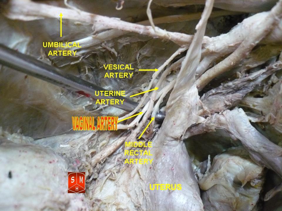
Vesical arteries are variable in number. They supply the bladder and terminal ureter. The two most prominent are the superior vesical artery and the inferior vesical artery. The superior vesical artery comes off of the internal iliac artery and sometimes the umbilical artery. The inferior vesical artery comes off of the internal iliac artery. The inferior vesical artery is a Pelvis, pelvic branch of the internal iliac artery in men; and in women it branches from the vaginal artery. This literature has been reviewed recently with observations of variation in pelvic vascularization and the close relationship between Vagina, vaginal and bladder vascularization in women. References Arteries of the abdomen {{circulatory-stub ... [...More Info...] [...Related Items...] OR: [Wikipedia] [Google] [Baidu] |
Bladder
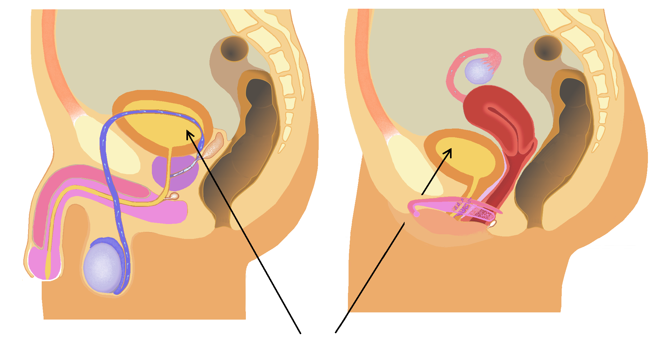
The bladder () is a hollow organ in humans and other vertebrates that stores urine from the kidneys. In placental mammals, urine enters the bladder via the ureters and exits via the urethra during urination. In humans, the bladder is a distensible organ that sits on the pelvic floor. The typical adult human bladder will hold between 300 and (10 and ) before the urge to empty occurs, but can hold considerably more. The Latin phrase for "urinary bladder" is ''vesica urinaria'', and the term ''vesical'' or prefix ''vesico-'' appear in connection with associated structures such as vesical veins. The modern Latin word for "bladder" – ''cystis'' – appears in associated terms such as cystitis (inflammation of the bladder). Structure In humans, the bladder is a hollow muscular organ situated at the base of the pelvis. In gross anatomy, the bladder can be divided into a broad (base), a body, an apex, and a neck. The apex (also called the vertex) is directed forward toward th ... [...More Info...] [...Related Items...] OR: [Wikipedia] [Google] [Baidu] |
Ureter
The ureters are tubes composed of smooth muscle that transport urine from the kidneys to the urinary bladder. In an adult human, the ureters typically measure 20 to 30 centimeters in length and about 3 to 4 millimeters in diameter. They are lined with urothelial cells, a form of transitional epithelium, and feature an extra layer of smooth muscle in the lower third to aid in peristalsis. The ureters can be affected by a number of diseases, including urinary tract infections and kidney stone. is when a ureter is narrowed, due to for example chronic inflammation. Congenital abnormalities that affect the ureters can include the development of two ureters on the same side or abnormally placed ureters. Additionally, reflux of urine from the bladder back up the ureters is a condition commonly seen in children. The ureters have been identified for at least two thousand years, with the word "ureter" stemming from the stem relating to urinating and seen in written records since at ... [...More Info...] [...Related Items...] OR: [Wikipedia] [Google] [Baidu] |
Superior Vesical Artery
The superior vesical artery supplies numerous branches to the upper part of the bladder. This artery often also gives branches to the vas deferens and can provide minor collateral circulation for the testicles. Structure The superior vesical artery, a vital component of the pelvic vascular system, stems from the umbilical artery, which serves as a conduit for oxygenated blood during fetal development. Emerging typically as a single vessel, the superior vesical artery exhibits consistent anatomical features across individuals. Its trajectory leads it towards the superior aspect of the bladder, where it branches intricately to supply blood to this organ and surrounding structures. In males, a noteworthy branch arising from the superior vesical artery is the vesiculo-prostatic artery. This branch extends its reach to the prostate gland, contributing to its vascularization. The consistent presence of this arterial branch underscores its significance in male pelvic anatomy and physi ... [...More Info...] [...Related Items...] OR: [Wikipedia] [Google] [Baidu] |
Inferior Vesical Artery
The inferior vesical artery (or inferior vesical artery) is an artery of the pelvis which arises from the internal iliac artery and supplies parts of the urinary bladder as well as other structures of the urinary system and structures of the male reproductive system. Some sources consider this vessel to be present only in males, and cite the vaginal artery as the homologous structure in females; others consider it to be present in both sexes, with the vessel taking the form of a small branch of a vaginal artery in females. Structure Origin The inferior vesical artery is a branch of the anterior division of the internal iliac artery. It frequently has a common origin with the middle rectal artery. Course The inferior vesical artery passes medially across the pelvic floor. Distribution The inferior vesical artery is distributed to the trigone and inferior portion of the urinary bladder, the ureter, prostate, vas deferens, and seminal vesicles.vas deferens The vas ... [...More Info...] [...Related Items...] OR: [Wikipedia] [Google] [Baidu] |
Internal Iliac Artery
The internal iliac artery (formerly known as the hypogastric artery) is the main artery of the pelvis. Structure The internal iliac artery supplies the walls and viscera of the pelvis, the buttock, the reproductive organs, and the medial compartment of the thigh. The vesicular branches of the internal iliac arteries supply the bladder. It is a short, thick vessel, smaller than the external iliac artery, and about 3 to 4 cm in length. Course The internal iliac artery arises at the bifurcation of the common iliac artery, opposite the lumbosacral articulation, and, passing downward to the upper margin of the greater sciatic foramen, divides into two large trunks, an anterior and a posterior. It is posterior to the ureter, anterior to the internal iliac vein, anterior to the lumbosacral trunk, and anterior to the piriformis muscle. Near its origin, it is medial to the external iliac vein, which lies between it and the psoas major muscle. It is above the obturator ... [...More Info...] [...Related Items...] OR: [Wikipedia] [Google] [Baidu] |
Umbilical Artery
The umbilical artery is a paired artery (with one for each half of the body) that is found in the abdominal and pelvic regions. In the fetus, it extends into the umbilical cord. Structure Development The umbilical arteries supply systemic arterial blood from the fetus to the placenta. Although this blood is sometimes referred to as deoxygenated blood it is not, and has the same oxygen saturation and nutrients as blood distributed to the other fetal tissues. There are usually two umbilical arteries present together with one umbilical vein in the umbilical cord. The umbilical arteries surround the urinary bladder and then carry all the deoxygenated blood out of the fetus through the umbilical cord. Inside the placenta, the umbilical arteries connect with each other at a distance of approximately 5 mm from the cord insertion in what is called the ''Hyrtl anastomosis''. Subsequently, they branch into chorionic arteries or ''intraplacental fetal arteries''. The umbilical arter ... [...More Info...] [...Related Items...] OR: [Wikipedia] [Google] [Baidu] |
Pelvis
The pelvis (: pelves or pelvises) is the lower part of an Anatomy, anatomical Trunk (anatomy), trunk, between the human abdomen, abdomen and the thighs (sometimes also called pelvic region), together with its embedded skeleton (sometimes also called bony pelvis or pelvic skeleton). The pelvic region of the trunk includes the bony pelvis, the pelvic cavity (the space enclosed by the bony pelvis), the pelvic floor, below the pelvic cavity, and the perineum, below the pelvic floor. The pelvic skeleton is formed in the area of the back, by the sacrum and the coccyx and anteriorly and to the left and right sides, by a pair of hip bones. The two hip bones connect the spine with the lower limbs. They are attached to the sacrum posteriorly, connected to each other anteriorly, and joined with the two femurs at the hip joints. The gap enclosed by the bony pelvis, called the pelvic cavity, is the section of the body underneath the abdomen and mainly consists of the reproductive organs and ... [...More Info...] [...Related Items...] OR: [Wikipedia] [Google] [Baidu] |
Vaginal Artery
The vaginal artery is an artery in females that supplies blood to the vagina and the base of the bladder. Structure The vaginal artery is usually a branch of the internal iliac artery. Some sources say that the vaginal artery can arise from the uterine artery, but the phrase vaginal branches of uterine artery is the term for blood supply to the vagina coming from the uterine artery. The vaginal artery is frequently represented by two or three branches. These descend to the vagina, supplying its mucous membrane. They anastomose with branches from the uterine artery. It can send branches to the bulb of the vestibule, the fundus of the bladder, and the contiguous part of the rectum. Function The vaginal artery supplies oxygenated blood to the muscular wall of the vagina, along with the uterine artery and the internal pudendal artery. It also supplies the cervix, along with the uterine artery. Other animals In horses, the vaginal artery may haemorrhage after birth, wh ... [...More Info...] [...Related Items...] OR: [Wikipedia] [Google] [Baidu] |
Vascularization
Vascularisation is the physiological process through which blood vessels form in tissues or organs. Vascularisation is crucial to supply the organs and tissues with an adequate supply of oxygen and nutrients and for removing waste products. Blood vessels transport blood, water, and nutrients needed to support body systems. When blood vessels lose efficiency, it may lead to serious diseases such as cancer, heart disease, and diabetes. Scientists are currently working on ways to grow new blood vessels to help with tissue engineering and healing injuries. This is why vascularisation is important in medicine. Mechanisms These are processes in which vascularisation happens and should not be confused with vascularisation itself: Angiogenesis It is the process where new blood vessels form from pre-existing ones. This happens naturally when the body needs to repair tissue or when a wound needs to heal. It is driven by signals from growth factors, such as Vascular Endothelial Growth ... [...More Info...] [...Related Items...] OR: [Wikipedia] [Google] [Baidu] |
Vagina
In mammals and other animals, the vagina (: vaginas or vaginae) is the elastic, muscular sex organ, reproductive organ of the female genital tract. In humans, it extends from the vulval vestibule to the cervix (neck of the uterus). The #Vaginal opening and hymen, vaginal introitus is normally partly covered by a thin layer of mucous membrane, mucosal tissue called the hymen. The vagina allows for Copulation (zoology), copulation and birth. It also channels Menstruation (mammal), menstrual flow, which occurs in humans and closely related primates as part of the menstrual cycle. To accommodate smoother penetration of the vagina during sexual intercourse or other sexual activity, vaginal moisture increases during sexual arousal in human females and other female mammals. This increase in moisture provides vaginal lubrication, which reduces friction. The texture of the vaginal walls creates friction for the penis during sexual intercourse and stimulates it toward ejaculation, en ... [...More Info...] [...Related Items...] OR: [Wikipedia] [Google] [Baidu] |


