|
Vertebral Veins
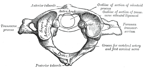
The vertebral vein is formed in the suboccipital triangle, from numerous small tributaries which spring from the internal vertebral venous plexuses and issue from the vertebral canal above the posterior arch of the atlas. They unite with small veins from the deep muscles at the upper part of the back of the neck, and form a vessel which enters the foramen in the transverse process of the atlas, and descends, forming a dense plexus around the vertebral artery, in the canal formed by the transverse foramina of the upper six cervical vertebrae. This plexus ends in a single trunk, which emerges from the transverse foramina of the sixth cervical vertebra, and opens at the root of the neck into the back part of the innominate vein near its origin, its mouth being guarded by a pair of valves. On the right side, it crosses the first part of the subclavian artery In human anatomy, the subclavian arteries are paired major arteries of the upper thorax, below the clavicle. They receive ... [...More Info...] [...Related Items...] OR: [Wikipedia] [Google] [Baidu] |
Deep Cervical Vein
The deep cervical vein (posterior vertebral vein or posterior deep cervical vein) is the vena comitans of the deep cervical artery. The vein is formed in the suboccipital region by the convergence of communicating branches of the occipital vein, veins draining the suboccipital muscles, and veins from the venous plexuses that surround cervical nerves. The vein and corresponding artery then pass in between the semispinalis capitis muscle and the semispinalis colli muscle. The vein passes anterior-ward in between the transverse process Each vertebra (: vertebrae) is an irregular bone with a complex structure composed of bone and some hyaline cartilage, that make up the vertebral column or spine, of vertebrates. The proportions of the vertebrae differ according to their spina ... of the 7th cervical vertebra and the neck of the first rib to terminate in the vertebral vein. References External links Veins of the head and neck {{circulatory-stub ... [...More Info...] [...Related Items...] OR: [Wikipedia] [Google] [Baidu] |
Brachiocephalic Vein
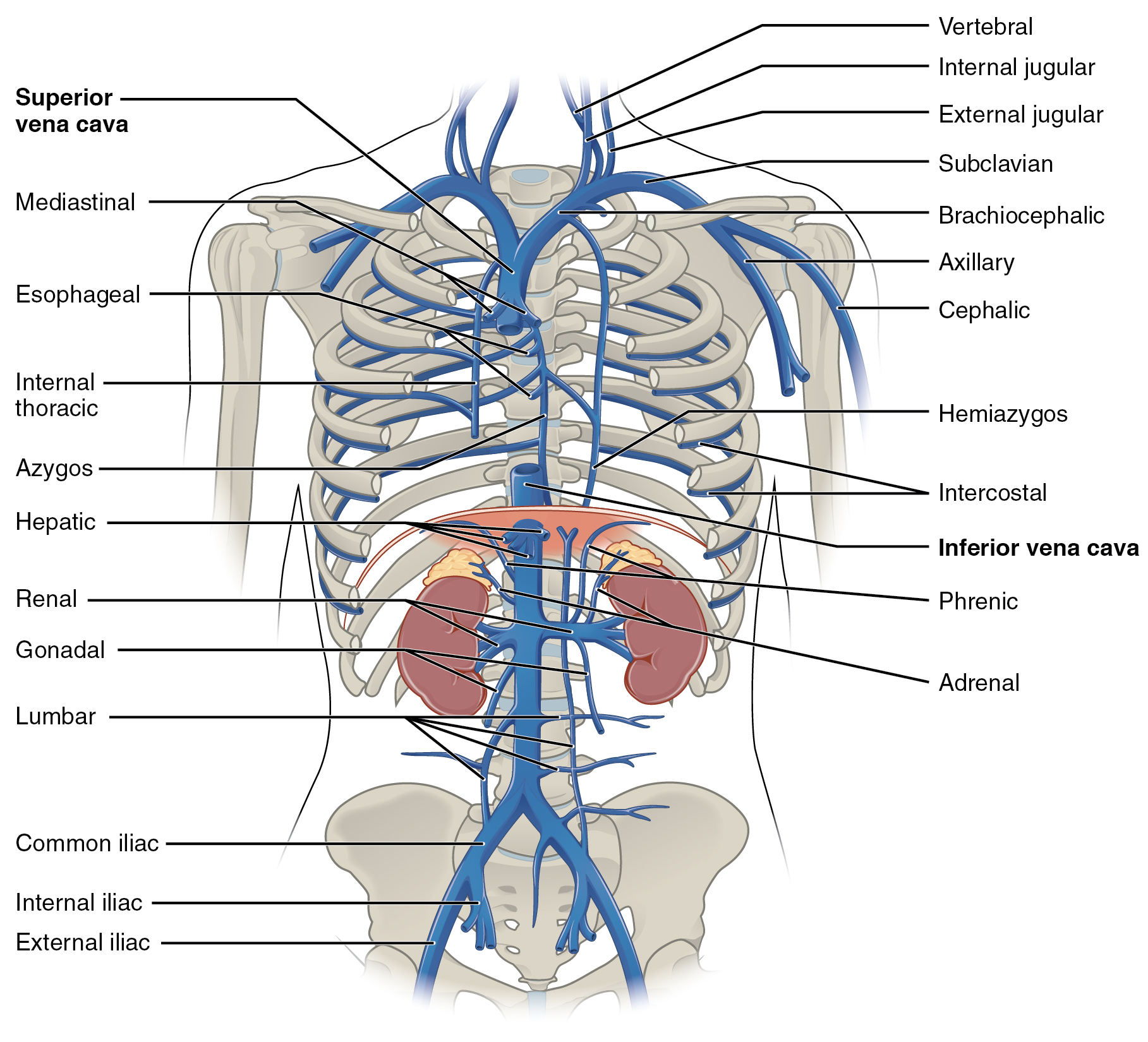
The left and right brachiocephalic veins (previously called innominate veins) are major veins in the Thorax, upper chest, formed by the union of the ipsilateral internal jugular vein and subclavian vein (the so-called venous angle) behind the sternoclavicular joint. The left brachiocephalic vein is more than twice the length of the right brachiocephalic vein. These veins merge to form the superior vena cava, a great vessel, posterior to the junction of the first costal cartilage with the Manubrium, manubrium of the sternum. The brachiocephalic veins are the major veins returning blood to the superior vena cava. Left and right veins Left brachiocephalic vein The left brachiocephalic vein is about 6cm, more than twice the length of the right brachiocephalic vein. and is formed by the confluence of the left subclavian vein, subclavian and left internal jugular veins. In addition the left vein receives drainage from the following tributaries: * The left vertebral vein, internal thor ... [...More Info...] [...Related Items...] OR: [Wikipedia] [Google] [Baidu] |
Vertebral Artery
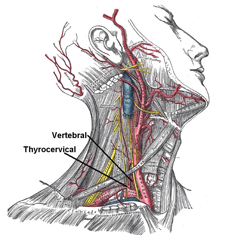
The vertebral arteries are major artery, arteries of the neck. Typically, the vertebral arteries originate from the subclavian arteries. Each vessel courses superiorly along each side of the neck, merging within the skull to form the single, midline basilar artery. As the supplying component of the ''vertebrobasilar vascular system'', the vertebral arteries supply blood to the upper spinal cord, brainstem, cerebellum, and Cerebral circulation#Posterior cerebral circulation, posterior part of brain. Structure The vertebral arteries usually arise from the posterosuperior aspect of the central subclavian arteries on each side of the body, then enter deep to the transverse process at the level of the 6th cervical vertebrae (C6), or occasionally (in 7.5% of cases) at the level of C7. They then proceed superiorly, in the transverse foramen of each cervical vertebra. Once they have passed through the transverse foramen of C1 (also known as the Atlas (anatomy), atlas), the vertebral ... [...More Info...] [...Related Items...] OR: [Wikipedia] [Google] [Baidu] |
Suboccipital Triangle
The suboccipital triangle is a region of the neck bounded by the following three muscles of the suboccipital group of muscles: * Rectus capitis posterior major - above and medially * Obliquus capitis superior - above and laterally * Obliquus capitis inferior - below and laterally (Rectus capitis posterior minor is also in this region but does not form part of the triangle) It is covered by a layer of dense fibro-fatty tissue, situated beneath the semispinalis capitis. The floor is formed by the posterior atlantooccipital membrane, and the posterior arch of the atlas. In the deep groove on the upper surface of the posterior arch of the atlas are the vertebral artery and the first cervical or suboccipital nerve. In the past, the vertebral artery was accessed here in order to conduct angiography of the circle of Willis. Presently, formal angiography of the circle of Willis is performed via catheter angiography, with access usually being acquired at the common femoral artery. Alte ... [...More Info...] [...Related Items...] OR: [Wikipedia] [Google] [Baidu] |
Internal Vertebral Venous Plexuses
The internal vertebral venous plexuses (intraspinal veins) lie within the vertebral canal in the epidural space, embedded within epidural fat. They receive tributaries from bones, red bone marrow, and spinal cord. They are arranged into four interconnected, vertically oriented vessels - two situated anteriorly, and two posteriorly: *The anterior internal vertebral venous plexus consists of two large plexiform veins situated upon the posterior surfaces of the vertebral bodies and intervertebral discs on either side of the posterior longitudinal ligament (underneath this ligament they are interconnected by transverse branches into which the basivertebral veins open). * The posterior internal vertebral venous plexus consists of two veins situated - one on either side - upon the anterior aspect of the vertebral arches and ligamenta flava. They form anastomoses with posterior external plexuses by way of veins passing through or between the ligamenta flava. The anterior and posterior i ... [...More Info...] [...Related Items...] OR: [Wikipedia] [Google] [Baidu] |
Vertebral Canal
In human anatomy, the spinal canal, vertebral canal or spinal cavity is an elongated body cavity enclosed within the dorsal bony arches of the vertebral column, which contains the spinal cord, spinal roots and dorsal root ganglia. It is a process of the dorsal body cavity formed by alignment of the vertebral foramina. Under the vertebral arches, the spinal canal is also covered anteriorly by the posterior longitudinal ligament and posteriorly by the ligamentum flavum. The potential space between these ligaments and the dura mater covering the spinal cord is known as the epidural space. Spinal nerves exit the spinal canal via the intervertebral foramina under the corresponding vertebral pedicles. In humans, the spinal cord gets outgrown by the vertebral column during development into adulthood, and the lower section of the spinal canal is occupied by the filum terminale and a bundle of spinal nerves known as the cauda equina instead of the actual spinal cord, which finis ... [...More Info...] [...Related Items...] OR: [Wikipedia] [Google] [Baidu] |
Atlas (anatomy)
In anatomy, the atlas (C1) is the most superior (first) cervical vertebra of the spine and is located in the neck. The bone is named for Atlas of Greek mythology, just as Atlas bore the weight of the heavens, the first cervical vertebra supports the head. However, the term atlas was first used by the ancient Romans for the seventh cervical vertebra (C7) due to its suitability for supporting burdens. In Greek mythology, Atlas was condemned to bear the weight of the heavens as punishment for rebelling against Zeus. Ancient depictions of Atlas show the globe of the heavens resting at the base of his neck, on C7. Sometime around 1522, anatomists decided to call the first cervical vertebra the atlas. Scholars believe that by switching the designation atlas from the seventh to the first cervical vertebra Renaissance anatomists were commenting that the point of man's burden had shifted from his shoulders to his head—that man's true burden was not a physical load, but rather, his m ... [...More Info...] [...Related Items...] OR: [Wikipedia] [Google] [Baidu] |
Neck
The neck is the part of the body in many vertebrates that connects the head to the torso. It supports the weight of the head and protects the nerves that transmit sensory and motor information between the brain and the rest of the body. Additionally, the neck is highly flexible, allowing the head to turn and move in all directions. Anatomically, the human neck is divided into four compartments: vertebral, visceral, and two vascular compartments. Within these compartments, the neck houses the cervical vertebrae, the cervical portion of the spinal cord, upper parts of the respiratory and digestive tracts, endocrine glands, nerves, arteries and veins. The muscles of the neck, which are separate from the compartments, form the boundaries of the neck triangles. In anatomy, the neck is also referred to as the or . However, when the term ''cervix'' is used alone, it often refers to the uterine cervix, the neck of the uterus. Therefore, the adjective ''cervical'' ... [...More Info...] [...Related Items...] OR: [Wikipedia] [Google] [Baidu] |
Vertebral Artery
The vertebral arteries are major artery, arteries of the neck. Typically, the vertebral arteries originate from the subclavian arteries. Each vessel courses superiorly along each side of the neck, merging within the skull to form the single, midline basilar artery. As the supplying component of the ''vertebrobasilar vascular system'', the vertebral arteries supply blood to the upper spinal cord, brainstem, cerebellum, and Cerebral circulation#Posterior cerebral circulation, posterior part of brain. Structure The vertebral arteries usually arise from the posterosuperior aspect of the central subclavian arteries on each side of the body, then enter deep to the transverse process at the level of the 6th cervical vertebrae (C6), or occasionally (in 7.5% of cases) at the level of C7. They then proceed superiorly, in the transverse foramen of each cervical vertebra. Once they have passed through the transverse foramen of C1 (also known as the Atlas (anatomy), atlas), the vertebral ... [...More Info...] [...Related Items...] OR: [Wikipedia] [Google] [Baidu] |
Cervical Vertebrae
In tetrapods, cervical vertebrae (: vertebra) are the vertebrae of the neck, immediately below the skull. Truncal vertebrae (divided into thoracic and lumbar vertebrae in mammals) lie caudal (toward the tail) of cervical vertebrae. In sauropsid species, the cervical vertebrae bear cervical ribs. In lizards and saurischian dinosaurs, the cervical ribs are large; in birds, they are small and completely fused to the vertebrae. The vertebral transverse processes of mammals are homologous to the cervical ribs of other amniotes. Most mammals have seven cervical vertebrae, with the only three known exceptions being the manatee with six, the two-toed sloth with five or six, and the three-toed sloth with nine. In humans, cervical vertebrae are the smallest of the true vertebrae and can be readily distinguished from those of the thoracic or lumbar regions by the presence of a transverse foramen, an opening in each transverse process, through which the vertebral artery, verteb ... [...More Info...] [...Related Items...] OR: [Wikipedia] [Google] [Baidu] |
Innominate Vein
The left and right brachiocephalic veins (previously called innominate veins) are major veins in the upper chest, formed by the union of the ipsilateral internal jugular vein and subclavian vein (the so-called venous angle) behind the sternoclavicular joint. The left brachiocephalic vein is more than twice the length of the right brachiocephalic vein. These veins merge to form the superior vena cava, a great vessel, posterior to the junction of the first costal cartilage with the manubrium of the sternum. The brachiocephalic veins are the major veins returning blood to the superior vena cava. Left and right veins Left brachiocephalic vein The left brachiocephalic vein is about 6cm, more than twice the length of the right brachiocephalic vein. and is formed by the confluence of the left subclavian and left internal jugular veins. In addition the left vein receives drainage from the following tributaries: * The left vertebral vein, internal thoracic vein, inferior thyroid vein ... [...More Info...] [...Related Items...] OR: [Wikipedia] [Google] [Baidu] |
Subclavian Artery
In human anatomy, the subclavian arteries are paired major arteries of the upper thorax, below the clavicle. They receive blood from the aortic arch. The left subclavian artery supplies blood to the left arm and the right subclavian artery supplies blood to the right arm, with some branches supplying the head and thorax. On the left side of the body, the subclavian comes directly off the aortic arch, while on the right side it arises from the relatively short brachiocephalic artery when it bifurcates into the subclavian and the right common carotid artery. The usual branches of the subclavian on both sides of the body are the vertebral artery, the internal thoracic artery, the thyrocervical trunk, the costocervical trunk and the dorsal scapular artery, which may branch off the transverse cervical artery, which is a branch of the thyrocervical trunk. The subclavian becomes the axillary artery at the lateral border of the first rib. Structure From its origin, the subclavian art ... [...More Info...] [...Related Items...] OR: [Wikipedia] [Google] [Baidu] |


