|
Venous Angle
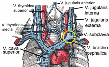
The venous angle (also known as Pirogoff's angle and in Latin as ''angulus venosus'') is the junction where the ipsilateral internal jugular vein and subclavian vein unite to form the ipsilateral brachiocephalic vein. The thoracic duct drains at the left venous angle, and the right lymphatic duct drains at the right venous angle. At the venous angle, the carotid sheath and axillary sheath intermingle, forming a continuous neurovascular ensheathment. The eponym is a reference to Nikolay Pirogov Nikolay Ivanovich Pirogov (Russian: Николай Иванович Пирогов; – ) was a Russian scientist, medical doctor, pedagogue, public figure, and corresponding member of the Russian Academy of Sciences (1847), one of the most wi .... References {{Reflist Cardiovascular system ... [...More Info...] [...Related Items...] OR: [Wikipedia] [Google] [Baidu] |
Ipsilateral
Standard anatomical terms of location are used to describe unambiguously the anatomy of humans and other animals. The terms, typically derived from Latin or Greek roots, describe something in its standard anatomical position. This position provides a definition of what is at the front ("anterior"), behind ("posterior") and so on. As part of defining and describing terms, the body is described through the use of anatomical planes and axes. The meaning of terms that are used can change depending on whether a vertebrate is a biped or a quadruped, due to the difference in the neuraxis, or if an invertebrate is a non-bilaterian. A non-bilaterian has no anterior or posterior surface for example but can still have a descriptor used such as proximal or distal in relation to a body part that is nearest to, or furthest from its middle. International organisations have determined vocabularies that are often used as standards for subdisciplines of anatomy. For example, '' Terminolog ... [...More Info...] [...Related Items...] OR: [Wikipedia] [Google] [Baidu] |
Internal Jugular Vein
The internal jugular vein is a paired jugular vein that collects blood from the brain and the superficial parts of the face and neck. This vein runs in the carotid sheath with the common carotid artery and vagus nerve. It begins in the posterior compartment of the jugular foramen, at the base of the skull. It is somewhat dilated at its origin, which is called the ''superior bulb''. This vein also has a common trunk into which drains the anterior branch of the retromandibular vein, the facial vein, and the lingual vein. It runs down the side of the neck in a vertical direction, being at one end lateral to the internal carotid artery, and then lateral to the common carotid artery, and at the root of the neck, it unites with the subclavian vein to form the brachiocephalic vein (innominate vein); a little above its termination is a second dilation, the ''inferior bulb''. Above, it lies upon the rectus capitis lateralis, behind the internal carotid artery and the nerves pa ... [...More Info...] [...Related Items...] OR: [Wikipedia] [Google] [Baidu] |
Subclavian Vein
The subclavian vein is a paired large vein, one on either side of the body, that is responsible for draining blood from the upper extremities, allowing this blood to return to the heart. The left subclavian vein plays a key role in the absorption of lipids, by allowing products that have been carried by lymph in the thoracic duct to enter the bloodstream. The diameter of the subclavian veins is approximately 1–2 cm, depending on the individual. Structure Each subclavian vein is a continuation of the axillary vein and runs from the outer border of the first rib to the medial border of anterior scalene muscle. From here it joins with the internal jugular vein to form the brachiocephalic vein (also known as "innominate vein"). The angle of union is termed the venous angle. The subclavian vein follows the subclavian artery and is separated from the subclavian artery by the insertion of anterior scalene. Thus, the subclavian vein lies anterior to the anterior scalene while ... [...More Info...] [...Related Items...] OR: [Wikipedia] [Google] [Baidu] |
Brachiocephalic Vein
The left and right brachiocephalic veins (previously called innominate veins) are major veins in the Thorax, upper chest, formed by the union of the ipsilateral internal jugular vein and subclavian vein (the so-called venous angle) behind the sternoclavicular joint. The left brachiocephalic vein is more than twice the length of the right brachiocephalic vein. These veins merge to form the superior vena cava, a great vessel, posterior to the junction of the first costal cartilage with the Manubrium, manubrium of the sternum. The brachiocephalic veins are the major veins returning blood to the superior vena cava. Left and right veins Left brachiocephalic vein The left brachiocephalic vein is about 6cm, more than twice the length of the right brachiocephalic vein. and is formed by the confluence of the left subclavian vein, subclavian and left internal jugular veins. In addition the left vein receives drainage from the following tributaries: * The left vertebral vein, internal thor ... [...More Info...] [...Related Items...] OR: [Wikipedia] [Google] [Baidu] |
Thoracic Duct
In human anatomy, the thoracic duct (also known as the ''left lymphatic duct'', ''alimentary duct'', ''chyliferous duct'', and ''Van Hoorne's canal'') is the larger of the two lymph ducts of the lymphatic system (the other being the right lymphatic duct). The thoracic duct usually begins from the upper aspect of the cisterna chyli, passing out of the abdomen through the aortic hiatus into first the posterior mediastinum and then the superior mediastinum, extending as high up as the root of the neck before descending to drain into the systemic (blood) circulation at the venous angle. The thoracic duct carries chyle, a liquid containing both lymph and emulsified fats, rather than pure lymph. It also collects most of the lymph in the body other than from the right thorax, arm, head, and neck (which are drained by the right lymphatic duct). When the duct ruptures, the resulting flood of liquid into the pleural cavity is known as chylothorax. Structure In adults, the thoraci ... [...More Info...] [...Related Items...] OR: [Wikipedia] [Google] [Baidu] |
Right Lymphatic Duct
The right lymphatic duct is an important lymphatic vessel that drains the right upper quadrant of the human body. It forms various combinations with the right subclavian vein and right internal jugular vein. Structure The right lymphatic duct courses along the medial border of the anterior scalene at the root of the neck. The right lymphatic duct forms various combinations with the right subclavian vein and right internal jugular vein. It is approximately 1.25 cm long. Variations A right lymphatic duct that enters directly into the junction of the internal jugular and subclavian veins is uncommon. Function The right duct drains lymph fluid from: * the upper right section of the trunk, (right thoracic cavity, via the right bronchomediastinal trunk) * the right arm (via the right subclavian trunk) * and right side of the head and neck (via the right jugular trunk) * also, in some individuals, the lower lobe of the left lung. All other sections of the human body are ... [...More Info...] [...Related Items...] OR: [Wikipedia] [Google] [Baidu] |
Carotid Sheath
The carotid sheath is a condensation of the deep cervical fascia enveloping multiple vital neurovascular structures of the neck, including the common and internal carotid arteries, the internal jugular vein, the vagus nerve (CN X), and ansa cervicalis. The carotid sheath helps protects the structures contained therein. Anatomy One carotid sheath is situated on each side of the neck, extending between the base of the skull superiorly and the thorax inferiorly. Superiorly, the carotid sheath encircles the margins of the carotid canal and jugular foramen. Inferiorly, it terminates at the arch of the aorta; it is continuous inferiorly with the axillary sheath at the venous angle. Its inferior end occurs at the level of the first rib and sternum inferiorly (varying between the levels of C7 and T4). Structure The carotid sheath is a fibrous connective tissue formation surrounding several important structures of the neck. It is thicker around the arteries than around the ... [...More Info...] [...Related Items...] OR: [Wikipedia] [Google] [Baidu] |
Axillary Sheath
The axillary sheath is a fibrous sheath that encloses the axillary artery and the three cords of the brachial plexus to form the neurovascular bundle. It is surrounded by the axillary fat.Last's Anatomuy, 9th Edt It is an extension of the prevertebral fascia of the deep cervical fascia and is continuous with the carotid sheath The carotid sheath is a condensation of the deep cervical fascia enveloping multiple vital neurovascular structures of the neck, including the common and internal carotid arteries, the internal jugular vein, the vagus nerve (CN X), and ansa c ... at the venous angle. A brachial plexus nerve block can be achieved by injecting anaesthetic into this area. References External links * Description at upstate.edu Arteries of the upper limb Fascia Axillas {{Portal bar, Anatomy ... [...More Info...] [...Related Items...] OR: [Wikipedia] [Google] [Baidu] |
Nikolay Pirogov
Nikolay Ivanovich Pirogov (Russian: Николай Иванович Пирогов; – ) was a Russian scientist, medical doctor, pedagogue, public figure, and corresponding member of the Russian Academy of Sciences (1847), one of the most widely recognized Russian physicians. Considered to be the founder of field surgery, he was the first surgeon to use anaesthesia in a field operation (1847) and one of the first surgeons in Europe to use diethyl ether, ether as an anaesthetic. He is credited with the invention of various kinds of surgical operations and developing his own technique of using orthopedic cast, plaster casts to treat fractured bones. Biography Childhood and training Nikolay Pirogov was born in Moscow, the 13th of 14 children of Ivan Ivanovich Pirogov (born around 1772), a major in the commissary service and a treasurer at the College of War, Moscow Food Depot whose own father came from peasants and served as a soldier in Peter the Great's army before retiring a ... [...More Info...] [...Related Items...] OR: [Wikipedia] [Google] [Baidu] |


