|
Vascular Bypass
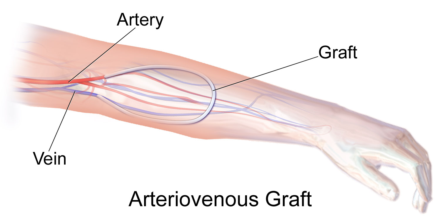
A vascular bypass is a surgical procedure performed to redirect blood flow from one area to another by reconnecting blood vessels. Often, this is done to bypass around a diseased artery, from an area of normal blood flow to another relatively normal area. It is commonly performed due to inadequate blood flow (ischemia) caused by atherosclerosis, as a part of organ transplantation, or for vascular access in hemodialysis. In general, someone's own vein (autograft) is the preferred graft material (or conduit) for a vascular bypass, but other types of grafts such as polytetrafluoroethylene (Teflon), polyethylene terephthalate (Dacron), or a different person's vein (allograft) are also commonly used. Arteries can also serve as vascular grafts. A surgeon sews the graft to the source and target vessels by hand using surgical suture, creating a surgical anastomosis. Common bypass sites include the heart (coronary artery bypass surgery) to treat coronary artery disease, and the legs ... [...More Info...] [...Related Items...] OR: [Wikipedia] [Google] [Baidu] |
Surgery
Surgery is a medical specialty that uses manual and instrumental techniques to diagnose or treat pathological conditions (e.g., trauma, disease, injury, malignancy), to alter bodily functions (e.g., malabsorption created by bariatric surgery such as gastric bypass), to reconstruct or alter aesthetics and appearance (cosmetic surgery), or to remove unwanted tissue (biology), tissues (body fat, glands, scars or skin tags) or foreign bodies. The act of performing surgery may be called a surgical procedure or surgical operation, or simply "surgery" or "operation". In this context, the verb "operate" means to perform surgery. The adjective surgical means pertaining to surgery; e.g. surgical instruments, operating theater, surgical facility or surgical nurse. Most surgical procedures are performed by a pair of operators: a surgeon who is the main operator performing the surgery, and a surgical assistant who provides in-procedure manual assistance during surgery. Modern surgical opera ... [...More Info...] [...Related Items...] OR: [Wikipedia] [Google] [Baidu] |
Peripheral Vascular Disease
Peripheral artery disease (PAD) is a vascular disorder that causes abnormal narrowing of arteries other than those that supply the heart or brain. PAD can happen in any blood vessel, but it is more common in the legs than the arms. When narrowing occurs in the heart, it is called coronary artery disease (CAD), and in the brain, it is called cerebrovascular disease. Peripheral artery disease most commonly affects the legs, but other arteries may also be involved, such as those of the arms, neck, or kidneys. Peripheral artery disease (PAD) is a form of peripheral vascular disease. Vascular refers to the arteries and veins within the body. PAD differs from peripheral veinous disease. PAD means the arteries are narrowed or blocked—the vessels that carry oxygen-rich blood as it moves from the heart to other parts of the body. Peripheral veinous disease, on the other hand, refers to problems with veins—the vessels that bring the blood back to the heart. The classic symptom is ... [...More Info...] [...Related Items...] OR: [Wikipedia] [Google] [Baidu] |
Anterior Tibial Artery
The anterior tibial artery is an artery of the leg. It carries blood to the anterior compartment of the leg and dorsal surface of the foot, from the popliteal artery. Structure Course The anterior tibial artery is a branch of the popliteal artery. It originates at the distal end of the popliteus muscle posterior to the tibia. The artery typically passes anterior to the popliteus muscle prior to passing between the tibia and fibula through an oval opening at the superior aspect of the interosseus membrane. The artery then descends between the tibialis anterior and extensor digitorum longus muscles. It is accompanied by the anterior tibial vein, and the deep peroneal nerve, along its course. It crosses the anterior aspect of the ankle joint, at which point it becomes the dorsalis pedis artery. Branches The branches of the anterior tibial artery are: * posterior tibial recurrent artery * anterior tibial recurrent artery * muscular branches * anterior medial malleolar ar ... [...More Info...] [...Related Items...] OR: [Wikipedia] [Google] [Baidu] |
EVAR
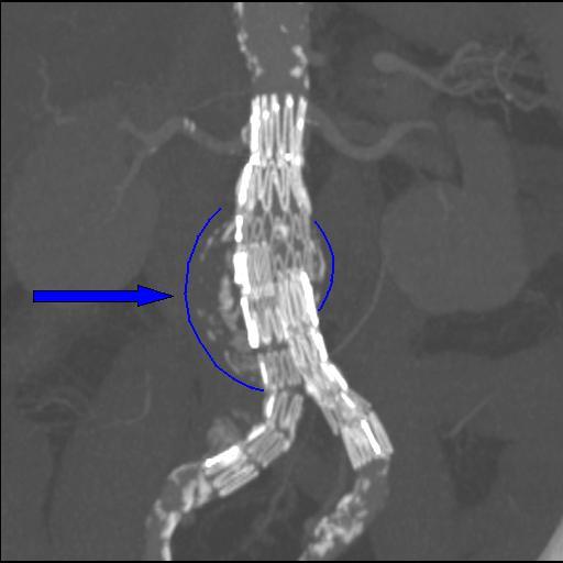
Endovascular aneurysm repair (EVAR) is a type of minimally-invasive endovascular surgery used to treat pathology of the aorta, most commonly an abdominal aortic aneurysm (AAA). When used to treat thoracic aortic disease, the procedure is then specifically termed TEVAR for "thoracic endovascular aortic/aneurysm repair." EVAR involves the placement of an expandable stent graft within the aorta to treat aortic disease without operating directly on the aorta. In 2003, EVAR surpassed open aortic surgery as the most common technique for repair of AAA, and in 2010, EVAR accounted for 78% of all intact AAA repair in the United States. Medical uses Standard EVAR is appropriate for aneurysms that begin below the renal arteries, where there exists an adequate length of normal aorta (the ''"proximal aortic neck"'') for reliable attachment of the endograft without leakage of blood around the device ("'' endoleak''"). If the proximal aortic neck is also involved with the aneurysm, the patient ... [...More Info...] [...Related Items...] OR: [Wikipedia] [Google] [Baidu] |
Axillary Artery
In human anatomy, the axillary artery is a large blood vessel that conveys oxygenated blood to the lateral aspect of the thorax, the axilla (armpit) and the upper limb. Its origin is at the lateral margin of the first rib, before which it is called the subclavian artery. After passing the lower margin of teres major muscle, teres major it becomes the brachial artery. Structure The axillary artery is often referred to as having three parts, with these divisions based on its location relative to the pectoralis minor muscle, which is superficial to the artery. * First part – the part of the artery superior to the pectoralis minor * Second part – the part of the artery posterior to the pectoralis minor * Third part – the part of the artery inferior to the pectoralis minor. Relations The axillary artery is accompanied by the axillary vein, which lies medial to the artery, along its length. In the axilla, the axillary artery is surrounded by the brachial plexus. The second ... [...More Info...] [...Related Items...] OR: [Wikipedia] [Google] [Baidu] |
Leriche Syndrome
In medicine, aortoiliac occlusive disease is a form of central artery disease involving the blockage of the abdominal aorta as it transitions into the common iliac arteries. Signs and symptoms Classically, it is described in male patients as a triad of the following signs and symptoms: # claudication of the buttocks and thighs # absent or decreased femoral pulses # erectile dysfunction Diagnosis The physical examination usually shows weakened femoral pulses and a reduced ankle-brachial index. The diagnosis can be verified by color duplex scanning, which reveals either a peak systolic velocity ratio ≥2.5 at the site of stenosis and/or a monophasic waveform. MRA and multidetector CTA are often used to determine the extent and type of obstruction. Another technique is digital subtraction angiography which allows verification of the diagnosis and endovascular treatment in a single session. Angiography provides important information regarding the perfusion and patency of distal ... [...More Info...] [...Related Items...] OR: [Wikipedia] [Google] [Baidu] |
Aortic Bifurcation
The aortic bifurcation is the point at which the abdominal aorta bifurcates (forks) into the left and right common iliac arteries. The aortic bifurcation is usually seen at the level of L4, just above the junction of the left and right common iliac veins. The right common iliac artery passes in front of the left common iliac vein. In some individuals, mainly women with lumbar lordosis, this vein can be compressed between the vertebra and the artery. This is the so-called Cockett syndrome or May–Thurner syndrome can cause a slower venous flow and the possibility of deep venous thrombosis in the left leg mainly in pregnancy. In surface anatomy, the bifurcation approximately corresponds to the umbilicus. Additional images Image:Gray531.png, The abdominal aorta and its branches. Image:Gray847.png, Abdominal portion of the sympathetic trunk, with the celiac and hypogastric plexuses. Image:Gray1121.png, Posterior abdominal wall, after removal of the peritoneum, showi ... [...More Info...] [...Related Items...] OR: [Wikipedia] [Google] [Baidu] |
Femoral Artery
The femoral artery is a large artery in the thigh and the main arterial supply to the thigh and leg. The femoral artery gives off the deep femoral artery and descends along the anteromedial part of the thigh in the femoral triangle. It enters and passes through the adductor canal, and becomes the popliteal artery as it passes through the adductor hiatus in the adductor magnus near the junction of the middle and distal thirds of the thigh. The femoral artery proximal to the origin of the deep femoral artery is referred to as the ''common femoral artery'', whereas the femoral artery distal to this origin is referred to as the ''superficial femoral artery''. Structure The femoral artery represents the continuation of the external iliac artery beyond the inguinal ligament underneath which the vessel passes to enter the thigh. The vessel passes under the inguinal ligament just medial of the midpoint of this ligament, midway between the anterior superior iliac spine and ... [...More Info...] [...Related Items...] OR: [Wikipedia] [Google] [Baidu] |
Trauma (medicine)
Injury is physiological damage to the living tissue of any organism, whether in humans, in other animals, or in plants. Injuries can be caused in many ways, including mechanically with penetration by sharp objects such as teeth or with blunt objects, by heat or cold, or by venoms and biotoxins. Injury prompts an inflammatory response in many taxa of animals; this prompts wound healing. In both plants and animals, substances are often released to help to occlude the wound, limiting loss of fluids and the entry of pathogens such as bacteria. Many organisms secrete antimicrobial chemicals which limit wound infection; in addition, animals have a variety of immune responses for the same purpose. Both plants and animals have regrowth mechanisms which may result in complete or partial healing over the injury. Cells too can repair damage to a certain degree. Taxonomic range Animals Injury in animals is sometimes defined as mechanical damage to anatomical struct ... [...More Info...] [...Related Items...] OR: [Wikipedia] [Google] [Baidu] |
Aneurysm
An aneurysm is an outward :wikt:bulge, bulging, likened to a bubble or balloon, caused by a localized, abnormal, weak spot on a blood vessel wall. Aneurysms may be a result of a hereditary condition or an acquired disease. Aneurysms can also be a wikt:Special:Search/nidus, nidus (starting point) for clot formation (thrombosis) and Embolism, embolization. As an aneurysm increases in size, the risk of rupture increases, which could lead to uncontrolled bleeding. Although they may occur in any blood vessel, particularly lethal examples include aneurysms of the circle of Willis in the brain, aortic aneurysms affecting the thoracic aorta, and abdominal aortic aneurysms. Aneurysms can arise in the heart itself following a Myocardial infarction, heart attack, including both Ventricular aneurysm, ventricular and atrial septal aneurysms. There are congenital atrial septal defect, atrial septal aneurysms, a rare heart defect. Etymology The word is from Greek language, Greek: ἀνεύρ� ... [...More Info...] [...Related Items...] OR: [Wikipedia] [Google] [Baidu] |
Acute Limb Ischemia
Acute limb ischaemia (ALI) occurs when there is a sudden lack of blood flow to a limb within 14 days of symptoms onset. On the other hand, when the symptoms exceed 14 days, it is called critical limb ischemia (CLI). CLI is the end stage of peripheral vascular disease where there is still some collateral circulation (alternate circulation pathways) that bring some blood flow (although inadequate) to the distal parts of the limbs. While limbs in both acute and chronic limb ischemia may be pulseless, a chronically ischemic limb is typically warm and pink due to a well-developed collateral artery network and does not need emergency intervention to avoid limb loss, whereas ALI is a vascular emergency. Acute limb ischaemia is usually caused by embolism or thrombosis, or rarely by dissection or trauma. Thrombosis is usually caused by peripheral vascular disease (atherosclerotic disease that leads to blood vessel blockage), while an embolism is usually of cardiac origin. In the United S ... [...More Info...] [...Related Items...] OR: [Wikipedia] [Google] [Baidu] |
Angina Pectoris
Angina, also known as angina pectoris, is chest pain or pressure, usually caused by insufficient blood flow to the heart muscle (myocardium). It is most commonly a symptom of coronary artery disease. Angina is typically the result of partial obstruction or spasm of the arteries that supply blood to the heart muscle. The main mechanism of coronary artery obstruction is atherosclerosis as part of coronary artery disease. Other causes of angina include abnormal heart rhythms, heart failure and, less commonly, anemia. The term derives , and can therefore be translated as "a strangling feeling in the chest". An urgent medical assessment is suggested to rule out serious medical conditions. There is a relationship between severity of angina and degree of oxygen deprivation in the heart muscle. However, the severity of angina does not always match the degree of oxygen deprivation to the heart or the risk of a heart attack (myocardial infarction). Some people may experience s ... [...More Info...] [...Related Items...] OR: [Wikipedia] [Google] [Baidu] |


