Axillary Artery on:
[Wikipedia]
[Google]
[Amazon]
In
 The axillary artery has several smaller branches. The branches can be remembered, in order, when traveling from the heart, with the
Image:Gray576.png, The veins of the right axilla, viewed from in front.
Image:Gray809.png, The right brachial plexus (infraclavicular portion) in the axillary fossa; viewed from below and in front.
Image:Gray810.png, Suprascapular and axillary nerves of right side, seen from behind.
File:PLEXUS BRACHIALIS.jpg, Brachial plexus and axillary artery
File:Slide3v.JPG, Axillary artery
File:Slide2bbbb.JPG, Axillary artery
File:Slide9JJJ.JPG, Axillary artery
File:Slide11OOO.JPG, Axillary artery
File:Slide15SSS.JPG, Axillary artery
File:Slide5EEEE.JPG, Axillary artery
human anatomy
Human anatomy (gr. ἀνατομία, "dissection", from ἀνά, "up", and τέμνειν, "cut") is primarily the scientific study of the morphology of the human body. Anatomy is subdivided into gross anatomy and microscopic anatomy. Gross ...
, the axillary artery is a large blood vessel
Blood vessels are the tubular structures of a circulatory system that transport blood throughout many Animal, animals’ bodies. Blood vessels transport blood cells, nutrients, and oxygen to most of the Tissue (biology), tissues of a Body (bi ...
that conveys oxygenated blood
Blood is a body fluid in the circulatory system of humans and other vertebrates that delivers necessary substances such as nutrients and oxygen to the cells, and transports metabolic waste products away from those same cells.
Blood is com ...
to the lateral aspect of the thorax
The thorax (: thoraces or thoraxes) or chest is a part of the anatomy of mammals and other tetrapod animals located between the neck and the abdomen.
In insects, crustaceans, and the extinct trilobites, the thorax is one of the three main di ...
, the axilla
The axilla (: axillae or axillas; also known as the armpit, underarm or oxter) is the area on the human body directly under the shoulder joint. It includes the axillary space, an anatomical space within the shoulder girdle between the arm a ...
(armpit) and the upper limb
The upper Limb (anatomy), limbs or upper extremities are the forelimbs of an upright posture, upright-postured tetrapod vertebrate, extending from the scapulae and clavicles down to and including the digit (anatomy), digits, including all the musc ...
. Its origin is at the lateral margin of the first rib
In vertebrate anatomy, ribs () are the long curved bones which form the rib cage, part of the axial skeleton. In most tetrapods, ribs surround the thoracic cavity, enabling the lungs to expand and thus facilitate breathing by expanding the ...
, before which it is called the subclavian artery
In human anatomy, the subclavian arteries are paired major arteries of the upper thorax, below the clavicle. They receive blood from the aortic arch. The left subclavian artery supplies blood to the left arm and the right subclavian artery suppli ...
.
After passing the lower margin of teres major
The teres major muscle is a muscle of the upper limb. It attaches to the scapula and the humerus and is one of the seven scapulohumeral muscles. It is a thick but somewhat flattened muscle.
The teres major muscle (from Latin ''teres'', meanin ...
it becomes the brachial artery
The brachial artery is the major blood vessel of the (upper) arm. It is the continuation of the axillary artery beyond the lower margin of teres major muscle. It continues down the ventral surface of the arm until it reaches the cubital fossa ...
.
Structure
The axillary artery is often referred to as having three parts, with these divisions based on its location relative to thepectoralis minor
Pectoralis minor muscle () is a thin, triangular muscle, situated at the upper part of the chest, beneath the pectoralis major in the human body. It arises from ribs III-V; it inserts onto the coracoid process of the scapula. It is innervated by ...
muscle, which is superficial to the artery.
* First part – the part of the artery superior to the pectoralis minor
* Second part – the part of the artery posterior to the pectoralis minor
* Third part – the part of the artery inferior to the pectoralis minor.
Relations
The axillary artery is accompanied by theaxillary vein
In human anatomy, the axillary vein is a large blood vessel that conveys blood from the lateral aspect of the thorax, axilla (armpit) and upper limb toward the heart. There is one axillary vein on each side of the body.
Structure
Its origin i ...
, which lies medial to the artery, along its length.
In the axilla, the axillary artery is surrounded by the brachial plexus
The brachial plexus is a network of nerves (nerve plexus) formed by the anterior rami of the lower four Spinal nerve#Cervical nerves, cervical nerves and first Spinal nerve#Thoracic nerves, thoracic nerve (cervical spinal nerve 5, C5, Cervical spi ...
. The second part of the axillary artery is the reference for the locational descriptions of the cords in the brachial plexus
The brachial plexus is a network of nerves (nerve plexus) formed by the anterior rami of the lower four Spinal nerve#Cervical nerves, cervical nerves and first Spinal nerve#Thoracic nerves, thoracic nerve (cervical spinal nerve 5, C5, Cervical spi ...
. For example, the posterior cord
The posterior cord is a part of the brachial plexus
The brachial plexus is a network of nerves (nerve plexus) formed by the anterior rami of the lower four Spinal nerve#Cervical nerves, cervical nerves and first Spinal nerve#Thoracic nerves, thor ...
of the brachial plexus is so named because it lies posterior to the second part of the artery.
Branches
mnemonics
A mnemonic device ( ), memory trick or memory device is any learning technique that aids information retention or retrieval in the human memory, often by associating the information with something that is easier to remember.
It makes use of e ...
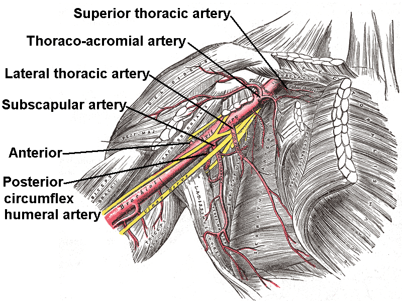
"Screw The Lawyers Save A Patient", "Summertime: The Lakers Schedule Another Parade", "Sixties Teens Love Sex And Pot", or "She Tastes Like Sweet Apple Pie." The origin of these branches is highly variable (e.g. the posterior and anterior circumflex arteries often have a common trunk). An arterial branch is named for its course, not its origin.
* First part (1 branch)
** Superior thoracic artery (Supreme thoracic artery)
* Second part (2 branches)
** Thoraco-acromial artery
** Lateral thoracic artery. If the lateral thoracic artery is not branching from the axillary artery, will most likely branch from the following (in order of likelihood): (1) thoracoacromial, (2) third part of axillary artery, (3) suprascapular artery, (4) subscapular artery
* Third part (3 branches)
** Subscapular artery
** Anterior humeral circumflex artery
** Posterior humeral circumflex artery
Continues as the brachial artery
The brachial artery is the major blood vessel of the (upper) arm. It is the continuation of the axillary artery beyond the lower margin of teres major muscle. It continues down the ventral surface of the arm until it reaches the cubital fossa ...
past the inferior border of the teres major
The teres major muscle is a muscle of the upper limb. It attaches to the scapula and the humerus and is one of the seven scapulohumeral muscles. It is a thick but somewhat flattened muscle.
The teres major muscle (from Latin ''teres'', meanin ...
.
Clinical significance
The axillary artery can be safely clamped without endangering the arm, but only in a location proximal to the origin of the subscapular artery (and distal to the thyrocervical trunk of the subclavian artery). The anastomotic network surrounding the scapula provides an alternate path for collateral circulation to the arm from arteries including thedorsal scapular artery
The transverse cervical artery (transverse artery of neck or transversa colli artery) is an artery in the neck and a branch of the thyrocervical trunk, running at a higher level than the suprascapular artery.
Structure
It passes transversely ...
and suprascapular artery
The suprascapular artery is a branch of the thyrocervical trunk on the neck.
Structure
At first, it passes downward and laterally across the scalenus anterior and phrenic nerve, being covered by the sternocleidomastoid muscle; it then crosses t ...
.
The right axillary artery is often used as an arterial cannula
A cannula (; Latin meaning 'little reed'; : cannulae or cannulas) is a tube that can be inserted into the body, often for the delivery or removal of fluid or for the gathering of samples. In simple terms, a cannula can surround the inner or out ...
tion site in cardiac surgery, particularly for repair of aortic dissection
Aortic dissection (AD) occurs when an injury to the innermost layer of the aorta allows blood to flow between the layers of the aortic wall, forcing the layers apart. In most cases, this is associated with a sudden onset of agonizing ches ...
and replacement of the ascending aorta
The ascending aorta (AAo) is a portion of the aorta commencing at the upper part of the base of the left ventricle, on a level with the lower border of the third costal cartilage behind the left half of the sternum.
Structure
It passes obliqu ...
and aortic arch
The aortic arch, arch of the aorta, or transverse aortic arch () is the part of the aorta between the ascending and descending aorta. The arch travels backward, so that it ultimately runs to the left of the trachea.
Structure
The aorta begins ...
.
Additional images
References
External links
* * * – "Axillary Region: Parts of the Axillary Artery" * – "The axillary artery and its major branches shown in relation to major landmarks." {{Authority control Arteries of the upper limb Axillas