|
Varicosities
A neuroeffector junction is a site where a motor neuron releases a neurotransmitter to affect a target—non-neuronal—cell. This junction functions like a synapse. However, unlike most neurons, somatic efferent motor neurons innervate skeletal muscle, and are always excitatory. Visceral efferent neurons innervate smooth muscle, cardiac muscle, and glands, and have the ability to be either excitatory or inhibitory in function. Neuroeffector junctions are known as neuromuscular junctions when the target cell is a muscle fiber. Non-synaptic transmission is characteristic of autonomic neuroeffector junctions. The structure of the autonomic neuromuscular junction consists of several essential features including that: the terminal portions of autonomic nerve fibers are varicose and mobile, transmitters being released 'en passage' from varying distances from the effector cells; while there is no structural post-junctional specialization on effector cells, receptors for neurotransmitters ... [...More Info...] [...Related Items...] OR: [Wikipedia] [Google] [Baidu] |
Vas Deferens
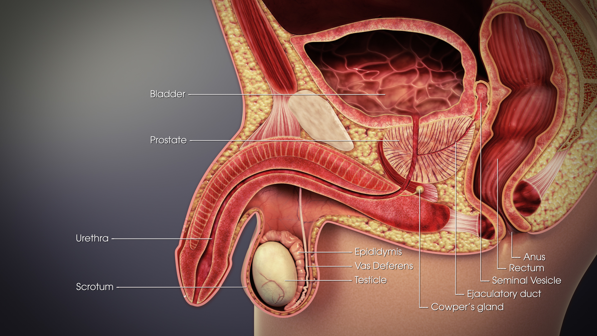
The vas deferens (: vasa deferentia), ductus deferens (: ductūs deferentes), or sperm duct is part of the male reproductive system of many vertebrates. In mammals, spermatozoa are produced in the seminiferous tubules and flow into the epididymal duct. The end of the epididymis is connected to the vas deferens. The vas deferens ends with an opening into the ejaculatory duct at a point where the duct of the seminal vesicle also joins the ejaculatory duct. The vas deferens is a partially coiled tube which exits the abdominal cavity through the inguinal canal. Etymology ''Vas deferens'' is Latin, meaning "carrying-away vessel" while ''ductus deferens'', also Latin, means "carrying-away duct". Structure The human vas deferens measures 30–35 cm in length, and 2–3 mm in diameter. It is continuous proximally with the tail of the epididymis, and exhibits a tortuous, convoluted initial/proximal section (which measures 2–3 cm in length). Distally, it forms a dilated ... [...More Info...] [...Related Items...] OR: [Wikipedia] [Google] [Baidu] |
Human Gastrointestinal Tract
The gastrointestinal tract (GI tract, digestive tract, alimentary canal) is the tract or passageway of the digestive system that leads from the mouth to the anus. The tract is the largest of the body's systems, after the cardiovascular system. The GI tract contains all the major organs of the digestive system, in humans and other animals, including the esophagus, stomach, and intestines. Food taken in through the mouth is digested to extract nutrients and absorb energy, and the waste expelled at the anus as feces. ''Gastrointestinal'' is an adjective meaning of or pertaining to the stomach and intestines. Most animals have a "through-gut" or complete digestive tract. Exceptions are more primitive ones: sponges have small pores ( ostia) throughout their body for digestion and a larger dorsal pore ( osculum) for excretion, comb jellies have both a ventral mouth and dorsal anal pores, while cnidarians and acoels have a single pore for both digestion and excretion. The ... [...More Info...] [...Related Items...] OR: [Wikipedia] [Google] [Baidu] |
Transduction (biophysics)
In biophysics, transduction is the conveyance of energy from one electron (a donor) to another (a receptor), at the same time that the class of energy changes. Photonic energy, the kinetic energy of a photon, may follow the following paths: * be released again as a photon of less energy; * be transferred to a recipient with no change in class; * be dissipated as heat; or * be transduced. See also *Homeostasis *Phosphate *Pigments * NAD * ATP *Cytochrome Cytochromes are redox-active proteins containing a heme, with a central iron (Fe) atom at its core, as a cofactor. They are involved in the electron transport chain and redox catalysis. They are classified according to the type of heme and its ... Biophysics Photonics {{biophysics-stub ... [...More Info...] [...Related Items...] OR: [Wikipedia] [Google] [Baidu] |
Action Potential
An action potential (also known as a nerve impulse or "spike" when in a neuron) is a series of quick changes in voltage across a cell membrane. An action potential occurs when the membrane potential of a specific Cell (biology), cell rapidly rises and falls. This depolarization then causes adjacent locations to similarly depolarize. Action potentials occur in several types of Membrane potential#Cell excitability, excitable cells, which include animal cells like neurons and myocyte, muscle cells, as well as some plant cells. Certain endocrine cells such as pancreatic beta cells, and certain cells of the anterior pituitary gland are also excitable cells. In neurons, action potentials play a central role in cell–cell interaction, cell–cell communication by providing for—or with regard to saltatory conduction, assisting—the propagation of signals along the neuron's axon toward axon terminal, synaptic boutons situated at the ends of an axon; these signals can then connect wit ... [...More Info...] [...Related Items...] OR: [Wikipedia] [Google] [Baidu] |
Ganglion
A ganglion (: ganglia) is a group of neuron cell bodies in the peripheral nervous system. In the somatic nervous system, this includes dorsal root ganglia and trigeminal ganglia among a few others. In the autonomic nervous system, there are both sympathetic and parasympathetic ganglia which contain the cell bodies of postganglionic sympathetic and parasympathetic neurons respectively. A pseudoganglion looks like a ganglion, but only has nerve fibers and has no nerve cell bodies. Structure Ganglia are primarily made up of somata and dendritic structures, which are bundled or connected. Ganglia often interconnect with other ganglia to form a complex system of ganglia known as a plexus. Ganglia provide relay points and intermediary connections between different neurological structures in the body, such as the peripheral and central nervous systems. Among vertebrates there are three major groups of ganglia: * Dorsal root ganglia (also known as the spinal ganglia) contai ... [...More Info...] [...Related Items...] OR: [Wikipedia] [Google] [Baidu] |
Micrometre
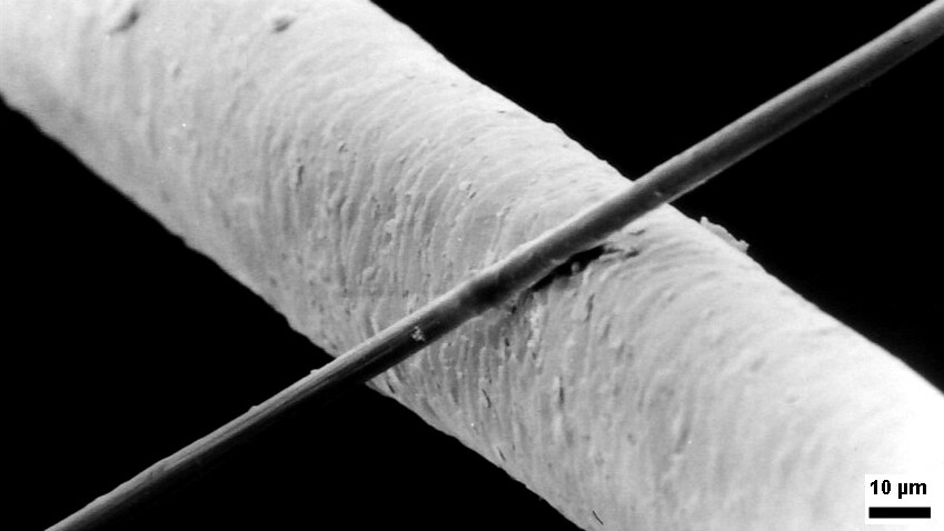
The micrometre (English in the Commonwealth of Nations, Commonwealth English as used by the International Bureau of Weights and Measures; SI symbol: μm) or micrometer (American English), also commonly known by the non-SI term micron, is a unit of length in the International System of Units (SI) equalling (SI standard prefix "micro-" = ); that is, one millionth of a metre (or one thousandth of a millimetre, , or about ). The nearest smaller common SI Unit, SI unit is the nanometre, equivalent to one thousandth of a micrometre, one millionth of a millimetre or one billionth of a metre (). The micrometre is a common unit of measurement for wavelengths of infrared radiation as well as sizes of biological cell (biology), cells and bacteria, and for grading wool by the diameter of the fibres. The width of a single human hair ranges from approximately 20 to . Examples Between 1 μm and 10 μm: * 1–10 μm – length of a typical bacterium * 3–8 μm – width of str ... [...More Info...] [...Related Items...] OR: [Wikipedia] [Google] [Baidu] |
Immunohistochemistry
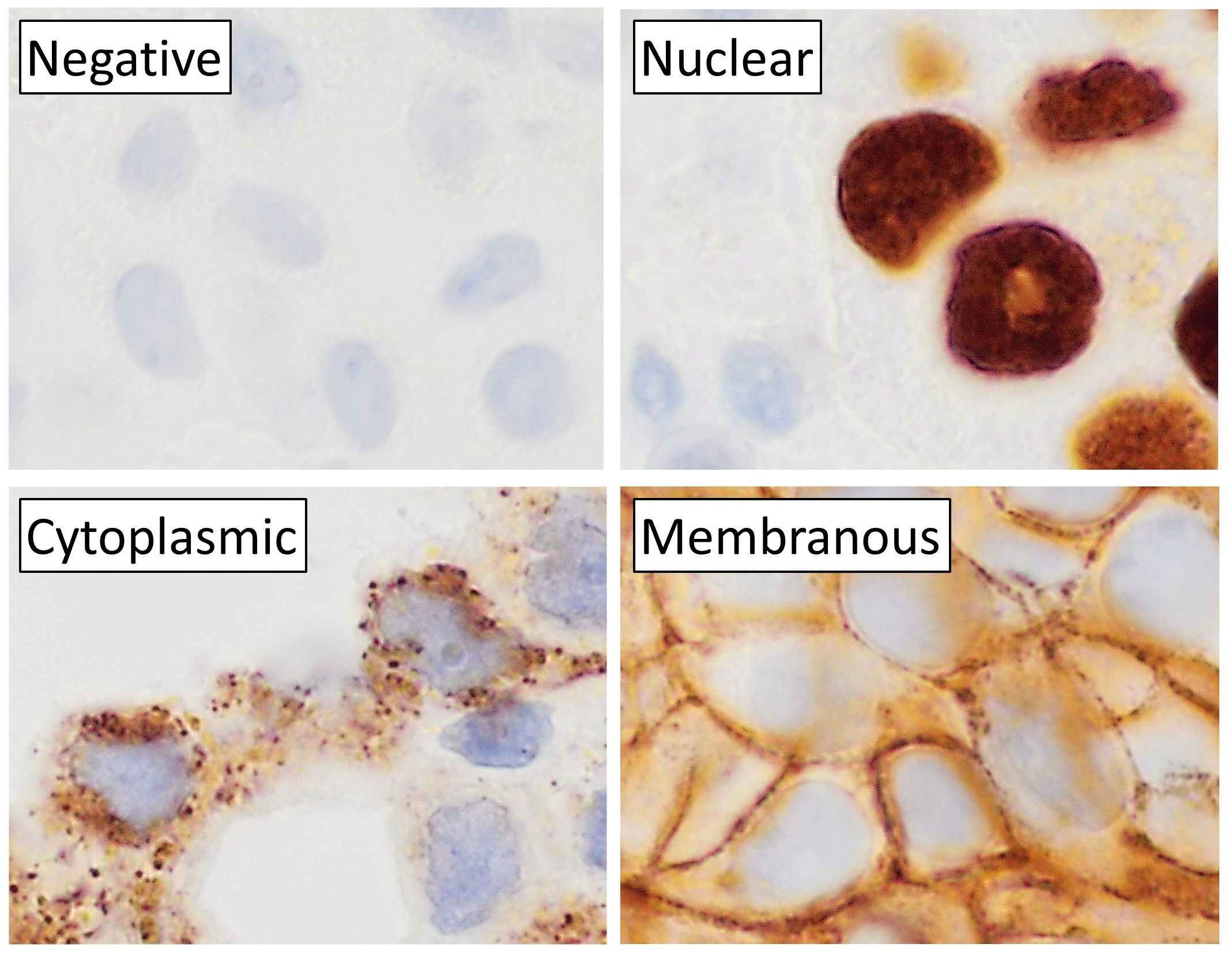
Immunohistochemistry is a form of immunostaining. It involves the process of selectively identifying antigens in cells and tissue, by exploiting the principle of Antibody, antibodies binding specifically to antigens in biological tissues. Albert Coons, Albert Hewett Coons, Ernst Berliner, Ernest Berliner, Norman Jones and Hugh J Creech was the first to develop immunofluorescence in 1941. This led to the later development of immunohistochemistry. Immunohistochemical staining is widely used in the diagnosis of abnormal cells such as those found in cancerous tumors. In some cancer cells certain tumor antigens are expressed which make it possible to detect. Immunohistochemistry is also widely used in basic research, to understand the distribution and localization of biomarkers and differentially expressed proteins in different parts of a biological tissue. Sample preparation Immunohistochemistry can be performed on tissue that has been fixed and embedded in Paraffin wax, paraffin, ... [...More Info...] [...Related Items...] OR: [Wikipedia] [Google] [Baidu] |
Adrenergic Receptor
The adrenergic receptors or adrenoceptors are a class of G protein-coupled receptors that are targets of many catecholamines like norepinephrine (noradrenaline) and epinephrine (adrenaline) produced by the body, but also many medications like beta blockers, beta-2 (β2) agonists and alpha-2 (α2) agonists, which are used to treat high blood pressure and asthma, for example. Many cells have these receptors, and the binding of a catecholamine to the receptor will generally stimulate the sympathetic nervous system (SNS). The SNS is responsible for the fight-or-flight response, which is triggered by experiences such as exercise or fear-causing situations. This response dilates pupils, increases heart rate, mobilizes energy, and diverts blood flow from non-essential organs to skeletal muscle. These effects together tend to increase physical performance momentarily. History By the turn of the 19th century, it was agreed that the stimulation of sympathetic nerves could cause ... [...More Info...] [...Related Items...] OR: [Wikipedia] [Google] [Baidu] |
Nanometre
330px, Different lengths as in respect to the Molecule">molecular scale. The nanometre (international spelling as used by the International Bureau of Weights and Measures; SI symbol: nm), or nanometer (American spelling), is a unit of length in the International System of Units (SI), equal to one billionth ( short scale) or one thousand million (long scale) of a meter (0.000000001 m) and to 1000 picometres. One nanometre can be expressed in scientific notation as 1 × 10⁻⁹ m and as m. History The nanometre was formerly known as the "''millimicrometre''" – or, more commonly, the "''millimicron''" for short – since it is of a micrometer. It was often denoted by the symbol ''mμ'' or, more rarely, as ''μμ'' (however, ''μμ'' should refer to a ''millionth'' of a micron). Etymology The name combines the SI prefix '' nano-'' (from the Ancient Greek , ', "dwarf") with the parent unit name ''metre'' (from Greek , ', "unit of measurement"). ... [...More Info...] [...Related Items...] OR: [Wikipedia] [Google] [Baidu] |
Paracrine Signalling
In cellular biology, paracrine signaling is a form of cell signaling, a type of cellular communication in which a cell produces a signal to induce changes in nearby cells, altering the behaviour of those cells. Signaling molecules known as paracrine factors diffuse over a relatively short distance (local action), as opposed to cell signaling by endocrine factors, hormones which travel considerably longer distances via the circulatory system; juxtacrine interactions; and autocrine signaling. Cells that produce paracrine factors secrete them into the immediate extracellular environment. Factors then travel to nearby cells in which the gradient of factor received determines the outcome. However, the exact distance that paracrine factors can travel is not certain. Although paracrine signaling elicits a diverse array of responses in the induced cells, most paracrine factors utilize a relatively streamlined set of receptors and pathways. In fact, different organs in the body - ... [...More Info...] [...Related Items...] OR: [Wikipedia] [Google] [Baidu] |
Nerve
A nerve is an enclosed, cable-like bundle of nerve fibers (called axons). Nerves have historically been considered the basic units of the peripheral nervous system. A nerve provides a common pathway for the Electrochemistry, electrochemical nerve impulses called action potentials that are transmitted along each of the axons to peripheral organs or, in the case of sensory nerves, from the periphery back to the central nervous system. Each axon is an extension of an individual neuron, along with other supportive cells such as some Schwann cells that coat the axons in myelin. Each axon is surrounded by a layer of connective tissue called the endoneurium. The axons are bundled together into groups called Nerve fascicle, fascicles, and each fascicle is wrapped in a layer of connective tissue called the perineurium. The entire nerve is wrapped in a layer of connective tissue called the epineurium. Nerve cells (often called neurons) are further classified as either Sensory neuron, sens ... [...More Info...] [...Related Items...] OR: [Wikipedia] [Google] [Baidu] |
Peripheral Nervous System
The peripheral nervous system (PNS) is one of two components that make up the nervous system of Bilateria, bilateral animals, with the other part being the central nervous system (CNS). The PNS consists of nerves and ganglia, which lie outside the brain and the spinal cord. The main function of the PNS is to connect the CNS to the Limb (anatomy), limbs and Organ (anatomy), organs, essentially serving as a relay between the brain and spinal cord and the rest of the body. Unlike the CNS, the PNS is not protected by the vertebral column and skull, or by the blood–brain barrier, which leaves it exposed to toxins. The peripheral nervous system can be divided into a somatic nervous system, somatic division and an autonomic nervous system, autonomic division. Each of these can further be differentiated into a sensory and a motor sector. In the somatic nervous system, the cranial nerves are part of the PNS with the exceptions of the olfactory nerve and epithelia and the optic nerve (c ... [...More Info...] [...Related Items...] OR: [Wikipedia] [Google] [Baidu] |


