|
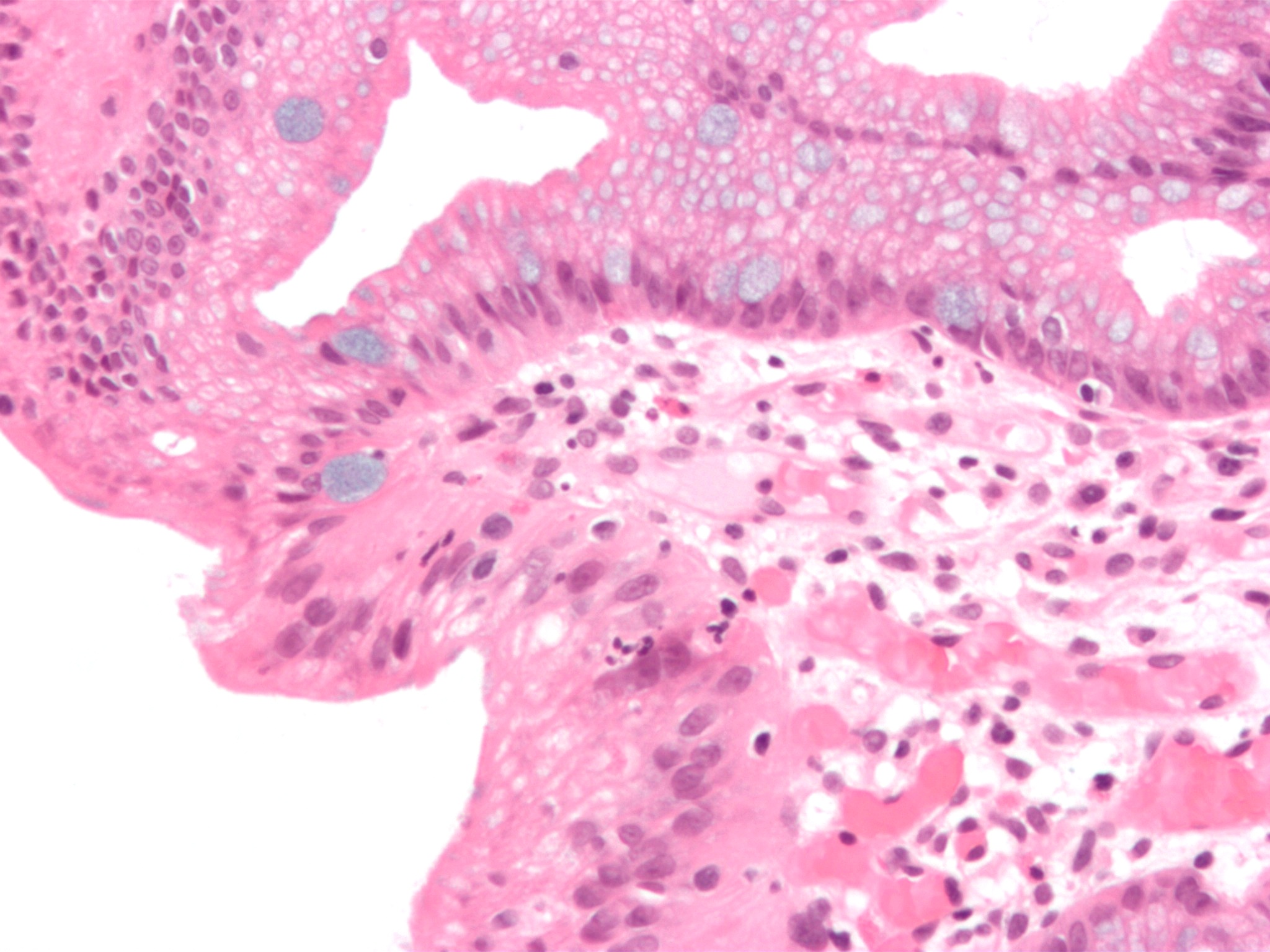
Uterine Carcinosarcoma
Carcinosarcomas are malignant tumors that consist of a mixture of carcinoma (or epithelial cancer) and sarcoma (or mesenchymal/connective tissue cancer). Carcinosarcomas are rare tumors, and can arise in diverse organs, such as the skin, salivary glands, lungs, the esophagus, pancreas, colon, uterus and ovaries. Cellular origins Four main hypotheses have been proposed for the cellular origins of carcinosarcoma, based largely on the pathology of the disease. First, the collision tumor hypothesis, which proposes the collision of two independent tumors resulting in a single neoplasm, based on the observation that skin cancers and superficial malignant fibrous histiocytomas are commonly seen in patients with sun-damaged skin; second, the composition hypothesis, which suggests that the mesenchymal component represents a pseudosarcomatous reaction to the epithelial malignancy; third, the combination hypothesis, which suggests that both the epithelial and mesenchymal components of t ... [...More Info...] [...Related Items...] OR: [Wikipedia] [Google] [Baidu] |
Micrograph
A micrograph or photomicrograph is a photograph or digital image taken through a microscope or similar device to show a magnify, magnified image of an object. This is opposed to a macrograph or photomacrograph, an image which is also taken on a microscope but is only slightly magnified, usually less than 10 times. Micrography is the practice or art of using microscopes to make photographs. A micrograph contains extensive details of microstructure. A wealth of information can be obtained from a simple micrograph like behavior of the material under different conditions, the phases found in the system, failure analysis, grain size estimation, elemental analysis and so on. Micrographs are widely used in all fields of microscopy. Types Photomicrograph A light micrograph or photomicrograph is a micrograph prepared using an optical microscope, a process referred to as ''photomicroscopy''. At a basic level, photomicroscopy may be performed simply by connecting a camera to a micros ... [...More Info...] [...Related Items...] OR: [Wikipedia] [Google] [Baidu] |
Large Intestine
The large intestine, also known as the large bowel, is the last part of the gastrointestinal tract and of the digestive system in tetrapods. Water is absorbed here and the remaining waste material is stored in the rectum as feces before being removed by defecation. The colon is the longest portion of the large intestine, and the terms are often used interchangeably but most sources define the large intestine as the combination of the cecum, colon, rectum, and anal canal. Some other sources exclude the anal canal. In humans, the large intestine begins in the right iliac region of the pelvis, just at or below the waist, where it is joined to the end of the small intestine at the cecum, via the ileocecal valve. It then continues as the colon ascending the abdomen, across the width of the abdominal cavity as the transverse colon, and then descending to the rectum and its endpoint at the anal canal. Overall, in humans, the large intestine is about long, which is about on ... [...More Info...] [...Related Items...] OR: [Wikipedia] [Google] [Baidu] |
Uterine Adenosarcoma
Uterine adenosarcoma is an uncommon form of cancer that arises from mesenchymal tissue of the uterus and has a benign glandular component. Signs and symptoms The most common presentation is vaginal bleeding. Other presentations include pelvic mass and uterine polyp. Generally, the clinical findings are non-specific. Pathology Uterine adenosarcoma have, by definition, a malignant stroma and benign glandular elements. The World Health Organization (WHO) criteria have a mitotic rate cut point; however, this is often disregarded, as bland-appearing tumours with a low mitotic rate are known to metastasize occasionally. Image: Uterine adenosarcoma - low mag.jpg , Low mag. Image: Uterine adenosarcoma - add - intermed mag.jpg , Intermed. mag. Image: Uterine adenosarcoma - very high mag.jpg , Very high mag. Treatment Uterine adenosarcomas are typically treated with a total abdominal hysterectomy and bilateral salpingoophorectomy (TAH-BSO). Ovary The ovary is an orga ... [...More Info...] [...Related Items...] OR: [Wikipedia] [Google] [Baidu] |
Adenosarcoma
Adenosarcoma (also Mullerian Adenosarcoma) is a rare malignant tumor that occurs in women of all age groups, but most commonly post- menopause. Adenosarcoma arises from mesenchymal tissue and has a mixture of the tumoral components of an adenoma, a tumor of epithelial origin, and a sarcoma, a tumor originating from connective tissue.NCI Dictionary of Cancer Terms: Adenosarcoma." National Cancer Institute, National Institutes of Health, www.cancer.gov/publications/dictionaries/cancer-terms/def/adenosarcoma. Carroll, A., Ramirez, P. T., Westin, S. N., Soliman, P. T., Munsell, M. F., Nick, A. M., ... & Fleming, N. D. (2014). Uterine adenosarcoma: an analysis on management, outcomes, and risk factors for recurrence. Gynecologic oncology, 135(3), 455-461. The adenoma, or epithelial component of the tumor, is benign, while the sarcomatous stroma is malignant.Podduturi, V., & Pinto, K. R. (2016, January). Mullerian adenosarcoma of the cervix with heterologous elements and sarcomatou ... [...More Info...] [...Related Items...] OR: [Wikipedia] [Google] [Baidu] |
Histogenesis
Histogenesis is the formation of different tissues from undifferentiated cells. These cells are constituents of three primary germ layers, the endoderm, mesoderm, and ectoderm. The science of the microscopic structures of the tissues formed within histogenesis is termed histology. Germ layers A germ layer is a collection of cells, formed during animal and mammalian embryogenesis. Germ layers are typically pronounced within vertebrate organisms; however, animals or mammals more complex than sponges (eumetazoans and agnotozoans) produce two or three primary tissue layers. Animals with radial symmetry, such as cnidarians, produce two layers, called the ectoderm and endoderm. They are diploblastic. Animals with bilateral symmetry produce a third layer in-between called mesoderm, making them triploblastic. Germ layers will eventually give rise to all of an animal's or mammal's tissues and organs through a process called organogenesis. Endoderm The endoderm is one of the g ... [...More Info...] [...Related Items...] OR: [Wikipedia] [Google] [Baidu] |
Mutation
In biology, a mutation is an alteration in the nucleic acid sequence of the genome of an organism, virus, or extrachromosomal DNA. Viral genomes contain either DNA or RNA. Mutations result from errors during DNA or viral replication, mitosis, or meiosis or other types of damage to DNA (such as pyrimidine dimers caused by exposure to ultraviolet radiation), which then may undergo error-prone repair (especially microhomology-mediated end joining), cause an error during other forms of repair, or cause an error during replication ( translesion synthesis). Mutations may also result from insertion or deletion of segments of DNA due to mobile genetic elements. Mutations may or may not produce detectable changes in the observable characteristics ( phenotype) of an organism. Mutations play a part in both normal and abnormal biological processes including: evolution, cancer, and the development of the immune system, including junctional diversity. Mutation is the ultima ... [...More Info...] [...Related Items...] OR: [Wikipedia] [Google] [Baidu] |
KRAS
''KRAS'' ( Kirsten rat sarcoma virus) is a gene that provides instructions for making a protein called K-Ras, a part of the RAS/MAPK pathway. The protein relays signals from outside the cell to the cell's nucleus. These signals instruct the cell to grow and divide ( proliferate) or to mature and take on specialized functions ( differentiate). It is called ''KRAS'' because it was first identified as a viral oncogene in the Kirsten RAt Sarcoma virus. The oncogene identified was derived from a cellular genome, so , when found in a cellular genome, is called a proto-oncogene. The K-Ras protein is a GTPase, a class of enzymes which convert the nucleotide guanosine triphosphate (GTP) into guanosine diphosphate (GDP). In this way the K-Ras protein acts like a switch that is turned on and off by the GTP and GDP molecules. To transmit signals, it must be turned on by attaching (binding) to a molecule of GTP. The K-Ras protein is turned off (inactivated) when it converts the GTP to G ... [...More Info...] [...Related Items...] OR: [Wikipedia] [Google] [Baidu] |
Metaplasia
Metaplasia ( gr, "change in form") is the transformation of one differentiated cell type to another differentiated cell type. The change from one type of cell to another may be part of a normal maturation process, or caused by some sort of abnormal stimulus. In simplistic terms, it is as if the original cells are not robust enough to withstand their environment, so they transform into another cell type better suited to their environment. If the stimulus causing metaplasia is removed or ceases, tissues return to their normal pattern of differentiation. Metaplasia is not synonymous with dysplasia, and is not considered to be an actual cancer. It is also contrasted with heteroplasia, which is the spontaneous abnormal growth of cytologic and histologic elements. Today, metaplastic changes are usually considered to be an early phase of carcinogenesis, specifically for those with a history of cancers or who are known to be susceptible to carcinogenic changes. Metaplastic change i ... [...More Info...] [...Related Items...] OR: [Wikipedia] [Google] [Baidu] |
Stem Cell
In multicellular organisms, stem cells are undifferentiated or partially differentiated cells that can differentiate into various types of cells and proliferate indefinitely to produce more of the same stem cell. They are the earliest type of cell in a cell lineage. They are found in both embryonic and adult organisms, but they have slightly different properties in each. They are usually distinguished from progenitor cells, which cannot divide indefinitely, and precursor or blast cells, which are usually committed to differentiating into one cell type. In mammals, roughly 50–150 cells make up the inner cell mass during the blastocyst stage of embryonic development, around days 5–14. These have stem-cell capability. '' In vivo'', they eventually differentiate into all of the body's cell types (making them pluripotent). This process starts with the differentiation into the three germ layers – the ectoderm, mesoderm and endoderm – at the gastrulation stage. Howev ... [...More Info...] [...Related Items...] OR: [Wikipedia] [Google] [Baidu] |
Malignant Fibrous Histiocytoma
Undifferentiated pleomorphic sarcoma (UPS), also termed pleomorphic myofibrosarcoma, high-grade myofibroblastic sarcoma, and high-grade myofibrosarcoma, is characterized by the World Health Organization (WHO), 2020, as a rare, poorly differentiated neoplasm, i.e. an abnormal growth of cells that have an unclear identity and/or cell of origin. WHO classified it as one of the undifferentiated/unclassified sarcomas in the category of tumors of uncertain differentiation. Sarcomas are cancers known or thought to derive from mesenchymal stem cells that typically develop in bone, muscle, fat, blood vessels, lymphatic vessels, tendons, and ligaments. More than 70 sarcoma subtypes have been described. The UPS subtype of these sarcomas consists of tumor cells that are poorly differentiated and may appear as spindle-shaped cells, histiocytes, and giant cells. UPS is considered a diagnosis that defies formal sub-classification after thorough histologic, immunohistochemical, and ultrastructura ... [...More Info...] [...Related Items...] OR: [Wikipedia] [Google] [Baidu] |
Skin Cancer
Skin cancers are cancers that arise from the skin. They are due to the development of abnormal cells that have the ability to invade or spread to other parts of the body. There are three main types of skin cancers: basal-cell skin cancer (BCC), squamous-cell skin cancer (SCC) and melanoma. The first two, along with a number of less common skin cancers, are known as nonmelanoma skin cancer (NMSC). Basal-cell cancer grows slowly and can damage the tissue around it but is unlikely to spread to distant areas or result in death. It often appears as a painless raised area of skin that may be shiny with small blood vessels running over it or may present as a raised area with an ulcer. Squamous-cell skin cancer is more likely to spread. It usually presents as a hard lump with a scaly top but may also form an ulcer. Melanomas are the most aggressive. Signs include a mole that has changed in size, shape, color, has irregular edges, has more than one color, is itchy or bleeds. More ... [...More Info...] [...Related Items...] OR: [Wikipedia] [Google] [Baidu] |
Neoplasm
A neoplasm () is a type of abnormal and excessive growth of tissue. The process that occurs to form or produce a neoplasm is called neoplasia. The growth of a neoplasm is uncoordinated with that of the normal surrounding tissue, and persists in growing abnormally, even if the original trigger is removed. This abnormal growth usually forms a mass, when it may be called a tumor. ICD-10 classifies neoplasms into four main groups: benign neoplasms, in situ neoplasms, malignant neoplasms, and neoplasms of uncertain or unknown behavior. Malignant neoplasms are also simply known as cancers and are the focus of oncology. Prior to the abnormal growth of tissue, as neoplasia, cells often undergo an abnormal pattern of growth, such as metaplasia or dysplasia. However, metaplasia or dysplasia does not always progress to neoplasia and can occur in other conditions as well. The word is from Ancient Greek 'new' and 'formation, creation'. Types A neoplasm can be benign, potentia ... [...More Info...] [...Related Items...] OR: [Wikipedia] [Google] [Baidu] |


