|
Tyrannotitan Remains 01
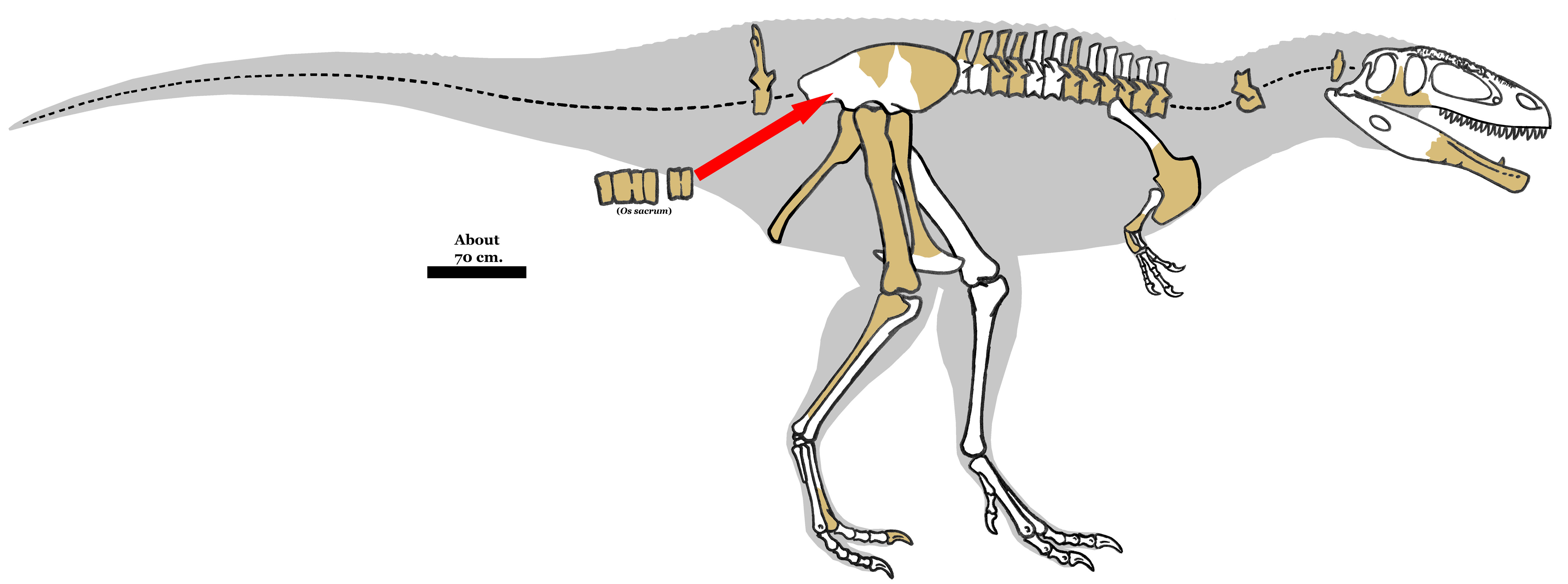
''Tyrannotitan'' (; ) is a genus of huge bipedal carnivorous dinosaur of the carcharodontosaurid family from the Aptian stage of the early Cretaceous period, discovered in Argentina. It is closely related to other giant predators like ''Carcharodontosaurus'' and especially ''Giganotosaurus'' as well as ''Mapusaurus''. Discovery and species ''Tyrannotitan chubutensis'' was described by Fernando E. Novas, Silvina de Valais, Pat Vickers-Rich, and Tom Rich in 2005. The fossils were found at La Juanita Farm, northeast of Paso de Indios, Chubut Province, Argentina. They are believed to have been from the Cerro Castaño Member, Cerro Barcino Formation (Aptian stage). The holotype material was designated MPEF-PV 1156 and included partial dentaries, teeth, back vertebrae 3–8 and 11–14, proximal tail vertebrae, ribs and chevrons, a fragmentary scapulocoracoid, humerus, ulna, partial ilium, a nearly complete femur, fibula, and left metatarsal 2. Additional material (designated M ... [...More Info...] [...Related Items...] OR: [Wikipedia] [Google] [Baidu] |
Early Cretaceous
The Early Cretaceous (geochronological name) or the Lower Cretaceous ( chronostratigraphic name), is the earlier or lower of the two major divisions of the Cretaceous. It is usually considered to stretch from 145 Ma to 100.5 Ma. Geology Proposals for the exact age of the Barremian-Aptian boundary ranged from 126 to 117 Ma until recently (as of 2019), but based on drillholes in Svalbard the defining early Aptian Oceanic Anoxic Event 1a (OAE1a) was carbon isotope dated to 123.1±0.3 Ma, limiting the possible range for the boundary to c. 122–121 Ma. There is a possible link between this anoxic event and a series of Early Cretaceous large igneous provinces (LIP). The Ontong Java- Manihiki- Hikurangi large igneous province, emplaced in the South Pacific at c. 120 Ma, is by far the largest LIP in Earth's history. The Ontong Java Plateau today covers an area of 1,860,000 km2. In the Indian Ocean another LIP began to form at c. 120 Ma, the Kergue ... [...More Info...] [...Related Items...] OR: [Wikipedia] [Google] [Baidu] |
Faunal Stage
In chronostratigraphy, a stage is a succession of rock strata laid down in a single age on the geologic timescale, which usually represents millions of years of deposition. A given stage of rock and the corresponding age of time will by convention have the same name, and the same boundaries. Rock series are divided into stages, just as geological epochs are divided into ages. Stages can be divided into smaller stratigraphic units called chronozones. (See chart at right for full terminology hierarchy.) Stages may also be divided into substages or indeed grouped as superstages. The term faunal stage is sometimes used, referring to the fact that the same fauna (animals) are found throughout the layer (by definition). Definition Stages are primarily defined by a consistent set of fossils (biostratigraphy) or a consistent magnetic polarity (see paleomagnetism) in the rock. Usually one or more index fossils that are common, found worldwide, easily recognized, and limited to a s ... [...More Info...] [...Related Items...] OR: [Wikipedia] [Google] [Baidu] |
Jugal
The jugal is a skull bone found in most reptiles, amphibians and birds. In mammals, the jugal is often called the malar or zygomatic. It is connected to the quadratojugal and maxilla, as well as other bones, which may vary by species. Anatomy The jugal bone is located on either side of the skull in the circumorbital region. It is the origin of several masticatory muscles in the skull. The jugal and lacrimal bones are the only two remaining from the ancestral circumorbital series: the prefrontal, postfrontal, postorbital, jugal, and lacrimal bones. During development, the jugal bone originates from dermal bone. In dinosaurs This bone is considered key in the determination of general traits in cases in which the entire skull has not been found intact (for instance, as with dinosaurs in paleontology). In some dinosaur genera the jugal also forms part of the lower margin of either the antorbital fenestra or the infratemporal fenestra, or both. Most commonly, this bone ar ... [...More Info...] [...Related Items...] OR: [Wikipedia] [Google] [Baidu] |
Metatarsal
The metatarsal bones, or metatarsus, are a group of five long bones in the foot, located between the tarsal bones of the hind- and mid-foot and the phalanges of the toes. Lacking individual names, the metatarsal bones are numbered from the medial side (the side of the great toe): the first, second, third, fourth, and fifth metatarsal (often depicted with Roman numerals). The metatarsals are analogous to the metacarpal bones of the hand. The lengths of the metatarsal bones in humans are, in descending order, second, third, fourth, fifth, and first. Structure The five metatarsals are dorsal convex long bones consisting of a shaft or body, a base (proximally), and a head ( distally).Platzer 2004, p. 220 The body is prismoid in form, tapers gradually from the tarsal to the phalangeal extremity, and is curved longitudinally, so as to be concave below, slightly convex above. The base or posterior extremity is wedge-shaped, articulating proximally with the tarsal bones, an ... [...More Info...] [...Related Items...] OR: [Wikipedia] [Google] [Baidu] |
Fibula
The fibula or calf bone is a human leg, leg bone on the Lateral (anatomy), lateral side of the tibia, to which it is connected above and below. It is the smaller of the two bones and, in proportion to its length, the most slender of all the long bones. Its upper extremity is small, placed toward the back of the Upper extremity of tibia, head of the tibia, below the knee, knee joint and excluded from the formation of this joint. Its lower extremity inclines a little forward, so as to be on a plane anterior to that of the upper end; it projects below the tibia and forms the lateral part of the ankle, ankle joint. Structure The bone has the following components: * Lateral malleolus * Interosseous membrane connecting the fibula to the tibia, forming a syndesmosis joint * The superior tibiofibular articulation is an arthrodial joint between the lateral condyle of tibia, lateral condyle of the tibia and the head of the fibula. * The inferior tibiofibular articulation (tibiofibular synd ... [...More Info...] [...Related Items...] OR: [Wikipedia] [Google] [Baidu] |
Femur
The femur (; ), or thigh bone, is the proximal bone of the hindlimb in tetrapod vertebrates. The head of the femur articulates with the acetabulum in the pelvic bone forming the hip joint, while the distal part of the femur articulates with the tibia (shinbone) and patella (kneecap), forming the knee joint. By most measures the two (left and right) femurs are the strongest bones of the body, and in humans, the largest and thickest. Structure The femur is the only bone in the upper leg. The two femurs converge medially toward the knees, where they articulate with the proximal ends of the tibiae. The angle of convergence of the femora is a major factor in determining the femoral-tibial angle. Human females have thicker pelvic bones, causing their femora to converge more than in males. In the condition ''genu valgum'' (knock knee) the femurs converge so much that the knees touch one another. The opposite extreme is ''genu varum'' (bow-leggedness). In the general pop ... [...More Info...] [...Related Items...] OR: [Wikipedia] [Google] [Baidu] |
Ilium (bone)
The ilium () (plural ilia) is the uppermost and largest part of the hip bone, and appears in most vertebrates including mammals and birds, but not bony fish. All reptiles have an ilium except snakes, although some snake species have a tiny bone which is considered to be an ilium. The ilium of the human is divisible into two parts, the body and the wing; the separation is indicated on the top surface by a curved line, the arcuate line, and on the external surface by the margin of the acetabulum. The name comes from the Latin (''ile'', ''ilis''), meaning "groin" or "flank". Structure The ilium consists of the body and wing. Together with the ischium and pubis, to which the ilium is connected, these form the pelvic bone, with only a faint line indicating the place of union. The body ( la, corpus) forms less than two-fifths of the acetabulum; and also forms part of the acetabular fossa. The internal surface of the body is part of the wall of the lesser pelvis and gives or ... [...More Info...] [...Related Items...] OR: [Wikipedia] [Google] [Baidu] |
Ulna
The ulna (''pl''. ulnae or ulnas) is a long bone found in the forearm that stretches from the elbow to the smallest finger, and when in anatomical position, is found on the medial side of the forearm. That is, the ulna is on the same side of the forearm as the little finger. It runs parallel to the radius, the other long bone in the forearm. The ulna is usually slightly longer than the radius, but the radius is thicker. Therefore, the radius is considered to be the larger of the two. Structure The ulna is a long bone found in the forearm that stretches from the elbow to the smallest finger, and when in anatomical position, is found on the medial side of the forearm. It is broader close to the elbow, and narrows as it approaches the wrist. Close to the elbow, the ulna has a bony process, the olecranon process, a hook-like structure that fits into the olecranon fossa of the humerus. This prevents hyperextension and forms a hinge joint with the trochlea of the humerus. There ... [...More Info...] [...Related Items...] OR: [Wikipedia] [Google] [Baidu] |
Humerus
The humerus (; ) is a long bone in the arm that runs from the shoulder to the elbow. It connects the scapula and the two bones of the lower arm, the radius and ulna, and consists of three sections. The humeral upper extremity consists of a rounded head, a narrow neck, and two short processes (tubercles, sometimes called tuberosities). The body is cylindrical in its upper portion, and more prismatic below. The lower extremity consists of 2 epicondyles, 2 processes (trochlea & capitulum), and 3 fossae (radial fossa, coronoid fossa, and olecranon fossa). As well as its true anatomical neck, the constriction below the greater and lesser tubercles of the humerus is referred to as its surgical neck due to its tendency to fracture, thus often becoming the focus of surgeons. Etymology The word "humerus" is derived from la, humerus, umerus meaning upper arm, shoulder, and is linguistically related to Gothic ''ams'' shoulder and Greek ''ōmos''. Structure Upper extremity The upper or pr ... [...More Info...] [...Related Items...] OR: [Wikipedia] [Google] [Baidu] |
Scapulocoracoid
The scapulocoracoid is the unit of the pectoral girdle that contains the coracoid and scapula. The coracoid itself is a beak-shaped bone that is commonly found in most vertebrates with a few exceptions. The scapula is commonly known as the ''shoulder blade''. The humerus is linked to the body via the scapula, and the clavicle is connected to the sternum via the scapula as well. Theria Theria (; Greek: , wild beast) is a subclass of mammals amongst the Theriiformes. Theria includes the eutherians (including the placental mammals) and the metatherians (including the marsupials) but excludes the egg-laying monotremes. C ...n mammals lack a scapulocoracoid. References * Vertebrates Comparative Anatomy, Function, Evolution by Kenneth V. Kardong. Page 325. Vertebrate anatomy {{Vertebrate anatomy-stub ... [...More Info...] [...Related Items...] OR: [Wikipedia] [Google] [Baidu] |
Chevron (anatomy)
A haemal arch also known as a chevron, is a bony arch on the ventral side of a tail vertebra of a vertebrate. The canal formed by the space between the arch and the vertebral body is the haemal canal. A spinous ventral process emerging from the haemal arch is referred to as the haemal spine. Blood vessels to and from the tail run through the arch. In reptiles, the caudofemoralis longus muscle, one of the main muscles involved in locomotion, attaches to the lateral sides of the haemal arches. In 1956, Alfred Sherwood Romer hypothesized that the position of the first haemal arch was sexually dimorphic in crocodilians and dinosaurs. However, subsequent research established that the size and position of the first haemal arch was not sexually dimorphic in crocodilians and found no evidence of significant variation in tyrannosaurid dinosaurs, indicating that haemal arches could not be used to distinguish between sexes after all. Haemal arches play an important role in the taxonomy of sa ... [...More Info...] [...Related Items...] OR: [Wikipedia] [Google] [Baidu] |
Anatomical Terms Of Location
Standard anatomical terms of location are used to unambiguously describe the anatomy of animals, including humans. The terms, typically derived from Latin or Greek roots, describe something in its standard anatomical position. This position provides a definition of what is at the front ("anterior"), behind ("posterior") and so on. As part of defining and describing terms, the body is described through the use of anatomical planes and anatomical axes. The meaning of terms that are used can change depending on whether an organism is bipedal or quadrupedal. Additionally, for some animals such as invertebrates, some terms may not have any meaning at all; for example, an animal that is radially symmetrical will have no anterior surface, but can still have a description that a part is close to the middle ("proximal") or further from the middle ("distal"). International organisations have determined vocabularies that are often used as standard vocabularies for subdisciplines o ... [...More Info...] [...Related Items...] OR: [Wikipedia] [Google] [Baidu] |

.jpg)


