|
Tunica Vasculosa Lentis
The tunica vasculosa lentis is an extensive capillary network, spreading over the posterior and lateral surfaces of the lens of the eye. It disappears normally shortly after birth, through apoptosis. The structure was not studied properly and in detail until the 1960s, when new technologies developed to allow the preservation of the networks in fetuses. The scanning electron microscope finally enabled researchers to see the network even in very small laboratory animals such as the mouse embryo. See also * Comparative vertebrate anatomy * Ophthalmology * Persistent tunica vasculosa lentis Persistent tunica vasculosa lentis is a congenital ocular anomaly. It is a form of persistent fetal vasculature (PFV). It is a developmental disorder of the vitreous. It is usually unilateral and first noticed in the neonatal period. It may be ... References Human eye anatomy {{Eye-stub ... [...More Info...] [...Related Items...] OR: [Wikipedia] [Google] [Baidu] |
Tunica (biology)
In biology, a tunica (, ; : tunicae) is a layer, coat, sheath, or similar covering. The word came to English from the Neo-Latin of science and medicine. Its literal sense is about the same as that of the word ''tunic'', with which it is cognate. In biology, one of its senses used to be the taxonomic name of a genus of plants, but the nomenclature has been revised and those plants are now included in the genus '' Petrorhagia''. In modern biology in general, ''tunica'' occurs as a technical or anatomical term mainly in botany and zoology. It usually refers to membranous structures that line or cover particular organs. In many such contexts, ''tunica'' is used interchangeably with ''tunic'' according to preference. An organ or organism that has a tunic(a) may be said to be ''tunicate'', as in a ''tunicate bulb''. This adjective ''tunicate'' is not to be confused with the noun ''tunicate'', which refers to a member of the subphylum '' Tunicata''. Botanical and related usages In botany ... [...More Info...] [...Related Items...] OR: [Wikipedia] [Google] [Baidu] |
Capillary
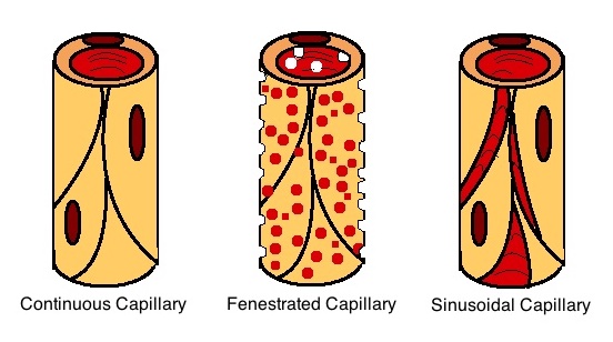
A capillary is a small blood vessel, from 5 to 10 micrometres in diameter, and is part of the microcirculation system. Capillaries are microvessels and the smallest blood vessels in the body. They are composed of only the tunica intima (the innermost layer of an artery or vein), consisting of a thin wall of simple squamous endothelial cells. They are the site of the exchange of many substances from the surrounding interstitial fluid, and they convey blood from the smallest branches of the arteries (arterioles) to those of the veins (venules). Other substances which cross capillaries include water, oxygen, carbon dioxide, urea, glucose, uric acid, lactic acid and creatinine. Lymph capillaries connect with larger lymph vessels to drain lymphatic fluid collected in microcirculation. Etymology ''Capillary'' comes from the Latin word , meaning "of or resembling hair", with use in English beginning in the mid-17th century. The meaning stems from the tiny, hairlike diameter of a capi ... [...More Info...] [...Related Items...] OR: [Wikipedia] [Google] [Baidu] |
Human Eye
The human eye is a sensory organ in the visual system that reacts to light, visible light allowing eyesight. Other functions include maintaining the circadian rhythm, and Balance (ability), keeping balance. The eye can be considered as a living optics, optical device. It is approximately spherical in shape, with its outer layers, such as the outermost, white part of the eye (the sclera) and one of its inner layers (the pigmented choroid) keeping the eye essentially stray light, light tight except on the eye's optic axis. In order, along the optic axis, the optical components consist of a first lens (the cornea, cornea—the clear part of the eye) that accounts for most of the optical power of the eye and accomplishes most of the Focus (optics), focusing of light from the outside world; then an aperture (the pupil) in a Diaphragm (optics), diaphragm (the Iris (anatomy), iris—the coloured part of the eye) that controls the amount of light entering the interior of the eye; then an ... [...More Info...] [...Related Items...] OR: [Wikipedia] [Google] [Baidu] |
Birth
Birth is the act or process of bearing or bringing forth offspring, also referred to in technical contexts as parturition. In mammals, the process is initiated by hormones which cause the muscular walls of the uterus to contract, expelling the fetus at a developmental stage when it is ready to feed and breathe. In some species, the offspring is precocial and can move around almost immediately after birth but in others, it is altricial and completely dependent on parenting. In marsupials, the fetus is born at a very immature stage after a short gestation and develops further in its mother's womb Pouch (marsupial), pouch. It is not only mammals that give birth. Some reptiles, amphibians, fish and invertebrates carry their developing young inside them. Some of these are Ovoviviparity, ovoviviparous, with the eggs being hatched inside the mother's body, and others are Viviparity, viviparous, with the embryo developing inside their body, as in the case of mammals. Human childbirth ... [...More Info...] [...Related Items...] OR: [Wikipedia] [Google] [Baidu] |
Apoptosis
Apoptosis (from ) is a form of programmed cell death that occurs in multicellular organisms and in some eukaryotic, single-celled microorganisms such as yeast. Biochemistry, Biochemical events lead to characteristic cell changes (Morphology (biology), morphology) and death. These changes include Bleb (cell biology), blebbing, Plasmolysis, cell shrinkage, Karyorrhexis, nuclear fragmentation, Pyknosis, chromatin condensation, Apoptotic DNA fragmentation, DNA fragmentation, and mRNA decay. The average adult human loses 50 to 70 1,000,000,000, billion cells each day due to apoptosis. For the average human child between 8 and 14 years old, each day the approximate loss is 20 to 30 billion cells. In contrast to necrosis, which is a form of traumatic cell death that results from acute cellular injury, apoptosis is a highly regulated and controlled process that confers advantages during an organism's life cycle. For example, the separation of fingers and toes in a developing human embryo ... [...More Info...] [...Related Items...] OR: [Wikipedia] [Google] [Baidu] |
Scanning Electron Microscope
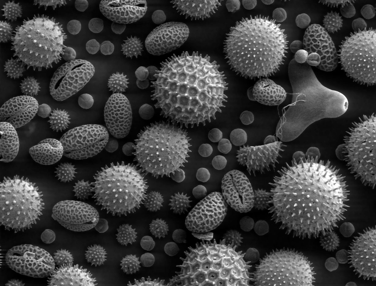
A scanning electron microscope (SEM) is a type of electron microscope that produces images of a sample by scanning the surface with a focused beam of electrons. The electrons interact with atoms in the sample, producing various signals that contain information about the surface topography and composition. The electron beam is scanned in a raster scan pattern, and the position of the beam is combined with the intensity of the detected signal to produce an image. In the most common SEM mode, secondary electrons emitted by atoms excited by the electron beam are detected using a secondary electron detector ( Everhart–Thornley detector). The number of secondary electrons that can be detected, and thus the signal intensity, depends, among other things, on specimen topography. Some SEMs can achieve resolutions better than 1 nanometer. Specimens are observed in high vacuum in a conventional SEM, or in low vacuum or wet conditions in a variable pressure or environmental SEM, an ... [...More Info...] [...Related Items...] OR: [Wikipedia] [Google] [Baidu] |
Laboratory Animal
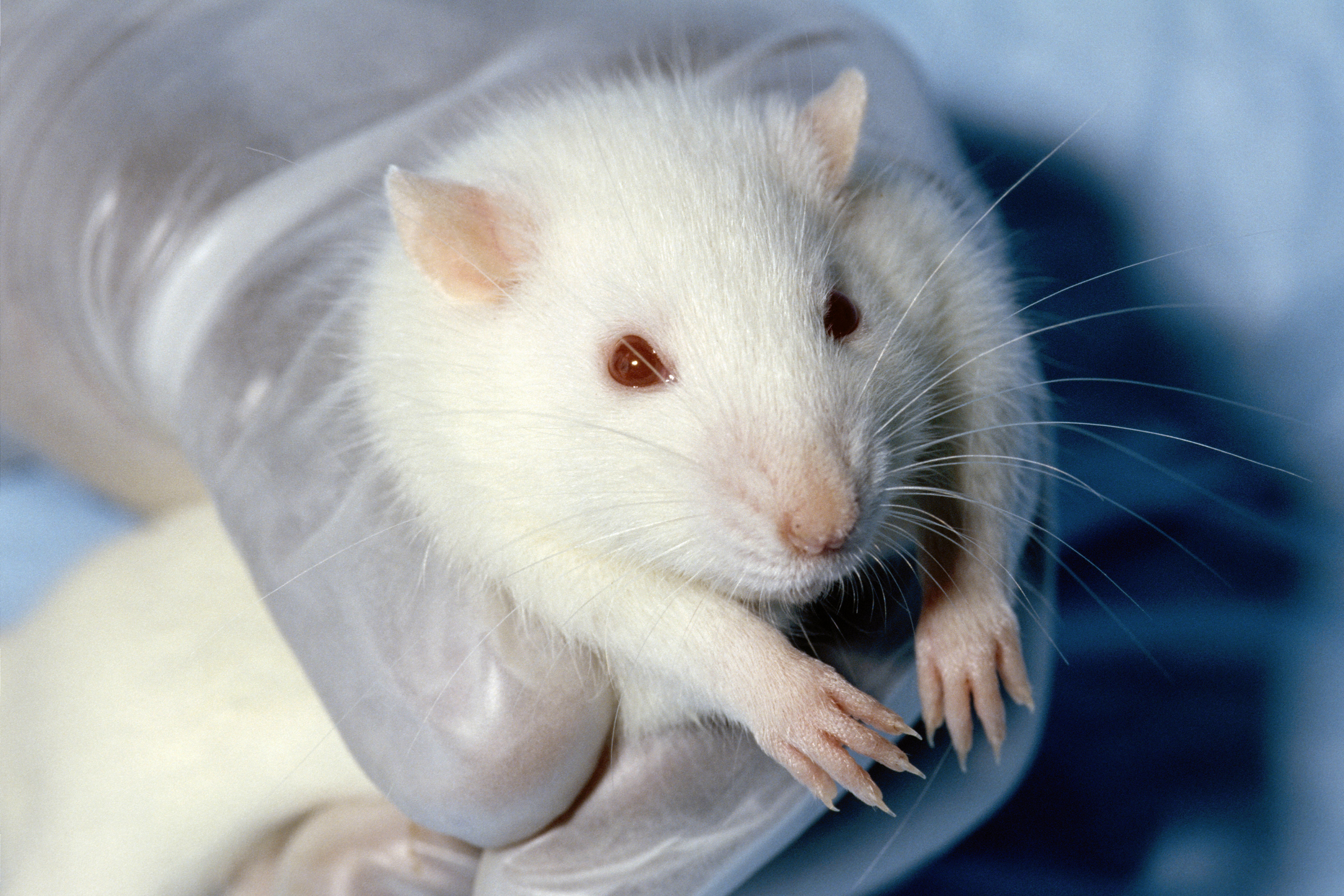
Animal testing, also known as animal experimentation, animal research, and ''in vivo'' testing, is the use of animals, as model organisms, in experiments that seek answers to scientific and medical questions. This approach can be contrasted with field studies in which animals are observed in their natural environments or habitats. Experimental research with animals is usually conducted in universities, medical schools, pharmaceutical companies, defense establishments, and commercial facilities that provide animal-testing services to the industry. The focus of animal testing varies on a continuum from Basic research, pure research, focusing on developing fundamental knowledge of an organism, to applied research, which may focus on answering some questions of great practical importance, such as finding a cure for a disease. Examples of applied research include testing disease treatments, breeding, defense research, and Toxicology testing, toxicology, including Testing cosmetics ... [...More Info...] [...Related Items...] OR: [Wikipedia] [Google] [Baidu] |
Comparative Vertebrate Anatomy
Comparative anatomy is the study of similarities and differences in the anatomy of different species. It is closely related to evolutionary biology and phylogeny (the evolution of species). The science began in the classical era, continuing in the early modern period with work by Pierre Belon who noted the similarities of the skeletons of birds and humans. Comparative anatomy has provided evidence of common descent, and has assisted in the classification of animals. History The first specifically anatomical investigation separate from a surgical or medical procedure is associated by Alcmaeon of Croton. Leonardo da Vinci made notes for a planned anatomical treatise in which he intended to compare the hands of various animals including bears. Pierre Belon, a French naturalist born in 1517, conducted research and held discussions on dolphin embryos as well as the comparisons between the skeletons of birds to the skeletons of humans. His research led to modern comparative anatom ... [...More Info...] [...Related Items...] OR: [Wikipedia] [Google] [Baidu] |
Ophthalmology
Ophthalmology (, ) is the branch of medicine that deals with the diagnosis, treatment, and surgery of eye diseases and disorders. An ophthalmologist is a physician who undergoes subspecialty training in medical and surgical eye care. Following a medical degree, a doctor specialising in ophthalmology must pursue additional postgraduate residency training specific to that field. In the United States, following graduation from medical school, one must complete a four-year residency in ophthalmology to become an ophthalmologist. Following residency, additional specialty training (or fellowship) may be sought in a particular aspect of eye pathology. Ophthalmologists prescribe medications to treat ailments, such as eye diseases, implement laser therapy, and perform surgery when needed. Ophthalmologists provide both primary and specialty eye care—medical and surgical. Most ophthalmologists participate in academic research on eye diseases at some point in their training and many inc ... [...More Info...] [...Related Items...] OR: [Wikipedia] [Google] [Baidu] |
Persistent Tunica Vasculosa Lentis
Persistent tunica vasculosa lentis is a congenital ocular anomaly. It is a form of persistent fetal vasculature (PFV). It is a developmental disorder of the vitreous. It is usually unilateral and first noticed in the neonatal period. It may be associated with microphthalmos, cataracts, and increased intraocular pressure. Elongated ciliary processes In the anatomy of the eye, the ciliary processes are formed by the inward folding of the various layers of the choroid, viz. the choroid proper and the lamina basalis, and are received between corresponding foldings of the suspensory ligam ... are visible through the dilated pupil. A USG B-scan confirms diagnosis in the presence of a cataract. See also * Persistent fetal vasculature * Tunica vasculosa lentis References * http://www.djo.harvard.edu/files/1568.pdf * Wright KW, Spiegel PH, Buckley EG. Pediatric Ophthalmology and Strabismus. Springer; US, New York: 2003. p. 17. Eye diseases {{eye-disease-stu ... [...More Info...] [...Related Items...] OR: [Wikipedia] [Google] [Baidu] |


