|
Trichoepithelioma
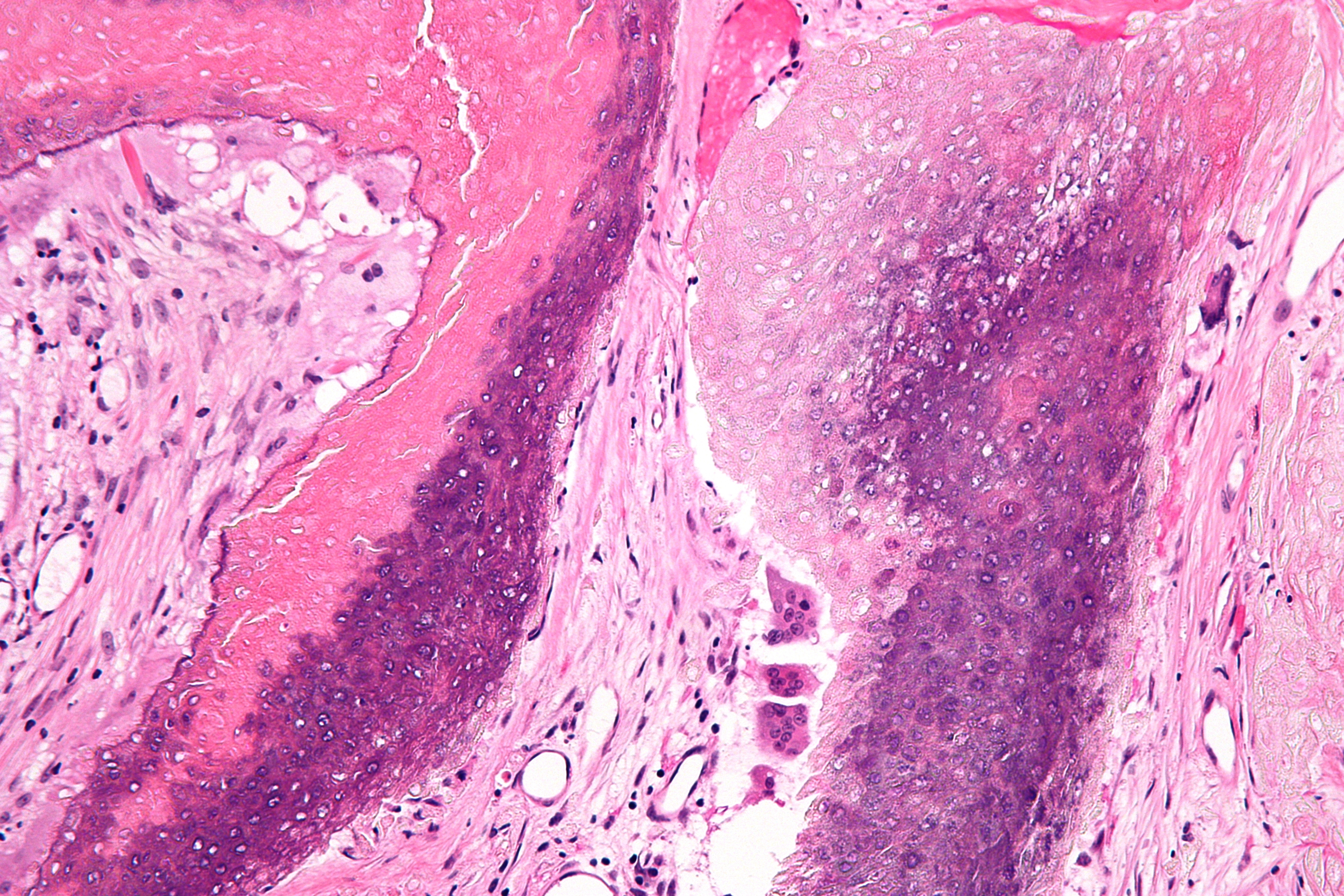
Trichoepithelioma is a neoplasm of the adnexa of the skin. Its appearance is similar to basal cell carcinoma. One form has been mapped to chromosome 9p21. Types Trichoepitheliomas may be divided into the following types: :* Multiple familial trichoepithelioma :* Solitary trichoepithelioma :* Desmoplastic trichoepithelioma Pathology Trichoepitheliomas consists of nests of basaloid cells, with palisading. They lack the myxoid stroma and artefactual clefting seen in basal cell carcinoma. Mitoses are uncommon when compared to basal cell carcinoma. Diagnosis Trichoepiteliomas often contain Merkel cells; an immunostain for CK20 can be used to demonstrate this. See also * Trichoblastoma * Pilomatricoma * CYLD cutaneous syndrome * List of cutaneous conditions Many skin conditions affect the human integumentary system—the organ system covering the entire surface of the Human body, body and composed of Human skin, skin, hair, Nail (anatomy), nails, and related mus ... [...More Info...] [...Related Items...] OR: [Wikipedia] [Google] [Baidu] |
CYLD Cutaneous Syndrome
CYLD cutaneous syndrome (CCS) encompasses three rare inherited cutaneous adnexal tumor syndromes: multiple familial trichoepithelioma (MFT1) (also termed epithelioma adenoides cysticum and epithelioma adenoides cysticum of Brooke), Brooke–Spiegler syndrome (BSS), and familial cylindromatosis (FC). Cutaneous adnexal tumors are a large group of skin tumors that consist of tissues that have differentiated (i.e. matured from stem cells) towards one of the four primary adnexal structures found in normal skin: hair follicles, sebaceous sweat glands, apocrine sweat glands, and eccrine sweat glands. CCS tumors are hair follicle tumors. Individuals with the MFT1, BSS, and FC forms of CCS carry a germline (i.e. present in the germ cells which give rise to an individual) mutation in one of their two '' CYLD'' (i.e. CYLD lysine 63 deubiquitinase) genes. These individuals have skin tumors that tend to cluster into MFT1, BSS, and/or FC types that differ from each other in their locations, o ... [...More Info...] [...Related Items...] OR: [Wikipedia] [Google] [Baidu] |
Desmoplastic Trichoepithelioma
Desmoplastic trichoepithelioma is a cutaneous condition characterized by a solitary, firm skin lesion on the face. Familial cases have been reported suggesting a possible genetic link. The diagnosis is made based on clinical and morphological features. Differential diagnosis includes trichoepithelioma, microcystic adnexal carcinoma, morpheaform basal cell carcinoma, and syringoma. Surgical excision is the treatment of choice. Desmoplastic trichoepithelioma is more common in females and usually affects children or adults. Signs and symptoms Desmoplastic trichoepithelioma manifests as a firm, soft, or white to yellowish annular nodule or papule on the cheeks or face with a central indentation. Causes Familial desmoplastic trichoepitheliomas have been reported suggesting a possible genetic link. Diagnosis Desmoplastic trichoepithelioma can be diagnosed based on clinical and morphological features. Desmoplastic trichoepithelioma may resemble other tumors such as conventiona ... [...More Info...] [...Related Items...] OR: [Wikipedia] [Google] [Baidu] |
List Of Cutaneous Conditions
Many skin conditions affect the human integumentary system—the organ system covering the entire surface of the Human body, body and composed of Human skin, skin, hair, Nail (anatomy), nails, and related muscle and glands. The major function of this system is as a barrier against the external environment. The skin weighs an average of four kilograms, covers an area of two square metres, and is made of three distinct layers: the epidermis (skin), epidermis, dermis, and subcutaneous tissue. The two main types of human skin are: glabrous skin, the hairless skin on the palms and soles (also referred to as the "palmoplantar" surfaces), and hair-bearing skin.Burns, Tony; ''et al''. (2006) ''Rook's Textbook of Dermatology CD-ROM''. Wiley-Blackwell. . Within the latter type, the hairs occur in structures called pilosebaceous units, each with hair follicle, sebaceous gland, and associated arrector pili muscle. Embryology, In the embryo, the epidermis, hair, and glands form from the ectod ... [...More Info...] [...Related Items...] OR: [Wikipedia] [Google] [Baidu] |
List Of Cutaneous Neoplasms Associated With Systemic Syndromes
Many cutaneous neoplasms occur in the setting of systemic syndromes. See also *List of cutaneous conditions *List of contact allergens *List of cutaneous conditions associated with increased risk of nonmelanoma skin cancer *List of cutaneous conditions associated with internal malignancy *List of cutaneous conditions caused by mutations in keratins *List of cutaneous conditions caused by problems with junctional proteins *List of dental abnormalities associated with cutaneous conditions *List of genes mutated in cutaneous conditions *List of genes mutated in pigmented cutaneous lesions *List of histologic stains that aid in diagnosis of cutaneous conditions *List of immunofluorescence findings for autoimmune bullous conditions *List of inclusion bodies that aid in diagnosis of cutaneous conditions *List of keratins expressed in the human integumentary system *List of radiographic findings associated with cutaneous conditions *List of specialized glands within the human integum ... [...More Info...] [...Related Items...] OR: [Wikipedia] [Google] [Baidu] |
Solitary Trichoepithelioma
Solitary trichoepithelioma is a cutaneous condition characterized by a firm dermal papules or nodules most commonly occurring on the face. See also * Trichoepithelioma * Skin lesion A skin condition, also known as cutaneous condition, is any medical condition that affects the integumentary system—the organ system that encloses the body and includes skin, nails, and related muscle and glands. The major function of this ... References Epidermal nevi, neoplasms, and cysts {{Epidermal-growth-stub ... [...More Info...] [...Related Items...] OR: [Wikipedia] [Google] [Baidu] |
Micrograph
A micrograph is an image, captured photographically or digitally, taken through a microscope or similar device to show a magnify, magnified image of an object. This is opposed to a macrograph or photomacrograph, an image which is also taken on a microscope but is only slightly magnified, usually less than 10 times. Micrography is the practice or art of using microscopes to make photographs. A photographic micrograph is a photomicrograph, and one taken with an electron microscope is an electron micrograph. A micrograph contains extensive details of microstructure. A wealth of information can be obtained from a simple micrograph like behavior of the material under different conditions, the phases found in the system, failure analysis, grain size estimation, elemental analysis and so on. Micrographs are widely used in all fields of microscopy. Types Photomicrograph A light micrograph or photomicrograph is a micrograph prepared using an optical microscope, a process referred to ... [...More Info...] [...Related Items...] OR: [Wikipedia] [Google] [Baidu] |
Merkel Cell
Merkel cells, also known as Merkel–Ranvier cells or tactile epithelial cells, are oval-shaped mechanoreceptors essential for light touch sensation and found in the skin of vertebrates. They are abundant in highly sensitive skin like that of the fingertips in humans, and make synaptic contacts with somatosensory afferent nerve fibers. It has been reported that Merkel cells are derived from neural crest cells, though more recent experiments in mammals have indicated that they are epithelial in origin. Merkel cells functionally resemble the enterochromaffin cell, the mechanosensory cell of the gastrointestinal epithelium.Chang W, Kanda H, Ikeda R, Ling J, DeBerry JJ, Gu JG. Merkel disc is a serotonergic synapse in the epidermis for transmitting tactile signals in mammals. Proc Natl Acad Sci U S A. 2016 Sep 13;113(37): E5491-500. doi: 10.1073/pnas.1610176113. Structure Merkel cells are found in the skin and some parts of the mucosa of all vertebrates. In mammalian skin, they ar ... [...More Info...] [...Related Items...] OR: [Wikipedia] [Google] [Baidu] |
Pilomatricoma
Pilomatricoma is a benign skin tumor derived from the hair matrix. These neoplasms are relatively uncommon and typically occur on the scalp, face, and upper extremities. Clinically, pilomatricomas present as a subcutaneous nodule or cyst with unremarkable overlying epidermis that can range in size from 0.5 to 3.0 cm, but the largest reported case was 24 cm. Presentation Associations Pilomatricomas have been observed in a variety of genetic disorders including Turner syndrome, myotonic dystrophy, Rubinstein–Taybi syndrome, trisomy 9, and Gardner syndrome. It has been reported that the prevalence of pilomatricomas in Turner syndrome is 2.6%. Hybrid cysts that are composed of epidermal inclusion cysts and pilomatricoma-like changes have been repeatedly observed in Gardner syndrome. This association has prognostic import, since cutaneous findings in children with Gardner syndrome generally precede colonic polyposis. Histologic features The characteristic components of ... [...More Info...] [...Related Items...] OR: [Wikipedia] [Google] [Baidu] |
Trichoblastoma
Trichoblastomas are a Human skin, skin condition characterized by benign neoplasms of the Hair follicle, follicular germinative cells known as ''trichoblasts''. ''Trichoblastic fibroma'' is a term used to describe small Nodule (medicine), nodular trichoblastomas that contain a conspicuous Fibrocyte, fibrocytic component, sometimes constituting over 50% of the lesion. Image at left shows a trichoblastoma from a 68-year-old Caucasian male. It shows a pseudo-encapsulated, multinodular, basaloid tumor with fibrocellular stroma spanning the reticular dermis extending into subcutaneous fat (A). No epidermal connection or retraction artifact was noted. Tumor lobules were arranged as monomorphous basaloid cells in a cribriform pattern with peripheral palisading some resembling abortive hair follicles (B, F). Focally, tumor lobules exhibited squamous eddies, papillary mesenchymal bodies, and a germinative component comprising basaloid cells admixed with distinct pales cells (Zellballen) (C ... [...More Info...] [...Related Items...] OR: [Wikipedia] [Google] [Baidu] |
Keratin 20
Keratin 20, often abbreviated CK20, is a protein that in humans is encoded by the ''KRT20'' gene. Keratin 20 is a type I cytokeratin. It is a major cellular protein of mature enterocytes and goblet cells and is specifically found in the gastric and intestinal mucosa. In immunohistochemistry, antibodies to CK20 can be used to identify a range of adenocarcinoma arising from epithelia that normally contain the CK20 protein. For example, the protein is commonly found in colorectal cancer, transitional cell carcinomas and in Merkel cell carcinoma, but is absent in lung cancer, prostate cancer Prostate cancer is the neoplasm, uncontrolled growth of cells in the prostate, a gland in the male reproductive system below the bladder. Abnormal growth of the prostate tissue is usually detected through Screening (medicine), screening tests, ..., and non-mucinous ovarian cancer. It is often used in combination with antibodies to CK7 to distinguish different types of glandular tumour ... [...More Info...] [...Related Items...] OR: [Wikipedia] [Google] [Baidu] |
Immunohistochemistry
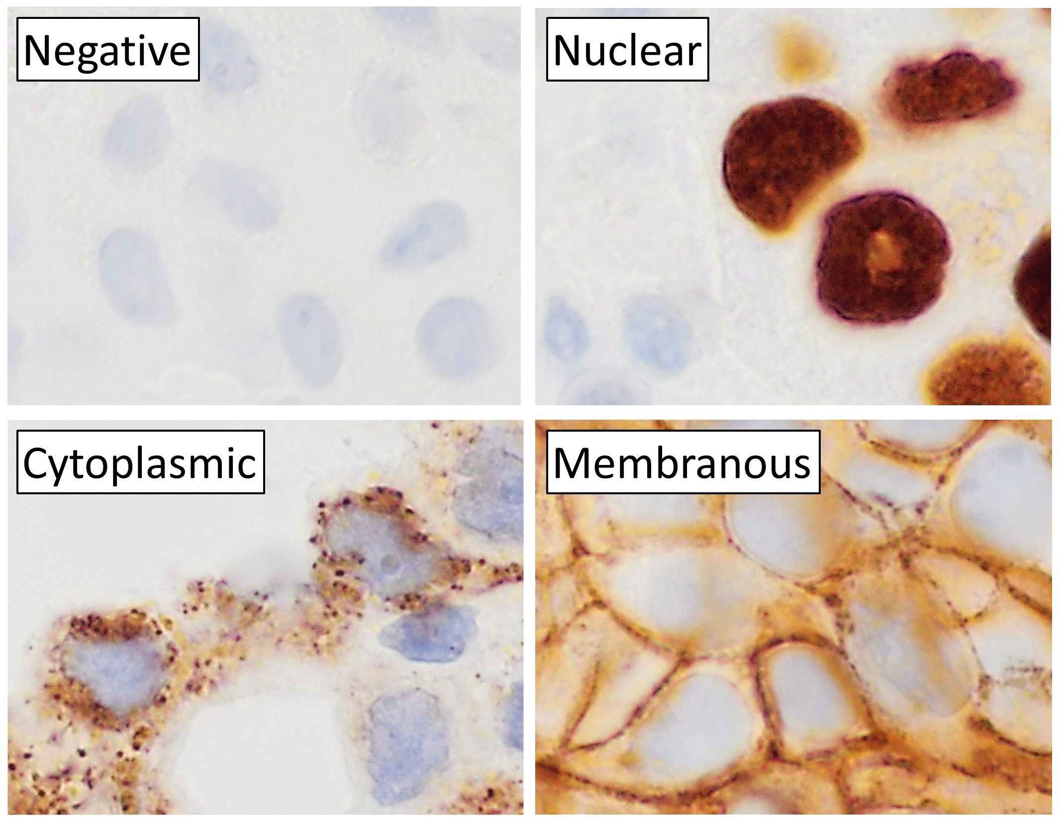
Immunohistochemistry is a form of immunostaining. It involves the process of selectively identifying antigens in cells and tissue, by exploiting the principle of Antibody, antibodies binding specifically to antigens in biological tissues. Albert Coons, Albert Hewett Coons, Ernst Berliner, Ernest Berliner, Norman Jones and Hugh J Creech was the first to develop immunofluorescence in 1941. This led to the later development of immunohistochemistry. Immunohistochemical staining is widely used in the diagnosis of abnormal cells such as those found in cancerous tumors. In some cancer cells certain tumor antigens are expressed which make it possible to detect. Immunohistochemistry is also widely used in basic research, to understand the distribution and localization of biomarkers and differentially expressed proteins in different parts of a biological tissue. Sample preparation Immunohistochemistry can be performed on tissue that has been fixed and embedded in Paraffin wax, paraffin, ... [...More Info...] [...Related Items...] OR: [Wikipedia] [Google] [Baidu] |
Palisade (pathology)
In histopathology, a palisade is a single layer of relatively long cells, arranged loosely perpendicular to a surface and parallel to each other. A rosette is a palisade in a halo or spoke-and-wheel arrangement, surrounding a central core or hub. A pseudorosette is a perivascular radial arrangement of neoplastic cells around a small blood vessel. Rosettes are characteristic of tumors. Rosette A ''rosette'' is a cell formation in a halo or spoke-and-wheel arrangement, surrounding a central core or hub. The central hub may consist of an empty-appearing lumen or a space filled with cytoplasmic processes. The cytoplasm of each of the cells in the rosette is often wedge-shaped with the apex directed toward the central core: the nuclei of the cells participating in the rosette are peripherally positioned and form a ring or halo around the hub. Pathogenesis Rosettes may be considered primary or secondary manifestations of tumor architecture. Primary rosettes form as a characteristic ... [...More Info...] [...Related Items...] OR: [Wikipedia] [Google] [Baidu] |


