|
Triangular Articular Disk
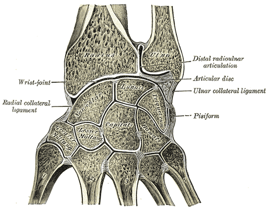
The triangular fibrocartilage complex (TFCC) is formed by the triangular fibrocartilage discus (TFC), the radioulnar ligaments (RULs) and the ulnocarpal ligaments (UCLs). Structure Triangular fibrocartilage disc The triangular fibrocartilage disc (TFC) is an articular discus that lies on the pole of the distal ulna. It has a triangular shape and a biconcave body; the periphery is thicker than its center. The central portion of the TFC is thin and consists of chondroid fibrocartilage; this type of tissue is often seen in structures that can bear compressive loads. This central area is often so thin that it is translucent and in some cases it is even absent. The peripheral portion of the TFC is well vascularized, while the central portion has no blood supply. This discus is attached by thick tissue to the base of the ulnar styloid and by thinner tissue to the edge of the radius just proximal to the radiocarpal articular surface. Radioulnar ligaments The radioulnar ligaments ... [...More Info...] [...Related Items...] OR: [Wikipedia] [Google] [Baidu] |
Radioulnar Ligament (other)
Radioulnar ligament can refer to: * Dorsal radioulnar ligament The dorsal radioulnar ligament (posterior radioulnar ligament) extends between corresponding surfaces on the dorsal aspect of the distal radioulnar articulation. References Ligaments of the upper limb {{Portal bar, Anatomy ... (ligamentum radioulnare dorsale) * Palmar radioulnar ligament (ligamentum radioulnare palmare) {{disambiguation ... [...More Info...] [...Related Items...] OR: [Wikipedia] [Google] [Baidu] |
Anatomical Terms Of Motion
Motion, the process of movement, is described using specific anatomical terms. Motion includes movement of organs, joints, limbs, and specific sections of the body. The terminology used describes this motion according to its direction relative to the anatomical position of the body parts involved. Anatomists and others use a unified set of terms to describe most of the movements, although other, more specialized terms are necessary for describing unique movements such as those of the hands, feet, and eyes. In general, motion is classified according to the anatomical plane it occurs in. ''Flexion'' and ''extension'' are examples of ''angular'' motions, in which two axes of a joint are brought closer together or moved further apart. ''Rotational'' motion may occur at other joints, for example the shoulder, and are described as ''internal'' or ''external''. Other terms, such as ''elevation'' and ''depression'', describe movement above or below the horizontal plane. Many ana ... [...More Info...] [...Related Items...] OR: [Wikipedia] [Google] [Baidu] |
Hypothenar Hammer Syndrome
Hypothenar hammer syndrome (HHS) is a vascular occlusion in humans in the region of the ulna. It is caused by repetitive trauma to the hand or wrist (such as that caused by the use of a hammer) by the vulnerable portion of the ulnar artery as it passes over the hamate bone, which may result in thrombosis, irregularity or aneurysm formation. HHS is a potentially curable cause of Raynaud's syndrome, distinct from hand–arm vibration syndrome. Cause Diagnosis A physical examination of the hand may show discoloration (blanching, mottling, and/ or cyanosis; gangrene may be present in advanced cases), unusual tenderness/ a callous over the hypothenar eminence, and fingertip ulcerations and splinter hemorrhages over ulnar digits; if an aneurysm is present, there may also be a pulsatile mass. Allen's test will be positive if an occlusion is present and negative if an aneurysm is present. An angiogram Angiography or arteriography is a medical imaging technique used to visualize the ... [...More Info...] [...Related Items...] OR: [Wikipedia] [Google] [Baidu] |
Wrist Arthroscopy
Wrist arthroscopy can be used to look inside the joint of the wrist. It is a minimally invasive technique which can be utilized for diagnostic purposes as well as for therapeutic interventions. Wrist arthroscopy has been used for diagnostic purposes since it was first introduced in 1979. However, it only became accepted as diagnostic tool around the mid-1980s. At that time, arthroscopy of the wrist was an innovative technique to determine whether a problem could be found in the wrist. A few years later, wrist arthroscopy could also be used as a therapeutic tool. Medical uses There are several therapeutic wrist arthroscopy indications, in this article the focus will be on the TFCC-lesion, the SL-lesion, the dorsal ganglion resection and the distal radius fracture. TFCC lesion The TFCC is a fibrous structure covering both the radiocarpal and distal radioulnar joint. Tears in this ligament occur commonly after a person falls or secondary to a wrist fracture. Abnormalities in the ... [...More Info...] [...Related Items...] OR: [Wikipedia] [Google] [Baidu] |
Arthrography
An arthrogram is a series of images of a joint after injection of a contrast medium, usually done by fluoroscopy or MRI. The injection is normally done under a local anesthetic such as Novocain or lidocaine. The radiologist or radiographer performs the study using fluoroscopy or x-ray to guide the placement of the needle into the joint and then injects around 10 ml of contrast based on age. There is some burning pain from the anesthetic and a painful bubbling feeling in the joint after the contrast is injected. This only lasts 20 – 30 hours until the Contrast is absorbed. During this time, while it is allowed, it is painful to use the limb for around 10 hours. After that the radiologist can more clearly see what is going on under your skin and can get results out within 24 to 48 hours. Types Conventional arthrography It is used primarily in the evaluation of menisci, cruciate ligaments, articular cartilage, and loose body within a joint. Fluoroscopic allows general view of the ... [...More Info...] [...Related Items...] OR: [Wikipedia] [Google] [Baidu] |
Radiography Radiography is an imaging technique using X-rays, gamma rays, or similar ionizing radiation and non-ionizing radiation to view the internal form of an object. Applications of radiography include medical radiography ("diagnostic" and "therapeutic") and industrial radiography. Similar techniques are used in airport security (where "body scanners" generally use bac |

