|
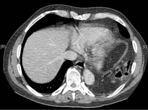
Traumatic Diaphragmatic Hernia
Diaphragmatic rupture (also called diaphragmatic injury or tear) is a tear of the diaphragm, the muscle across the bottom of the ribcage that plays a crucial role in breathing. Most commonly, acquired diaphragmatic tears result from physical trauma. Diaphragmatic rupture can result from blunt or penetrating trauma and occurs in about 0.5% of all people with trauma. Diagnostic techniques include X-ray, computed tomography, and surgical techniques such as an explorative surgery. Diagnosis is often difficult because signs may not show up on X-ray, or signs that do show up appear similar to other conditions. Signs and symptoms include chest and abdominal pain, difficulty breathing, and decreased lung sounds. When a tear is discovered, surgery is needed to repair it. Injuries to the diaphragm are usually accompanied by other injuries, and they indicate that more severe injury may have occurred. The outcome often depends more on associated injuries than on the diaphragmatic injury it ... [...More Info...] [...Related Items...] OR: [Wikipedia] [Google] [Baidu] |
X-ray
X-rays (or rarely, ''X-radiation'') are a form of high-energy electromagnetic radiation. In many languages, it is referred to as Röntgen radiation, after the German scientist Wilhelm Conrad Röntgen, who discovered it in 1895 and named it ''X-radiation'' to signify an unknown type of radiation.Novelline, Robert (1997). ''Squire's Fundamentals of Radiology''. Harvard University Press. 5th edition. . X-ray wavelengths are shorter than those of ultraviolet rays and longer than those of gamma rays. There is no universally accepted, strict definition of the bounds of the X-ray band. Roughly, X-rays have a wavelength ranging from 10 nanometers to 10 picometers, corresponding to frequencies in the range of 30 petahertz to 30 exahertz ( to ) and photon energies in the range of 100 eV to 100 keV, respectively. X-rays can penetrate many solid substances such as construction materials and living tissue, so X-ray radiography is widely used in medi ... [...More Info...] [...Related Items...] OR: [Wikipedia] [Google] [Baidu] |
Stabbing
A stabbing is penetration or rough contact with a sharp or pointed object at close range. ''Stab'' connotes purposeful action, as by an assassin or murderer, but it is also possible to accidentally stab oneself or others. Stabbing differs from slashing or cutting in that the motion of the object used in a stabbing generally moves perpendicular to and directly into the victim's body, rather than being drawn across it. Stabbings today are common among gangs and in prisons because knives are cheap, easy to acquire (or manufacture), easily concealable and relatively effective. In 2013, about 8 million stabbings occurred. History Stabbings have been common throughout human history and were the means used to assassinate a number of distinguished historical figures, such as Second Caliph Umar and Roman dictator Julius Caesar and emperor Caligula. In Japan, the historical practice of stabbing oneself deliberately in ritual suicide is known as '' seppuku'' (more colloquiall ... [...More Info...] [...Related Items...] OR: [Wikipedia] [Google] [Baidu] |
Pneumothorax
A pneumothorax is an abnormal collection of air in the pleural space between the lung and the chest wall. Symptoms typically include sudden onset of sharp, one-sided chest pain and shortness of breath. In a minority of cases, a one-way valve is formed by an area of damaged tissue, and the amount of air in the space between chest wall and lungs increases; this is called a tension pneumothorax. This can cause a steadily worsening oxygen shortage and low blood pressure. This leads to a type of shock called obstructive shock, which can be fatal unless reversed. Very rarely, both lungs may be affected by a pneumothorax. It is often called a "collapsed lung", although that term may also refer to atelectasis. A primary spontaneous pneumothorax is one that occurs without an apparent cause and in the absence of significant lung disease. A secondary spontaneous pneumothorax occurs in the presence of existing lung disease. Smoking increases the risk of primary spontaneous pneumot ... [...More Info...] [...Related Items...] OR: [Wikipedia] [Google] [Baidu] |
Hemothorax
A hemothorax (derived from hemo- lood+ thorax hest plural ''hemothoraces'') is an accumulation of blood within the pleural cavity. The symptoms of a hemothorax may include chest pain and difficulty breathing, while the clinical signs may include reduced breath sounds on the affected side and a rapid heart rate. Hemothoraces are usually caused by an injury, but they may occur spontaneously due to cancer invading the pleural cavity, as a result of a blood clotting disorder, as an unusual manifestation of endometriosis, in response to a collapsed lung, or rarely in association with other conditions. Hemothoraces are usually diagnosed using a chest X-ray, but they can be identified using other forms of imaging including ultrasound, a CT scan, or an MRI. They can be differentiated from other forms of fluid within the pleural cavity by analysing a sample of the fluid, and are defined as having a hematocrit of greater than 50% that of the person's blood. Hemothoraces may be tr ... [...More Info...] [...Related Items...] OR: [Wikipedia] [Google] [Baidu] |
Pulmonary Contusion
A pulmonary contusion, also known as lung contusion, is a bruise of the lung, caused by chest trauma. As a result of damage to capillaries, blood and other fluids accumulate in the lung tissue. The excess fluid interferes with gas exchange, potentially leading to inadequate oxygen levels ( hypoxia). Unlike pulmonary laceration, another type of lung injury, pulmonary contusion does not involve a cut or tear of the lung tissue. A pulmonary contusion is usually caused directly by blunt trauma but can also result from explosion injuries or a shock wave associated with penetrating trauma. With the use of explosives during World Wars I and II, pulmonary contusion resulting from blasts gained recognition. In the 1960s its occurrence in civilians began to receive wider recognition, in which cases it is usually caused by traffic accidents. The use of seat belts and airbags reduces the risk to vehicle occupants. Diagnosis is made by studying the cause of the injury, physical examina ... [...More Info...] [...Related Items...] OR: [Wikipedia] [Google] [Baidu] |
Sensitivity And Specificity
In medicine and statistics, sensitivity and specificity mathematically describe the accuracy of a test that reports the presence or absence of a medical condition. If individuals who have the condition are considered "positive" and those who do not are considered "negative", then sensitivity is a measure of how well a test can identify true positives and specificity is a measure of how well a test can identify true negatives: * Sensitivity (true positive rate) is the probability of a positive test result, conditioned on the individual truly being positive. * Specificity (true negative rate) is the probability of a negative test result, conditioned on the individual truly being negative. If the true status of the condition cannot be known, sensitivity and specificity can be defined relative to a " gold standard test" which is assumed correct. For all testing, both diagnoses and screening, there is usually a trade-off between sensitivity and specificity, such that higher sensiti ... [...More Info...] [...Related Items...] OR: [Wikipedia] [Google] [Baidu] |
Chest X-ray
A chest radiograph, called a chest X-ray (CXR), or chest film, is a projection radiograph of the chest used to diagnose conditions affecting the chest, its contents, and nearby structures. Chest radiographs are the most common film taken in medicine. Like all methods of radiography, chest radiography employs ionizing radiation in the form of X-rays to generate images of the chest. The mean radiation dose to an adult from a chest radiograph is around 0.02 mSv (2 mrem) for a front view (PA, or posteroanterior) and 0.08 mSv (8 mrem) for a side view (LL, or latero-lateral). Together, this corresponds to a background radiation equivalent time of about 10 days. Medical uses Conditions commonly identified by chest radiography * Pneumonia * Pneumothorax * Interstitial lung disease * Heart failure * Bone fracture * Hiatal hernia Chest radiographs are used to diagnose many conditions involving the chest wall, including its bones, and also structures contained within the tho ... [...More Info...] [...Related Items...] OR: [Wikipedia] [Google] [Baidu] |
Hypoxemia
Hypoxemia is an abnormally low level of oxygen in the blood. More specifically, it is oxygen deficiency in arterial blood. Hypoxemia has many causes, and often causes hypoxia as the blood is not supplying enough oxygen to the tissues of the body. Definition ''Hypoxemia'' refers to the low level of oxygen in blood, and the more general term ''hypoxia'' is an abnormally low oxygen content in any tissue or organ, or the body as a whole. Hypoxemia can cause hypoxia (hypoxemic hypoxia), but hypoxia can also occur via other mechanisms, such as anemia. Hypoxemia is usually defined in terms of reduced partial pressure of oxygen (mm Hg) in arterial blood, but also in terms of reduced content of oxygen (ml oxygen per dl blood) or percentage saturation of hemoglobin (the oxygen-binding protein within red blood cells) with oxygen, which is either found singly or in combination. While there is general agreement that an arterial blood gas measurement which shows that the partial pressure ... [...More Info...] [...Related Items...] OR: [Wikipedia] [Google] [Baidu] |
Ventilation (physiology)
Breathing (or ventilation) is the process of moving air into and from the lungs to facilitate gas exchange with the internal environment, mostly to flush out carbon dioxide and bring in oxygen. All aerobic creatures need oxygen for cellular respiration, which extracts energy from the reaction of oxygen with molecules derived from food and produces carbon dioxide as a waste product. Breathing, or "external respiration", brings air into the lungs where gas exchange takes place in the alveoli through diffusion. The body's circulatory system transports these gases to and from the cells, where "cellular respiration" takes place. The breathing of all vertebrates with lungs consists of repetitive cycles of inhalation and exhalation through a highly branched system of tubes or airways which lead from the nose to the alveoli. The number of respiratory cycles per minute is the breathing or respiratory rate, and is one of the four primary vital signs of life. Under normal conditi ... [...More Info...] [...Related Items...] OR: [Wikipedia] [Google] [Baidu] |
Pleural Cavity
The pleural cavity, pleural space, or interpleural space is the potential space between the pleurae of the pleural sac that surrounds each lung. A small amount of serous pleural fluid is maintained in the pleural cavity to enable lubrication between the membranes, and also to create a pressure gradient. The serous membrane that covers the surface of the lung is the visceral pleura and is separated from the outer membrane the parietal pleura by just the film of pleural fluid in the pleural cavity. The visceral pleura follows the fissures of the lung and the root of the lung structures. The parietal pleura is attached to the mediastinum, the upper surface of the diaphragm, and to the inside of the ribcage. Structure In humans, the left and right lungs are completely separated by the mediastinum, and there is no communication between their pleural cavities. Therefore, in cases of a unilateral pneumothorax, the contralateral lung will remain functioning normally unless ther ... [...More Info...] [...Related Items...] OR: [Wikipedia] [Google] [Baidu] |
Radiofrequency Ablation
Radiofrequency ablation (RFA), also called fulguration, is a medical procedure in which part of the electrical conduction system of the heart, tumor or other dysfunctional tissue is ablated using the heat generated from medium frequency alternating current (in the range of 350–500 kHz). RFA is generally conducted in the outpatient setting, using either local anesthetics or conscious sedation anesthesia. When it is delivered via catheter, it is called radiofrequency catheter ablation. Two important advantages of radio frequency current (over previously used low frequency AC or pulses of DC) are that it does not directly stimulate nerves or heart muscle and therefore can often be used without the need for general anesthesia, and that it is very specific for treating the desired tissue without significant collateral damage; due to this, it is gaining in popularity as an alternative for eligible patients who do not want to undergo surgery. Documented benefits have led to RFA ... [...More Info...] [...Related Items...] OR: [Wikipedia] [Google] [Baidu] |
Thoracentesis
Thoracentesis , also known as thoracocentesis (from Greek ''thōrax'' 'chest, thorax'— GEN ''thōrakos''—and ''kentēsis'' 'pricking, puncture'), pleural tap, needle thoracostomy, or needle decompression (often used term), is an invasive medical procedure to remove fluid or air from the pleural space for diagnostic or therapeutic purposes. A cannula, or hollow needle, is carefully introduced into the thorax, generally after administration of local anesthesia. The procedure was first performed by Morrill Wyman in 1850 and then described by Henry Ingersoll Bowditch in 1852. The recommended location varies depending upon the source. Some sources recommend the midaxillary line, in the eighth, ninth, or tenth intercostal space. Whenever possible, the procedure should be performed under ultrasound guidance, which has shown to reduce complications. Tension pneumothorax is a medical emergency that requires emergent needle decompression before a chest tube is placed. Indications ... [...More Info...] [...Related Items...] OR: [Wikipedia] [Google] [Baidu] |


.jpg)


