|
Tiffany Schmidt
Tiffany M. Schmidt is an American researcher and chronobiologist, currently working as an associate professor of Neurobiology at Northwestern University. Schmidt, who works in Evanston, Illinois, studies the role of retinal ganglion cells (RGC) to determine how light can affect behavior, hormonal changes, vision, sleep, and circadian entrainment. Education and career Schmidt completed her undergraduate studies at Luther College in Decorah, Iowa in 2006, receiving a Bachelor of Arts in Biology and Honors Psychology. Schmidt then pursued her PhD at the University of Minnesota from 2006 until 2010. She studied under Paulo Kofuji, researching a subset of retinal ganglion cells that are intrinsically photosensitive. Schmidt then performed postdoctoral research at Johns Hopkins University from 2011 until 2014. Schmidt worked in the Hattar Lab under Samer Hattar, continuing to study intrinsically photosensitive retinal ganglion cells ( ipRGC). Schmidt then began as an assistant prof ... [...More Info...] [...Related Items...] OR: [Wikipedia] [Google] [Baidu] |
Chronobiology
Chronobiology is a field of biology that examines timing processes, including periodic (cyclic) phenomena in living organisms, such as their adaptation to solar- and lunar-related rhythms. These cycles are known as biological rhythms. Chronobiology comes from the ancient Greek χρόνος (''chrónos'', meaning "time"), and biology, which pertains to the study, or science, of life. The related terms ''chronomics'' and ''chronome'' have been used in some cases to describe either the molecular mechanisms involved in chronobiological phenomena or the more quantitative aspects of chronobiology, particularly where comparison of cycles between organisms is required. Chronobiological studies include but are not limited to comparative anatomy, physiology, genetics, molecular biology and behavior of organisms related to their biological rhythms. Other aspects include epigenetics, development, reproduction, ecology and evolution. The subject Chronobiology studies variations of t ... [...More Info...] [...Related Items...] OR: [Wikipedia] [Google] [Baidu] |
Inner Plexiform Layer
The inner plexiform layer is an area of the retina that is made up of a dense reticulum of fibrils formed by interlaced dendrites A dendrite (from Greek δένδρον ''déndron'', "tree") or dendron is a branched cytoplasmic process that extends from a nerve cell that propagates the electrochemical stimulation received from other neural cells to the cell body, or soma ... of retinal ganglion cells and cells of the inner nuclear layer. Within this reticulum a few branched spongioblasts are sometimes embedded. References External links Overview at utah.edu * Human eye anatomy {{eye-stub ... [...More Info...] [...Related Items...] OR: [Wikipedia] [Google] [Baidu] |
Single-cell RNA-sequencing
Single-cell sequencing examines the nucleic acid sequence information from individual cells with optimized next-generation sequencing technologies, providing a higher resolution of cellular differences and a better understanding of the function of an individual cell in the context of its microenvironment. For example, in cancer, sequencing the DNA of individual cells can give information about mutations carried by small populations of cells. In development, sequencing the RNAs expressed by individual cells can give insight into the existence and behavior of different cell types. In microbial systems, a population of the same species can appear genetically clonal. Still, single-cell sequencing of RNA or epigenetic modifications can reveal cell-to-cell variability that may help populations rapidly adapt to survive in changing environments. Background A typical human cell consists of about 2 x 3.3 billion base pairs of DNA and 600 million mRNA bases. Usually, a mix of millions of ce ... [...More Info...] [...Related Items...] OR: [Wikipedia] [Google] [Baidu] |
National Eye Institute
The National Eye Institute (NEI) is part of the National Institutes of Health, U.S. National Institutes of Health (NIH), an agency of the United States Department of Health and Human Services, U.S. Department of Health and Human Services. The mission of NEI is "to eliminate vision loss and improve quality of life through vision research." NEI consists of two major branches for research: an extramural branch that funds studies outside NIH and an intramural branch that funds research on the NIH campus in Bethesda, Maryland. Most of the NEI budget funds extramural research. NEI was established in 1968 as the nation's leading supporter of eye health and vision research projects. These projects include Basic research, basic science research into the fundamental biology of the eye and the visual system. Translational research, NEI also funds translational and clinical research aimed at developing and testing therapies for eye diseases and disorders. This research is focused on developi ... [...More Info...] [...Related Items...] OR: [Wikipedia] [Google] [Baidu] |
National Institutes Of Health
The National Institutes of Health (NIH) is the primary agency of the United States government responsible for biomedical and public health research. It was founded in 1887 and is part of the United States Department of Health and Human Services (HHS). Many NIH facilities are located in Bethesda, Maryland, and other nearby suburbs of the Washington metropolitan area, with other primary facilities in the Research Triangle Park in North Carolina and smaller satellite facilities located around the United States. The NIH conducts its scientific research through the NIH Intramural Research Program (IRP) and provides significant biomedical research funding to non-NIH research facilities through its Extramural Research Program. , the IRP had 1,200 principal investigators and more than 4,000 postdoctoral fellows in basic, translational, and clinical research, being the largest biomedical research institution in the world, while, as of 2003, the extramural arm provided 28% of biomedical ... [...More Info...] [...Related Items...] OR: [Wikipedia] [Google] [Baidu] |
Chronobiology
Chronobiology is a field of biology that examines timing processes, including periodic (cyclic) phenomena in living organisms, such as their adaptation to solar- and lunar-related rhythms. These cycles are known as biological rhythms. Chronobiology comes from the ancient Greek χρόνος (''chrónos'', meaning "time"), and biology, which pertains to the study, or science, of life. The related terms ''chronomics'' and ''chronome'' have been used in some cases to describe either the molecular mechanisms involved in chronobiological phenomena or the more quantitative aspects of chronobiology, particularly where comparison of cycles between organisms is required. Chronobiological studies include but are not limited to comparative anatomy, physiology, genetics, molecular biology and behavior of organisms related to their biological rhythms. Other aspects include epigenetics, development, reproduction, ecology and evolution. The subject Chronobiology studies variations of t ... [...More Info...] [...Related Items...] OR: [Wikipedia] [Google] [Baidu] |
Society For Research On Biological Rhythms
The Society for Research on Biological Rhythms (SRBR) is a learned society and professional association headquartered in the United States created to advance the interests of chronobiology in academia, industry, education, and research. Formed in 1986, the society has around 1,000 members, and runs the associated academic journal, the Journal of Biological Rhythms. In addition to communicating with academic and public audiences on matters related to chronobiology, the society seeks to foster interdisciplinary exchange of ideas and advocates for the need for funding and research in biological rhythms to guide the development of related policies. Organisation The society holds biennial meetings and informal gatherings, and participates in peer-reviewed science and evidence-based policy making. It is one of four prominent existing Chronology Research Societies and one of the 14 societies that make up ''The World Federation of Societies for Chronobiology''. Through its journal, the ... [...More Info...] [...Related Items...] OR: [Wikipedia] [Google] [Baidu] |
Immunohistochemistry
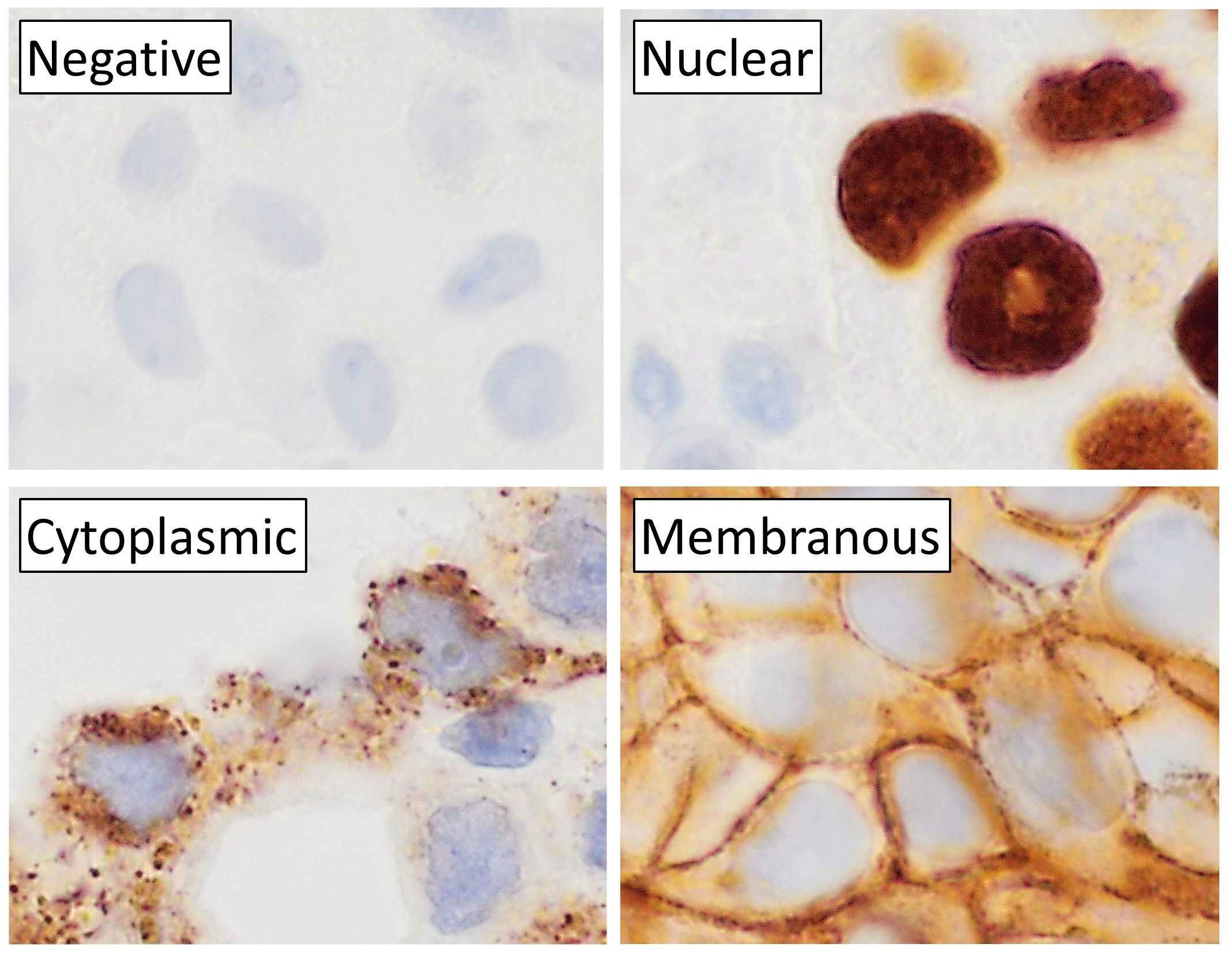
Immunohistochemistry is a form of immunostaining. It involves the process of selectively identifying antigens in cells and tissue, by exploiting the principle of Antibody, antibodies binding specifically to antigens in biological tissues. Albert Coons, Albert Hewett Coons, Ernst Berliner, Ernest Berliner, Norman Jones and Hugh J Creech was the first to develop immunofluorescence in 1941. This led to the later development of immunohistochemistry. Immunohistochemical staining is widely used in the diagnosis of abnormal cells such as those found in cancerous tumors. In some cancer cells certain tumor antigens are expressed which make it possible to detect. Immunohistochemistry is also widely used in basic research, to understand the distribution and localization of biomarkers and differentially expressed proteins in different parts of a biological tissue. Sample preparation Immunohistochemistry can be performed on tissue that has been fixed and embedded in Paraffin wax, paraffin, ... [...More Info...] [...Related Items...] OR: [Wikipedia] [Google] [Baidu] |
Photosensitivity
Photosensitivity is the amount to which an object reacts upon receiving photons, especially visible light. In medicine, the term is principally used for abnormal reactions of the skin, and two types are distinguished, photoallergy and phototoxicity. The photosensitive ganglion cells in the mammalian eye are a separate class of light-detecting cells from the photoreceptor cells that function in vision. Skin reactions Human medicine Sensitivity of the skin to a light source can take various forms. People with particular skin types are more sensitive to sunburn. Particular medications make the skin more sensitive to sunlight; these include most of the tetracycline antibiotics, heart drugs amiodarone, and sulfonamides. Some dietary supplements, such as St. John's Wort, include photosensitivity as a possible side effect. Particular conditions lead to increased light sensitivity. Patients with systemic lupus erythematosus experience skin symptoms after sunlight exposure; some typ ... [...More Info...] [...Related Items...] OR: [Wikipedia] [Google] [Baidu] |
Patch Clamp
The patch clamp technique is a laboratory technique in electrophysiology used to study ionic currents in individual Cell isolation, isolated living cells, tissue sections, or patches of cell membrane. The technique is especially useful in the study of excitable cells such as neurons, cardiomyocytes, muscle fibers, and pancreas, pancreatic beta cells, and can also be applied to the study of bacterial ion channels in specially prepared giant spheroplasts. Patch clamping can be performed using the voltage clamp technique. In this case, the voltage across the cell membrane is controlled by the experimenter and the resulting currents are recorded. Alternatively, the Electrophysiology, current clamp technique can be used. In this case, the current passing across the membrane is controlled by the experimenter and the resulting changes in voltage are recorded, generally in the form of action potentials. Erwin Neher and Bert Sakmann developed the patch clamp in the late 1970s and earl ... [...More Info...] [...Related Items...] OR: [Wikipedia] [Google] [Baidu] |
Voltage Clamp
The voltage clamp is an experimental method used by electrophysiologists to measure the ion currents through the membranes of excitable cells, such as neurons, while holding the membrane voltage at a set level. A basic voltage clamp will iteratively measure the membrane potential, and then change the membrane potential (voltage) to a desired value by adding the necessary current. This "clamps" the cell membrane at a desired constant voltage, allowing the voltage clamp to record what currents are delivered. Because the currents applied to the cell must be equal to (and opposite in charge to) the current going across the cell membrane at the set voltage, the recorded currents indicate how the cell reacts to changes in membrane potential. Cell membranes of excitable cells contain many different kinds of ion channels, some of which are voltage-gated. The voltage clamp allows the membrane voltage to be manipulated independently of the ionic currents, allowing the current–voltag ... [...More Info...] [...Related Items...] OR: [Wikipedia] [Google] [Baidu] |
Current Clamp
In electrical and electronic engineering, a current clamp, also known as current probe, is an electrical device with jaws which open to allow clamping around an electrical conductor. This allows measurement of the current in a conductor without the need to make physical contact with it, or to disconnect it for insertion through the probe. Current clamps are typically used to read the magnitude of alternating current (AC) and, with additional instrumentation, the phase and waveform can also be measured. Some clamp meters can measure currents of 1000 A and more. Hall effect and vane type clamps can also measure direct current (DC). Types of current clamp Current transformer A common form of current clamp comprises a split ring made of ferrite or soft iron. A wire coil is wound round one or both halves, forming one winding of a current transformer. The conductor it is clamped around forms the other winding. Like any transformer this type works only with AC or pulse waveform ... [...More Info...] [...Related Items...] OR: [Wikipedia] [Google] [Baidu] |


