|
Tibial Plateau Fracture
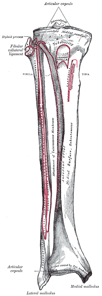
A tibial plateau fracture is a bone fracture, break of the upper part of the tibia (shinbone) that involves the knee joint. This could involve the medial, lateral, central, or bicondylar (medial and lateral). Symptoms include pain, knee effusion, swelling, and a decreased ability to move the knee. People are generally unable to walk. Complication may include injury to the artery or nerve, arthritis, and compartment syndrome. The cause is typically Major trauma, trauma such as a fall or motor vehicle collision. Risk factors include osteoporosis and certain sports such as skiing. Diagnosis is typically suspected based on symptoms and confirmed with radiography, X-rays and a CT scan. Some fractures may not be seen on plain X-rays. Pain may be managed with NSAIDs, opioids, and splint (medicine), splinting. In those who are otherwise healthy, treatment is generally by surgery. Occasionally, if the bones are well aligned and the ligaments of the knee are intact, people may be treated ... [...More Info...] [...Related Items...] OR: [Wikipedia] [Google] [Baidu] |
Fibular
The fibula (: fibulae or fibulas) or calf bone is a leg bone on the lateral side of the tibia, to which it is connected above and below. It is the smaller of the two bones and, in proportion to its length, the most slender of all the long bones. Its upper extremity is small, placed toward the back of the head of the tibia, below the knee joint and excluded from the formation of this joint. Its lower extremity inclines a little forward, so as to be on a plane anterior to that of the upper end; it projects below the tibia and forms the lateral part of the ankle joint. Structure The bone has the following components: * Lateral malleolus * Interosseous membrane connecting the fibula to the tibia, forming a syndesmosis joint * The superior tibiofibular articulation is an arthrodial joint between the lateral condyle of the tibia and the head of the fibula. * The inferior tibiofibular articulation (tibiofibular syndesmosis) is formed by the rough, convex surface of the medial side of ... [...More Info...] [...Related Items...] OR: [Wikipedia] [Google] [Baidu] |
Knee Effusion
Knee effusion, informally known as water on the knee, occurs when excess synovial fluid accumulates in or around the knee joint. It has many common causes, including arthritis, injury to the ligaments or meniscus, or fluid collecting in the bursa, a condition known as prepatellar bursitis. Signs and symptoms Signs and symptoms of water on the knee depend on the cause of excess synovial fluid build-up in the knee joint. While important in lubrication, shock absorption, and nutrient transportation, too much can often be the culprit of a variety of symptoms. Some of which include: Pain Osteoarthritis knee pain usually occurs while the joint is bearing weight, so the pain typically subsides with rest; some patients experience severe pain, while others report no discomfort. Even if one knee is much larger than the other, pain is not guaranteed. Swelling One knee may appear larger than the other. Puffiness around the bony parts of the knee appear prominent when compared with the oth ... [...More Info...] [...Related Items...] OR: [Wikipedia] [Google] [Baidu] |
Medial Collateral Ligament
The medial collateral ligament (MCL), also called the superficial medial collateral ligament (sMCL) or tibial collateral ligament (TCL), is one of the major ligaments of the knee. It is on the medial (inner) side of the knee joint and occurs in humans and other primates. Its primary function is to resist valgus (inward bending) forces on the knee. Structure It is a broad, flat, membranous band, situated slightly posterior on the medial side of the knee joint. It is attached proximally to the medial epicondyle of the femur, immediately below the adductor tubercle; below to the medial condyle of the tibia and medial surface of its body. It resists forces that would push the knee medially, which would otherwise produce valgus deformity. It provides up to 78% of the restraining force that resists valgus (inward pressing) loads on the knee. The fibers of the posterior part of the ligament are short and incline backward as they descend; they are inserted into the tibia above t ... [...More Info...] [...Related Items...] OR: [Wikipedia] [Google] [Baidu] |
Fibula
The fibula (: fibulae or fibulas) or calf bone is a leg bone on the lateral side of the tibia, to which it is connected above and below. It is the smaller of the two bones and, in proportion to its length, the most slender of all the long bones. Its upper extremity is small, placed toward the back of the head of the tibia, below the knee joint and excluded from the formation of this joint. Its lower extremity inclines a little forward, so as to be on a plane anterior to that of the upper end; it projects below the tibia and forms the lateral part of the ankle joint. Structure The bone has the following components: * Lateral malleolus * Interosseous membrane connecting the fibula to the tibia, forming a syndesmosis joint * The superior tibiofibular articulation is an arthrodial joint between the lateral condyle of the tibia and the head of the fibula. * The inferior tibiofibular articulation (tibiofibular syndesmosis) is formed by the rough, convex surface of the medial si ... [...More Info...] [...Related Items...] OR: [Wikipedia] [Google] [Baidu] |
EMedicine
eMedicine is an online clinical medical knowledge base founded in 1996 by doctors Scott Plantz and Jonathan Adler, and computer engineers Joanne Berezin and Jeffrey Berezin. The eMedicine website consists of approximately 6,800 medical topic review articles, each of which is associated with a clinical subspecialty "textbook". The knowledge base includes over 25,000 clinically multimedia files. Each article is authored by board certified specialists in the subspecialty to which the article belongs and undergoes three levels of physician peer-review, plus review by a Doctor of Pharmacy. The article's authors are identified with their current faculty appointments. Each article is updated yearly, or more frequently as changes in practice occur, and the date is published on the article. eMedicine.com was sold to WebMD in January, 2006 and is available as the Medscape Reference. History Plantz, Adler and Berezin evolved the concept for eMedicine.com in 1996 and deployed the initia ... [...More Info...] [...Related Items...] OR: [Wikipedia] [Google] [Baidu] |
Bumper (automobile)
A bumper is a structure attached to or integrated with the front and rear ends of a motor vehicle, to absorb impact in a minor collision, ideally minimizing repair costs. Stiff metal bumpers appeared on automobiles as early as 1904 that had a mainly ornamental function. Numerous developments, improvements in materials and technologies, as well as greater focus on functionality for protecting vehicle components and improving safety have changed bumpers over the years. Bumpers ideally minimize height mismatches between vehicles and Pedestrian safety through vehicle design, protect pedestrians from injury. Regulatory measures have been enacted to reduce vehicle repair costs and, more recently, impact on pedestrians. History Bumpers were, at first, just rigid metal bars. George Albert Lyon invented the earliest car bumper. The first bumper appeared on a vehicle in 1897, and Nesselsdorfer Wagenbau-Fabriksgesellschaft, an Austrian carmaker, installed it. The construction of these bum ... [...More Info...] [...Related Items...] OR: [Wikipedia] [Google] [Baidu] |
Femur
The femur (; : femurs or femora ), or thigh bone is the only long bone, bone in the thigh — the region of the lower limb between the hip and the knee. In many quadrupeds, four-legged animals the femur is the upper bone of the hindleg. The Femoral head, top of the femur fits into a socket in the pelvis called the hip joint, and the bottom of the femur connects to the shinbone (tibia) and kneecap (patella) to form the knee. In humans the femur is the largest and thickest bone in the body. Structure The femur is the only bone in the upper Human leg, leg. The two femurs converge Anatomical terms of location, medially toward the knees, where they articulate with the Anatomical terms of location, proximal ends of the tibiae. The angle at which the femora converge is an important factor in determining the femoral-tibial angle. In females, thicker pelvic bones cause the femora to converge more than in males. In the condition genu valgum, ''genu valgum'' (knock knee), the femurs conve ... [...More Info...] [...Related Items...] OR: [Wikipedia] [Google] [Baidu] |
Human Anatomical Terms
Anatomical terminology is a specialized system of terms used by anatomists, zoologists, and health professionals, such as doctors, surgeons, and pharmacists, to describe the structures and functions of the body. This terminology incorporates a range of unique terms, prefixes, and suffixes derived primarily from Ancient Greek and Latin. While these terms can be challenging for those unfamiliar with them, they provide a level of precision that reduces ambiguity and minimizes the risk of errors. Because anatomical terminology is not commonly used in everyday language, its meanings are less likely to evolve or be misinterpreted. For example, everyday language can lead to confusion in descriptions: the phrase "a scar above the wrist" could refer to a location several inches away from the hand, possibly on the forearm, or it could be at the base of the hand, either on the palm or dorsal (back) side. By using precise anatomical terms, such as "proximal," "distal," "palmar," or "do ... [...More Info...] [...Related Items...] OR: [Wikipedia] [Google] [Baidu] |
Valgus Deformity
A valgus deformity is a condition in which the bone segment distal to a joint is angled outward, that is, angled laterally, away from the body's midline. The opposite deformation, where the twist or angulation is directed medially, toward the center of the body, is called varus. Knee arthritis with valgus knee Rheumatoid knee commonly presents as valgus knee. Osteoarthritis knee may also sometimes present with valgus deformity though varus deformity is common. Total knee arthroplasty (TKA) to correct valgus deformity is surgically difficult and requires specialized implants called constrained condylar knees. Examples * Ankle: ''talipes valgus'' (from Latin ''talus'' = ankle and ''pes'' = foot) – outward turning of the heel, resulting in a 'flat foot' presentation. * Elbows: '' cubitus valgus'' (from Latin ''cubitus'' = elbow) – forearm is angled away from the body. * Foot: ''pes valgus'' (from Latin ''pes'' = foot) – a medial deviation of the foot at subtalar join ... [...More Info...] [...Related Items...] OR: [Wikipedia] [Google] [Baidu] |
Varus Deformity
A varus deformity is an excessive inward angulation ( medial angulation, that is, towards the body's midline) of the distal segment of a bone or joint. The opposite of varus is called valgus. The terms varus and valgus always refer to the direction that the distal segment of the joint points. For example, in a valgus deformity of the knee, the distal part of the leg below the knee is deviated ''outward, in relation to the femur,'' resulting in a '' knock-kneed'' appearance. Conversely, a ''varus'' deformity at the knee results in a '' bowlegged'' with the distal part of the leg deviated ''inward, in relation to the femur''. However, in relation to the mid-line of the body, the knee joint is deviated towards the mid-line. Terminology The terminology is made confusing by the etymology of these words. * The terms ''varus'' and ''valgus'' are both Latin, but confusingly, their Latin meanings conflict with their current usage. In current usage, as noted above, a varus deformity ... [...More Info...] [...Related Items...] OR: [Wikipedia] [Google] [Baidu] |
Necrosis
Necrosis () is a form of cell injury which results in the premature death of cells in living tissue by autolysis. The term "necrosis" came about in the mid-19th century and is commonly attributed to German pathologist Rudolf Virchow, who is often regarded as one of the founders of modern pathology. Necrosis is caused by factors external to the cell or tissue, such as infection, or trauma which result in the unregulated digestion of cell components. In contrast, ''apoptosis'' is a naturally occurring programmed and targeted cause of cellular death. While apoptosis often provides beneficial effects to the organism, necrosis is almost always detrimental and can be fatal. Cellular death due to necrosis does not follow the apoptotic signal transduction pathway, but rather various receptors are activated and result in the loss of cell membrane integrity and an uncontrolled release of products of cell death into the extracellular space. This initiates an inflammatory response in ... [...More Info...] [...Related Items...] OR: [Wikipedia] [Google] [Baidu] |
Hemarthrosis
Hemarthrosis is a bleeding into joint spaces. It is a common feature of hemophilia. Causes It usually follows injury but occurs mainly in patients with a predisposition to hemorrhage such as those being treated with warfarin (or other anticoagulants) and patients with hemophilia. It can be associated with knee joint arthroplasty. It has also been reported as a part of hemorrhagic syndrome in the Crimean-Congo Hemorrhagic Fever, suggesting a viral cause to the bleeding in a joint space. Diagnosis Hemarthrosis is diagnosed through the methods listed below: A physical examination is the first step, with the joints of the patient moved and bent to study possible loss of functioning. Synovial fluid analysis is another method to diagnose Hemarthrosis. It involves a small needle being inserted into the joint to draw the fluid. Reddish-colored hue of the sample is an indication of the blood being present. Imaging tests are normally done. The tests also include MRI, ultrasound an ... [...More Info...] [...Related Items...] OR: [Wikipedia] [Google] [Baidu] |


