|
Thalassodraco Scale
''Thalassodraco'' (meaning "sea dragon") is an extinct genus of ophthalmosaurid ichthyosaur from the Late Jurassic (Tithonian) Kimmeridge Clay Formation of England. The type species, ''T. etchesi'', was named in 2020, with the epithet in honour of the discoverer of the holotype, Steve Etches. Discovery and naming The holotype, MJML K1885 and the isolated slab MJML K1886, and referred specimen, MJML K1174, were discovered by plumber turned palaeontologist Steve Etches in 2009 and he added it to his personal fossil collection. He put the fossil on display alongside the rest of his collection when his museum, The Etches Collection, opened to the public in 2016. According to Jacobs & Martill (2020), Etches' museum contains "''many ichthyosaurs, including several articulated specimens and numerous isolated skull bones, vertebrae, girdle elements and fore and hind limbs. The majority of these specimens remain unstudied and several appear, at first glance, new to science''." Photogr ... [...More Info...] [...Related Items...] OR: [Wikipedia] [Google] [Baidu] |
Late Jurassic
The Late Jurassic is the third epoch of the Jurassic Period, and it spans the geologic time from 163.5 ± 1.0 to 145.0 ± 0.8 million years ago (Ma), which is preserved in Upper Jurassic strata.Owen 1987. In European lithostratigraphy, the name "Malm" indicates rocks of Late Jurassic age. In the past, ''Malm'' was also used to indicate the unit of geological time, but this usage is now discouraged to make a clear distinction between lithostratigraphic and geochronologic/chronostratigraphic units. Subdivisions The Late Jurassic is divided into three ages, which correspond with the three (faunal) stages of Upper Jurassic rock: Paleogeography During the Late Jurassic Epoch, Pangaea broke up into two supercontinents, Laurasia to the north, and Gondwana to the south. The result of this break-up was the spawning of the Atlantic Ocean. However, at this time, the Atlantic Ocean was relatively narrow. Life forms of the epoch This epoch is well known for many famous types of din ... [...More Info...] [...Related Items...] OR: [Wikipedia] [Google] [Baidu] |
Orbit (anatomy)
In anatomy, the orbit is the cavity or socket of the skull in which the eye and its appendages are situated. "Orbit" can refer to the bony socket, or it can also be used to imply the contents. In the adult human, the volume of the orbit is , of which the eye occupies . The orbital contents comprise the eye, the orbital and retrobulbar fascia, extraocular muscles, cranial nerves II, III, IV, V, and VI, blood vessels, fat, the lacrimal gland with its sac and duct, the eyelids, medial and lateral palpebral ligaments, cheek ligaments, the suspensory ligament, septum, ciliary ganglion and short ciliary nerves. Structure The orbits are conical or four-sided pyramidal cavities, which open into the midline of the face and point back into the head. Each consists of a base, an apex and four walls."eye, human."Encyclopædia Britannica from Encyclopædia Britannica 2006 Ultimate Reference Suite DVD 2009 Openings There are two important foramina, or windows, two important f ... [...More Info...] [...Related Items...] OR: [Wikipedia] [Google] [Baidu] |
Pectoral Girdle
The shoulder girdle or pectoral girdle is the set of bones in the appendicular skeleton which connects to the arm on each side. In humans it consists of the clavicle and scapula; in those species with three bones in the shoulder, it consists of the clavicle, scapula, and coracoid. Some mammalian species (such as the dog and the horse) have only the scapula. The pectoral girdles are to the upper limbs as the pelvic girdle is to the lower limbs; the girdles are the parts of the appendicular skeleton that anchor the appendages to the axial skeleton. In humans, the only true anatomical joints between the shoulder girdle and the axial skeleton are the sternoclavicular joints on each side. No anatomical joint exists between each scapula and the rib cage; instead the muscular connection or physiological joint between the two permits great mobility of the shoulder girdle compared to the compact pelvic girdle; because the upper limb is not usually involved in weight bearing, ... [...More Info...] [...Related Items...] OR: [Wikipedia] [Google] [Baidu] |
Anterior Zygapophysis
Standard anatomical terms of location are used to unambiguously describe the anatomy of animals, including humans. The terms, typically derived from Latin or Greek roots, describe something in its standard anatomical position. This position provides a definition of what is at the front ("anterior"), behind ("posterior") and so on. As part of defining and describing terms, the body is described through the use of anatomical planes and anatomical axes. The meaning of terms that are used can change depending on whether an organism is bipedal or quadrupedal. Additionally, for some animals such as invertebrates, some terms may not have any meaning at all; for example, an animal that is radially symmetrical will have no anterior surface, but can still have a description that a part is close to the middle ("proximal") or further from the middle ("distal"). International organisations have determined vocabularies that are often used as standard vocabularies for subdisciplines of anatomy ... [...More Info...] [...Related Items...] OR: [Wikipedia] [Google] [Baidu] |
Axis (anatomy)
In anatomy, the axis (from Latin ''axis'', "axle") or epistropheus is the second cervical vertebra (C2) of the spine, immediately inferior to the atlas, upon which the head rests. The axis' defining feature is its strong odontoid process (bony protrusion) known as the dens, which rises dorsally from the rest of the bone. Structure The body is deeper in front or in the back and is prolonged downward anteriorly to overlap the upper and front part of the third vertebra. It presents a median longitudinal ridge in front, separating two lateral depressions for the attachment of the longus colli muscles. Odontoid Process of Axis (Dens) The dens, also called the odontoid process or the peg, is the most pronounced projecting feature of the axis. The dens exhibits a slight constriction where it joins the main body of the vertebra. The condition where the dens is separated from the body of the axis is called ''os odontoideum'' and may cause nerve and circulation compression syndrom ... [...More Info...] [...Related Items...] OR: [Wikipedia] [Google] [Baidu] |
Atlas (anatomy)
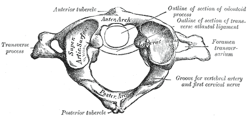
In anatomy, the atlas (C1) is the most superior (first) cervical vertebra of the spine and is located in the neck. It is named for Atlas of Greek mythology because, just as Atlas supported the globe, it supports the entire head. The atlas is the topmost vertebra and, with the axis (the vertebra below it), forms the joint connecting the skull and spine. The atlas and axis are specialized to allow a greater range of motion than normal vertebrae. They are responsible for the nodding and rotation movements of the head. The atlanto-occipital joint allows the head to nod up and down on the vertebral column. The dens acts as a pivot that allows the atlas and attached head to rotate on the axis, side to side. The atlas's chief peculiarity is that it has no body. It is ring-like and consists of an anterior and a posterior arch and two lateral masses. The atlas and axis are important neurologically because the brainstem extends down to the axis. Structure Anterior arch The a ... [...More Info...] [...Related Items...] OR: [Wikipedia] [Google] [Baidu] |
Tooth Enamel
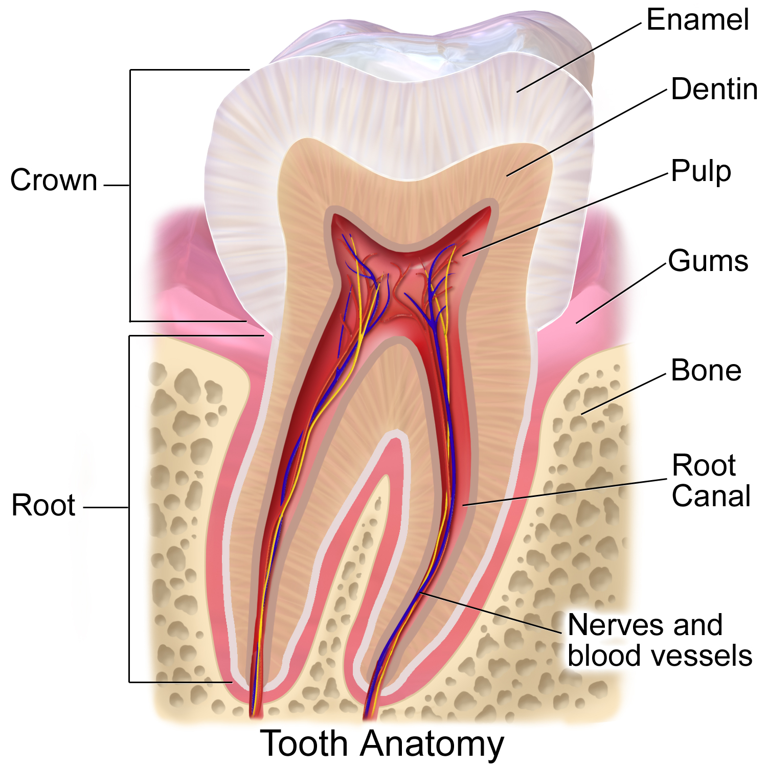
Tooth enamel is one of the four major tissues that make up the tooth in humans and many other animals, including some species of fish. It makes up the normally visible part of the tooth, covering the crown. The other major tissues are dentin, cementum, and dental pulp. It is a very hard, white to off-white, highly mineralised substance that acts as a barrier to protect the tooth but can become susceptible to degradation, especially by acids from food and drink. Calcium hardens the tooth enamel. In rare circumstances enamel fails to form, leaving the underlying dentin exposed on the surface. Features Enamel is the hardest substance in the human body and contains the highest percentage of minerals (at 96%),Ross ''et al.'', p. 485 with water and organic material composing the rest.Ten Cate's Oral Histology, Nancy, Elsevier, pp. 70–94 The primary mineral is hydroxyapatite, which is a crystalline calcium phosphate. Enamel is formed on the tooth while the tooth develops wit ... [...More Info...] [...Related Items...] OR: [Wikipedia] [Google] [Baidu] |
Ichthyopterygia
Ichthyopterygia ("fish flippers") was a designation introduced by Sir Richard Owen in 1840 to designate the Jurassic ichthyosaurs that were known at the time, but the term is now used more often for both true Ichthyosauria and their more primitive early and middle Triassic ancestors. Basal ichthyopterygians (prior to and ancestral to true Ichthyosauria) were mostly small (a meter or less in length) with elongated bodies and long, spool-shaped vertebrae, indicating that they swam in a sinuous, eel-like manner. This allowed for quick movements and maneuverability that were advantages in shallow-water hunting. Even at this early stage, they were already very specialised animals with proper flippers, and would have been incapable of movement on land. These animals seem to have been widely distributed around the coast of the northern half of Pangea, as they are known the Late Olenekian and Early Anisian (early part of the Triassic period) of Japan, China, Canada, and Spitsbergen ... [...More Info...] [...Related Items...] OR: [Wikipedia] [Google] [Baidu] |
Rostrum (anatomy)
Rostrum (from Latin ', meaning ''beak'') is a term used in anatomy for a number of phylogenetically unrelated structures in different groups of animals. Invertebrates * In crustaceans, the rostrum is the forward extension of the carapace in front of the eyes. It is generally a rigid structure, but can be connected by a hinged joint, as seen in Leptostraca. * Among insects, the rostrum is the name for the piercing mouthparts of the order Hemiptera as well as those of the snow scorpionflies, among many others. The long snout of weevils is also called a rostrum. * Gastropod molluscs have a rostrum or proboscis. * Cephalopod molluscs have hard beak-like mouthparts referred to as the rostrum. File:Washington DC Zoo - Macrobrachium rosenbergii 6.jpg, Crustacean: the rostrum of the shrimp '' Macrobrachium rosenbergii'' is serrated along both edges. File:Gminatus australis with Beetle.jpg, Insect: assassin bug piercing its prey with its rostrum File:Architeuthis beak.jpg, Ceph ... [...More Info...] [...Related Items...] OR: [Wikipedia] [Google] [Baidu] |
Teeth
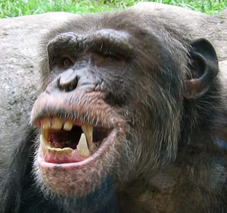
A tooth ( : teeth) is a hard, calcified structure found in the jaws (or mouths) of many vertebrates and used to break down food. Some animals, particularly carnivores and omnivores, also use teeth to help with capturing or wounding prey, tearing food, for defensive purposes, to intimidate other animals often including their own, or to carry prey or their young. The roots of teeth are covered by gums. Teeth are not made of bone, but rather of multiple tissues of varying density and hardness that originate from the embryonic germ layer, the ectoderm. The general structure of teeth is similar across the vertebrates, although there is considerable variation in their form and position. The teeth of mammals have deep roots, and this pattern is also found in some fish, and in crocodilians. In most teleost fish, however, the teeth are attached to the outer surface of the bone, while in lizards they are attached to the inner surface of the jaw by one side. In cartilaginous fish, ... [...More Info...] [...Related Items...] OR: [Wikipedia] [Google] [Baidu] |
Dentary
In anatomy, the mandible, lower jaw or jawbone is the largest, strongest and lowest bone in the human facial skeleton. It forms the lower jaw and holds the lower teeth in place. The mandible sits beneath the maxilla. It is the only movable bone of the skull (discounting the ossicles of the middle ear). It is connected to the temporal bones by the temporomandibular joints. The bone is formed in the fetus from a fusion of the left and right mandibular prominences, and the point where these sides join, the mandibular symphysis, is still visible as a faint ridge in the midline. Like other symphyses in the body, this is a midline articulation where the bones are joined by fibrocartilage, but this articulation fuses together in early childhood.Illustrated Anatomy of the Head and Neck, Fehrenbach and Herring, Elsevier, 2012, p. 59 The word "mandible" derives from the Latin word ''mandibula'', "jawbone" (literally "one used for chewing"), from '' mandere'' "to chew" and ''-bula'' ( ... [...More Info...] [...Related Items...] OR: [Wikipedia] [Google] [Baidu] |
Maxilla
The maxilla (plural: ''maxillae'' ) in vertebrates is the upper fixed (not fixed in Neopterygii) bone of the jaw formed from the fusion of two maxillary bones. In humans, the upper jaw includes the hard palate in the front of the mouth. The two maxillary bones are fused at the intermaxillary suture, forming the anterior nasal spine. This is similar to the mandible (lower jaw), which is also a fusion of two mandibular bones at the mandibular symphysis. The mandible is the movable part of the jaw. Structure In humans, the maxilla consists of: * The body of the maxilla * Four processes ** the zygomatic process ** the frontal process of maxilla ** the alveolar process ** the palatine process * three surfaces – anterior, posterior, medial * the Infraorbital foramen * the maxillary sinus * the incisive foramen Articulations Each maxilla articulates with nine bones: * two of the cranium: the frontal and ethmoid * seven of the face: the nasal, zygomatic, lacrimal, ... [...More Info...] [...Related Items...] OR: [Wikipedia] [Google] [Baidu] |


.jpg)


