|
Tephromela Gigantea
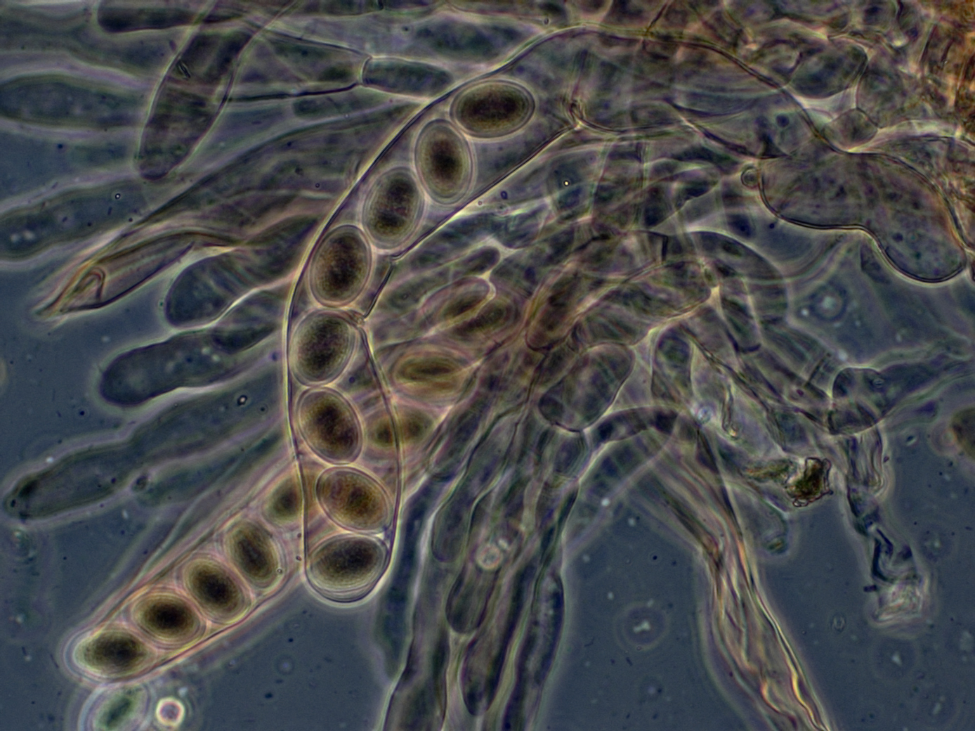
''Tephromela'' is a genus of lichens in the family Tephromelataceae. There are about 50 species in this widespread genus. The genus was established in 1929 by the French lichenologist Maurice Choisy, who separated these species from the broader genus ''Lecanora'' based on their distinctive straight asexual spores and dark violet spore-bearing layers. These rock and bark-dwelling lichens are characterized by their white to pale grey crusty growth and black -shaped reproductive structures with purple-tinted interiors. Taxonomy The genus was circumscribed in 1929 by the French lichenologist Maurice Choisy. He assigned '' Tephromela atra'' as the type species. In his original description, Choisy established ''Tephromela'' to accommodate species that had previously been classified under ''Lecanora'' but had distinctive characteristics that set them apart from the core ''Lecanora'' group. Specifically, he recognized that ''Lecanora atra'' (originally described by Acharius) represe ... [...More Info...] [...Related Items...] OR: [Wikipedia] [Google] [Baidu] |
Morphology (biology)
Morphology (from Ancient Greek μορφή (morphḗ) "form", and λόγος (lógos) "word, study, research") is the study of the form and structure of organisms and their specific structural features. This includes aspects of the outward appearance (shape, structure, color, pattern, size), as well as the form and structure of internal parts like bones and organs, i.e., anatomy. This is in contrast to physiology, which deals primarily with function. Morphology is a branch of life science dealing with the study of the overall structure of an organism or taxon and its component parts. History The etymology of the word "morphology" is from the Ancient Greek (), meaning "form", and (), meaning "word, study, research". While the concept of form in biology, opposed to function, dates back to Aristotle (see Aristotle's biology), the field of morphology was developed by Johann Wolfgang von Goethe (1790) and independently by the German anatomist and physiologist Karl Fried ... [...More Info...] [...Related Items...] OR: [Wikipedia] [Google] [Baidu] |
Septum
In biology, a septum (Latin language, Latin for ''something that encloses''; septa) is a wall, dividing a Body cavity, cavity or structure into smaller ones. A cavity or structure divided in this way may be referred to as septate. Examples Human anatomy * Interatrial septum, the wall of tissue that is a sectional part of the left and right atria of the heart * Interventricular septum, the wall separating the left and right ventricles of the heart * Lingual septum, a vertical layer of fibrous tissue that separates the halves of the tongue *Nasal septum: the cartilage wall separating the nostrils of the nose * Alveolar septum: the thin wall which separates the Pulmonary alveolus, alveoli from each other in the lungs * Orbital septum, a palpebral ligament in the upper and lower eyelids * Septum pellucidum or septum lucidum, a thin structure separating two fluid pockets in the brain * Uterine septum, a malformation of the uterus * Septum of the penis, Penile septum, a fibrous w ... [...More Info...] [...Related Items...] OR: [Wikipedia] [Google] [Baidu] |
Ellipsoid
An ellipsoid is a surface that can be obtained from a sphere by deforming it by means of directional Scaling (geometry), scalings, or more generally, of an affine transformation. An ellipsoid is a quadric surface; that is, a Surface (mathematics), surface that may be defined as the zero set of a polynomial of degree two in three variables. Among quadric surfaces, an ellipsoid is characterized by either of the two following properties. Every planar Cross section (geometry), cross section is either an ellipse, or is empty, or is reduced to a single point (this explains the name, meaning "ellipse-like"). It is Bounded set, bounded, which means that it may be enclosed in a sufficiently large sphere. An ellipsoid has three pairwise perpendicular Rotational symmetry, axes of symmetry which intersect at a Central symmetry, center of symmetry, called the center of the ellipsoid. The line segments that are delimited on the axes of symmetry by the ellipsoid are called the ''principal ax ... [...More Info...] [...Related Items...] OR: [Wikipedia] [Google] [Baidu] |
Ascospore
In fungi, an ascospore is the sexual spore formed inside an ascus—the sac-like cell that defines the division Ascomycota, the largest and most diverse Division (botany), division of fungi. After two parental cell nucleus, nuclei fuse, the ascus undergoes meiosis (halving of genetic material) followed by a mitosis (cell division), ordinarily producing eight genetically distinct haploid spores; most yeasts stop at four ascospores, whereas some moulds carry out extra post-meiotic divisions to yield dozens. Many asci build turgor, internal pressure and shoot their spores clear of the calm boundary layer, thin layer of still air enveloping the fruit body, whereas subterranean truffles depend on animals for biological dispersal, dispersal. Ontogeny, Development shapes both form and endurance of ascospores. A hook-shaped crozier aligns the paired nuclei; a double-biological membrane, membrane system then parcels each daughter nucleus, and successive wall layers of β-glucan, chitosan ... [...More Info...] [...Related Items...] OR: [Wikipedia] [Google] [Baidu] |
Bacidia
''Bacidia'' is a genus of lichen-forming fungi in the family Ramalinaceae. Taxonomy The genus was circumscribed by Giuseppe De Notaris in 1846. Description ''Bacidia'' is characterised by its crustose (crust-like) growth form. The main body (thallus) of these lichens typically appears as a thin layer that can be smooth, cracked, warty, or in texture. The thallus may sometimes develop specialised structures such as soredia (powdery propagules), isidia (small outgrowths), or tiny scale-like features. Its colour usually ranges from whitish to pale green, greenish-grey, pale grey, or fawn. Like all lichens, ''Bacidia'' species represent a symbiotic partnership with algae. Their (algal partner) belongs to the group, featuring spherical or broadly oval-shaped cells. The fungal component produces distinctive reproductive structures called apothecia, which are disc-shaped and typically measure up to 1 mm across (occasionally reaching 1.3 mm). These apothecia sit directly ... [...More Info...] [...Related Items...] OR: [Wikipedia] [Google] [Baidu] |
Ascus
An ascus (; : asci) is the sexual spore-bearing cell produced in ascomycete fungi. Each ascus usually contains eight ascospores (or octad), produced by meiosis followed, in most species, by a mitotic cell division. However, asci in some genera or species can occur in numbers of one (e.g. '' Monosporascus cannonballus''), two, four, or multiples of four. In a few cases, the ascospores can bud off conidia that may fill the asci (e.g. '' Tympanis'') with hundreds of conidia, or the ascospores may fragment, e.g. some '' Cordyceps'', also filling the asci with smaller cells. Ascospores are nonmotile, usually single celled, but not infrequently may be coenocytic (lacking a septum), and in some cases coenocytic in multiple planes. Mitotic divisions within the developing spores populate each resulting cell in septate ascospores with nuclei. The term ocular chamber, or oculus, refers to the epiplasm (the portion of cytoplasm not used in ascospore formation) that is surrounded by the ... [...More Info...] [...Related Items...] OR: [Wikipedia] [Google] [Baidu] |
Paraphyses
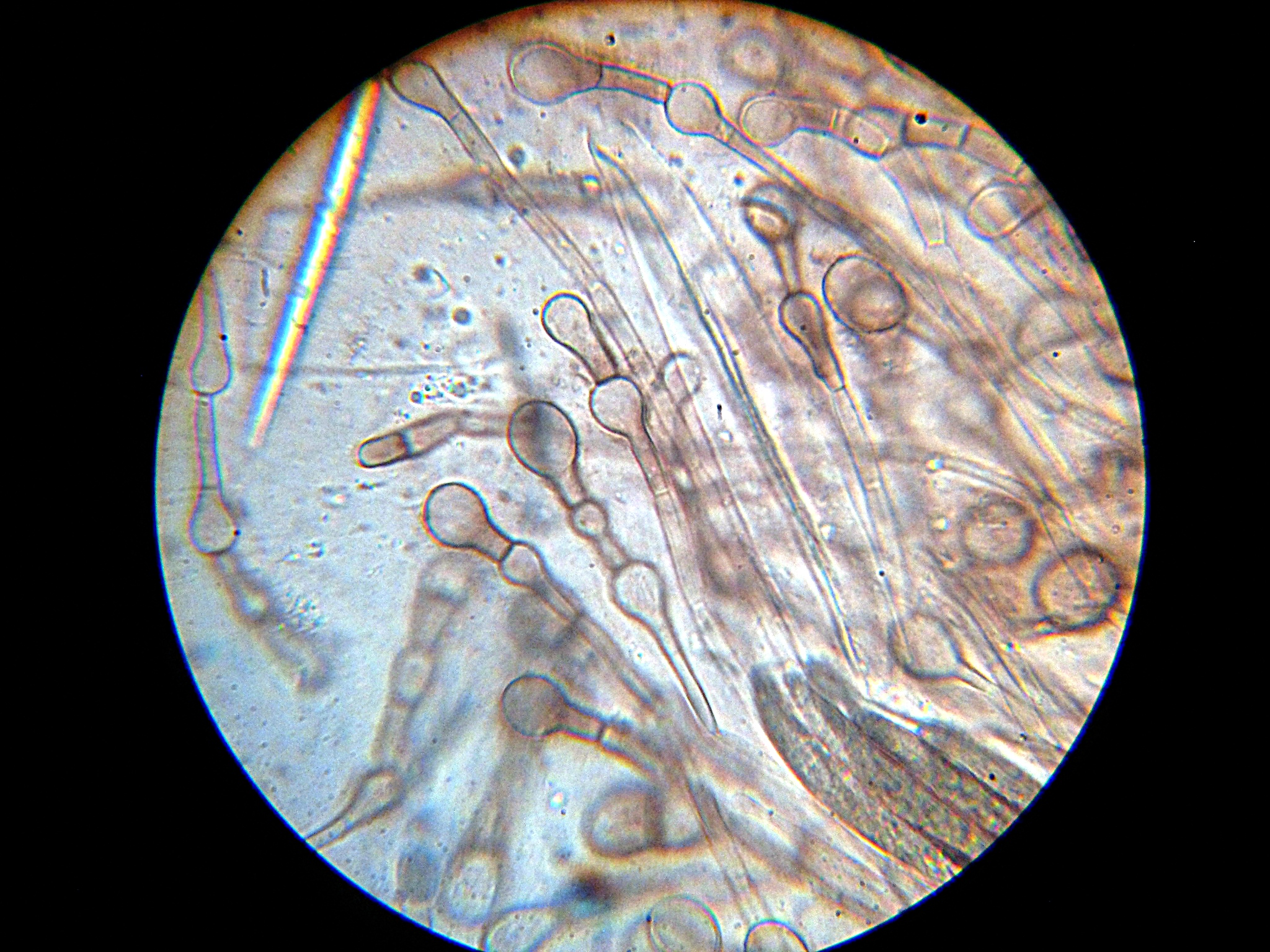
Paraphyses are erect sterile filament-like support structures occurring among the reproductive apparatuses of fungi, ferns, bryophytes and some thallophytes. The singular form of the word is paraphysis. In certain fungi, they are part of the fertile spore-bearing layer. More specifically, paraphyses are sterile filamentous hyphal end cells composing part of the hymenium of Ascomycota and Basidiomycota interspersed among either the asci or basidia respectively, and not sufficiently differentiated to be called cystidia A cystidium (: cystidia) is a relatively large cell found on the sporocarp of a basidiomycete (for example, on the surface of a mushroom gill), often between clusters of basidia. Since cystidia have highly varied and distinct shapes that are o ..., which are specialized, swollen, often protruding cells. The tips of paraphyses may contain the pigments which colour the hymenium. In ferns and mosses, they are filament-like structures that are found on sporangi ... [...More Info...] [...Related Items...] OR: [Wikipedia] [Google] [Baidu] |
Apothecia
An ascocarp, or ascoma (: ascomata), is the fruiting body ( sporocarp) of an ascomycete phylum fungus. It consists of very tightly interwoven hyphae and millions of embedded asci, each of which typically contains four to eight ascospores. Ascocarps are most commonly bowl-shaped (apothecia) but may take on a spherical or flask-like form that has a pore opening to release spores (perithecia) or no opening (cleistothecia). Classification The ascocarp is classified according to its placement (in ways not fundamental to the basic taxonomy). It is called ''epigeous'' if it grows above ground, as with the morels, while underground ascocarps, such as truffles, are termed ''hypogeous''. The structure enclosing the hymenium is divided into the types described below (apothecium, cleistothecium, etc.) and this character ''is'' important for the taxonomic classification of the fungus. Apothecia can be relatively large and fleshy, whereas the others are microscopic—about the s ... [...More Info...] [...Related Items...] OR: [Wikipedia] [Google] [Baidu] |
Staining
Staining is a technique used to enhance contrast in samples, generally at the Microscope, microscopic level. Stains and dyes are frequently used in histology (microscopic study of biological tissue (biology), tissues), in cytology (microscopic study of cell (biology), cells), and in the medical fields of histopathology, hematology, and cytopathology that focus on the study and diagnoses of diseases at the microscopic level. Stains may be used to define biological tissues (highlighting, for example, muscle fibers or connective tissue), cell (biology), cell populations (classifying different blood cells), or organelles within individual cells. In biochemistry, it involves adding a class-specific (DNA, proteins, lipids, carbohydrates) dye to a substrate to qualify or quantify the presence of a specific compound. Staining and fluorescent tagging can serve similar purposes. Biological staining is also used to mark cells in flow cytometry, and to flag proteins or nucleic acids in gel ... [...More Info...] [...Related Items...] OR: [Wikipedia] [Google] [Baidu] |
Medulla (lichenology) zone, but above the lower cortex.Galloway, D.J. (1992). Flora of Australia - ''Lichen Glossary'' The medulla generally has a cottony appearance. It is the widest layer of a heteromerous lichen thallus.
The medulla is a horizontal layer within a lichen thallus. It is a loosely arranged layer of interlaced hyphae below the upper cortex and photobiont A lichen ( , ) is a hybrid colony of algae or cyanobacteria living symbiotically among filaments of multiple fungus species, along with yeasts and bacteria embedded in the cortex or "skin", in a mutualistic relationship. References Fungal morphology and anatomy Lichenology {{lichen-stub ...[...More Info...] [...Related Items...] OR: [Wikipedia] [Google] [Baidu] |
Green Alga
The green algae (: green alga) are a group of chlorophyll-containing autotrophic eukaryotes consisting of the phylum Prasinodermophyta and its unnamed sister group that contains the Chlorophyta and Charophyta/ Streptophyta. The land plants ( Embryophytes) have emerged deep within the charophytes as a sister of the Zygnematophyceae. Since the realization that the Embryophytes emerged within the green algae, some authors are starting to include them. The completed clade that includes both green algae and embryophytes is monophyletic and is referred to as the clade Viridiplantae and as the kingdom Plantae. The green algae include unicellular and colonial flagellates, most with two flagella per cell, as well as various colonial, coccoid (spherical), and filamentous forms, and macroscopic, multicellular seaweeds. There are about 22,000 species of green algae, many of which live most of their lives as single cells, while other species form coenobia (colonies), long filaments, or ... [...More Info...] [...Related Items...] OR: [Wikipedia] [Google] [Baidu] |


