|
Tensor Fasciae Latae
The tensor fasciae latae (or tensor fasciæ latæ or, formerly, tensor vaginae femoris) is a muscle of the thigh. Together with the gluteus maximus, it acts on and is continuous with the iliotibial band, which attaches to the tibia. The muscle assists in keeping the balance of the pelvis while standing, walking, or running. Structure The tensor fasciae latae arises from the anterior part of the outer lip of the iliac crest; from the outer surface of the anterior superior iliac spine, and part of the outer border of the notch below it, between the gluteus medius and sartorius; and from the deep surface of the fascia lata. The tensor fasciae latae is inserted between the two layers of the iliotibial tract of the fascia lata about the junction of the middle and upper thirds of the thigh. It tautens the iliotibial tract and braces the knee, especially when the opposite foot is lifted.Saladin, Kenneth. Anatomy and Physiology. 6th ed. Mc-Graw Hill. 2010. The terminal insertion point ... [...More Info...] [...Related Items...] OR: [Wikipedia] [Google] [Baidu] |
Iliac Crest
The crest of the ilium (or iliac crest) is the superior border of the wing of ilium and the superolateral margin of the greater pelvis. Structure The iliac crest stretches posteriorly from the anterior superior iliac spine (ASIS) to the posterior superior iliac spine (PSIS). Behind the ASIS, it divides into an outer and inner lip separated by the intermediate zone. The outer lip bulges laterally into the iliac tubercle.Platzer (2004), p 186 Palpation, Palpable in its entire length, the crest is convex superiorly but is sinuously curved, being concave inward in front, concave outward behind.Palastanga (2006), p 243 It is thinner at the center than at the extremities. Development The iliac crest is derived from endochondral bone. Function To the external lip are attached the ''Tensor fasciae latae'', ''abdominal external oblique muscle, Obliquus externus abdominis'', and ''Latissimus dorsi muscle, Latissimus dorsi'', and along its whole length the ''fascia lata''; to the int ... [...More Info...] [...Related Items...] OR: [Wikipedia] [Google] [Baidu] |
Anatomical Terms Of Muscle
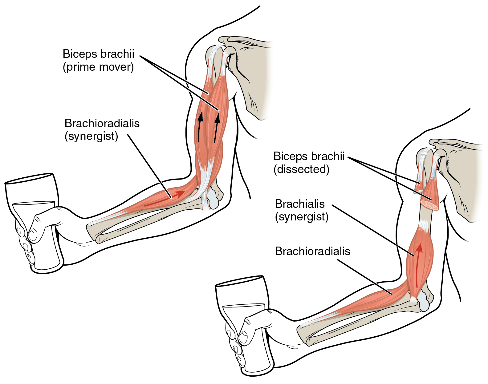
Anatomical terminology is used to uniquely describe aspects of skeletal muscle, cardiac muscle, and smooth muscle such as their actions, structure, size, and location. Types There are three types of muscle tissue in the body: skeletal, smooth, and cardiac. Skeletal muscle Skeletal muscle, or "voluntary muscle", is a striated muscle tissue that primarily joins to bone with tendons. Skeletal muscle enables movement of bones, and maintains posture. The widest part of a muscle that pulls on the tendons is known as the belly. Muscle slip A muscle slip is a slip of muscle that can either be an anatomical variant, or a branching of a muscle as in rib connections of the serratus anterior muscle. Smooth muscle Smooth muscle is involuntary and found in parts of the body where it conveys action without conscious intent. The majority of this type of muscle tissue is found in the digestive and urinary systems where it acts by propelling forward food, chyme, and feces in the former and u ... [...More Info...] [...Related Items...] OR: [Wikipedia] [Google] [Baidu] |
Hurdling
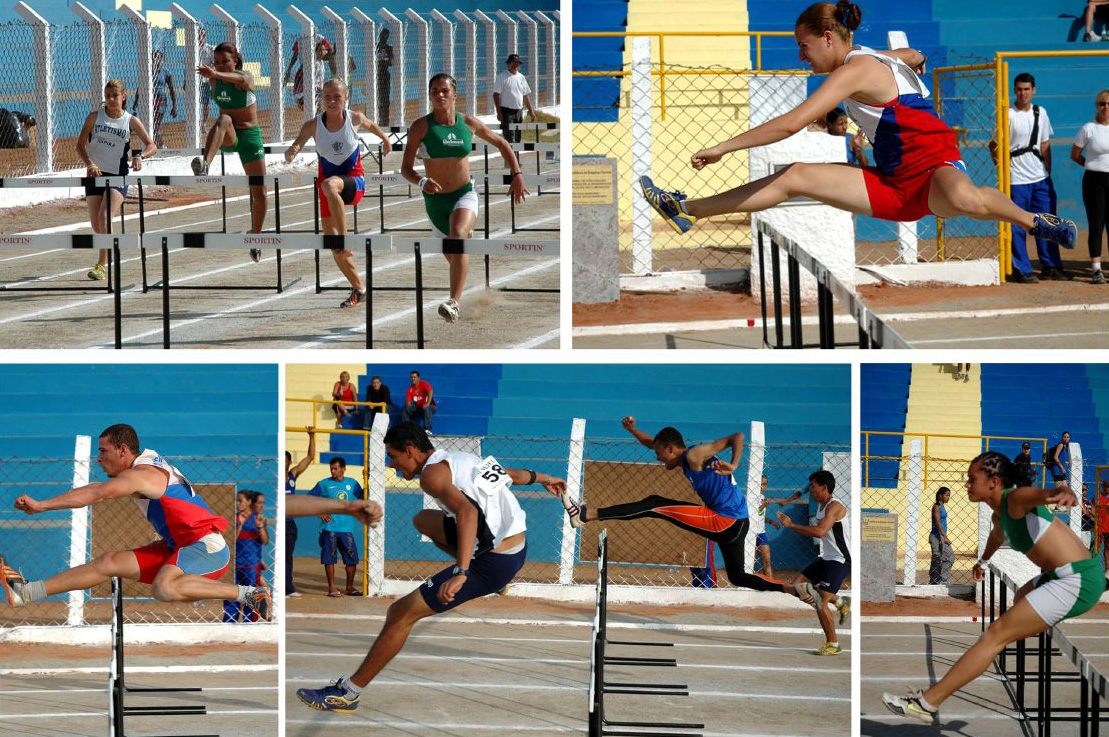
Hurdling is the act of jumping over an obstacle at a high speed or in a sprint. In the early 19th century, hurdlers ran at and jumped over each hurdle (sometimes known as 'burgles'), landing on both feet and checking their forward motion. Today, the dominant step patterns are the 3-step for high hurdles, 7-step for low hurdles, and 15-step for intermediate hurdles. Hurdling is a highly specialized form of obstacle racing, and is part of the sport of athletics (sport), athletics. In hurdling events, barriers known as hurdles are set at precisely measured heights and distances. Each athlete must pass over the hurdles; passing under or intentionally knocking over hurdles will result in disqualification. Accidental knocking over of hurdles is not cause for disqualification, but the hurdles are weighted to make doing so disadvantageous. In 1902 Spalding equipment company sold the Foster Patent Safety Hurdle, a wood hurdle. In 1923 some of the wood hurdles weighed each. Hurdle des ... [...More Info...] [...Related Items...] OR: [Wikipedia] [Google] [Baidu] |
Equestrianism
Equestrianism (from Latin , , , 'horseman', 'horse'), commonly known as horse riding ( Commonwealth English) or horseback riding (American English), includes the disciplines of riding, driving, and vaulting. This broad description includes the use of horses for practical working purposes, transportation, recreational activities, artistic or cultural exercises, and competitive sport. Overview of equestrian activities Horses are trained and ridden for practical working purposes, such as in police work or for controlling herd animals on a ranch. They are also used in competitive sports including dressage, endurance riding, eventing, reining, show jumping, tent pegging, vaulting, polo, horse racing, driving, and rodeo (see additional equestrian sports listed later in this article for more examples). Some popular forms of competition are grouped together at horse shows where horses perform in a wide variety of disciplines. Horses (and other equids such as mules ... [...More Info...] [...Related Items...] OR: [Wikipedia] [Google] [Baidu] |
Femur
The femur (; : femurs or femora ), or thigh bone is the only long bone, bone in the thigh — the region of the lower limb between the hip and the knee. In many quadrupeds, four-legged animals the femur is the upper bone of the hindleg. The Femoral head, top of the femur fits into a socket in the pelvis called the hip joint, and the bottom of the femur connects to the shinbone (tibia) and kneecap (patella) to form the knee. In humans the femur is the largest and thickest bone in the body. Structure The femur is the only bone in the upper Human leg, leg. The two femurs converge Anatomical terms of location, medially toward the knees, where they articulate with the Anatomical terms of location, proximal ends of the tibiae. The angle at which the femora converge is an important factor in determining the femoral-tibial angle. In females, thicker pelvic bones cause the femora to converge more than in males. In the condition genu valgum, ''genu valgum'' (knock knee), the femurs conve ... [...More Info...] [...Related Items...] OR: [Wikipedia] [Google] [Baidu] |
Condyle (anatomy)
A condyle (; in Merriam-Webster Online Dictionary '. , from ; κόνδυλος knuckle) is the round prominence at the end of a , most often part of a joint – an articulation with another bone. It is one of the markings or features of bones, and can refer to: * On the , in the joint: ** [...More Info...] [...Related Items...] OR: [Wikipedia] [Google] [Baidu] |
Femoral Head
The femoral head (femur head or head of the femur) is the highest part of the thigh bone (femur The femur (; : femurs or femora ), or thigh bone is the only long bone, bone in the thigh — the region of the lower limb between the hip and the knee. In many quadrupeds, four-legged animals the femur is the upper bone of the hindleg. The Femo ...). It is supported by the femoral neck. Structure The head is globular and forms rather more than a hemisphere, is directed upward, medialward, and a little forward, the greater part of its convexity being above and in front. The femoral head's surface is smooth. It is coated with cartilage in the fresh state, except over an ovoid depression, the fovea capitis, which is situated a little below and behind the center of the femoral head, and gives attachment to the ligament of head of femur. The thickest region of the articular cartilage is at the centre of the femoral head, measuring up to 2.8 mm. The diameter of the femoral hea ... [...More Info...] [...Related Items...] OR: [Wikipedia] [Google] [Baidu] |
Cutaneous Innervation
Cutaneous innervation refers to an area of the skin which is supplied by a specific cutaneous nerve. Dermatomes are similar; however, a dermatome only specifies the area served by a spinal nerve. In some cases, the dermatome is less specific (when a spinal nerve is the source for more than one cutaneous nerve), and in other cases it is more specific (when a cutaneous nerve is derived from multiple spinal nerves.) Modern texts are in agreement about which areas of the skin are served by which nerves, but there are minor variations in some of the details. The borders designated by the diagrams in the 1918 edition of Gray's Anatomy are similar, but not identical, to those generally accepted today. In the peripheral nervous system The peripheral nervous system (PNS) is divided into the somatic nervous system, the autonomic nervous system, and the enteric nervous system. However, it is the somatic nervous system, responsible for body movement and the reception of external stimuli, ... [...More Info...] [...Related Items...] OR: [Wikipedia] [Google] [Baidu] |
Superior Gluteal Nerve
The superior gluteal nerve is a mixed (motor and sensory) nerve of the sacral plexus that originates in the pelvis. It provides motor innervation to the gluteus medius, gluteus minimus, tensor fasciae latae; it also has a cutaneous branch. Structure Origin The superior gluteal nerve originates in the sacral plexus. It arises from the posterior divisions of L4, L5 and S1. Course It exits the pelvis through the greater sciatic foramen superior to the piriformis muscle. It is accompanied by the superior gluteal artery and the superior gluteal vein.''Thieme Atlas of Anatomy'' (2006), p 476 It passes lateral-ward in between the gluteus medius muscle and the gluteus minimus muscle, accompanied by the deep branch of the superior gluteal artery. It divides into a superior branch and an inferior branch. The inferior branch continues to pass between the two muscles to end in the tensor fasciae latae muscle. Distribution Motor * tensor fasciae latae musclePlatzer (2004) ... [...More Info...] [...Related Items...] OR: [Wikipedia] [Google] [Baidu] |
Lateral Condyle Of Tibia
The lateral condyle is the lateral portion of the upper extremity of tibia. It serves as the insertion for the biceps femoris muscle (small slip). Most of the tendon of the biceps femoris inserts on the fibula. See also * Gerdy's tubercle * Medial condyle of tibia Additional images File:Gray258.png, Bones of the right leg. Anterior surface. File:Slide2bib.JPG, Right knee in extension. Deep dissection. Posterior view. File:Slide2cocc.JPG, Right knee in extension. Deep dissection. Posterior view. References External links * * * () Bones of the lower limb Tibia {{musculoskeletal-stub ... [...More Info...] [...Related Items...] OR: [Wikipedia] [Google] [Baidu] |
Fascia Lata
The fascia lata is the deep fascia of the thigh. It encloses the thigh muscles and forms the outer limit of the fascial compartments of thigh, which are internally separated by the medial intermuscular septum and the lateral intermuscular septum. The fascia lata is thickened at its lateral side where it forms the iliotibial tract, a structure that runs to the tibia and serves as a site of muscle attachment. Structure The fascia lata is an investment for the whole of the thigh, but varies in thickness in different parts. It is thicker in the upper and lateral part of the thigh, where it receives a fibrous expansion from the gluteus maximus, and where the tensor fasciae latae is inserted between its layers; it is very thin behind and at the upper and medial part, where it covers the adductor muscles, and again becomes stronger around the knee, receiving fibrous expansions from the tendon of the biceps femoris laterally, from the sartorius medially, and from the quadriceps fem ... [...More Info...] [...Related Items...] OR: [Wikipedia] [Google] [Baidu] |
Sartorius Muscle
The sartorius muscle () is the longest muscle in the human body. It is a long, thin, superficial muscle that runs down the length of the thigh in the anterior compartment. Structure The sartorius muscle originates from the anterior superior iliac spine, and part of the notch between the anterior superior iliac spine and anterior inferior iliac spine. It runs obliquely across the upper and anterior part of the thigh in an inferomedial direction. It passes behind the medial condyle of the femur to end in a tendon. This tendon curves anteriorly to join the tendons of the gracilis and semitendinosus muscles in the pes anserinus, where it inserts into the superomedial surface of the tibia. Its upper portion forms the lateral border of the femoral triangle, and the point where it crosses adductor longus marks the apex of the triangle. Deep to sartorius and its fascia is the adductor canal, through which the saphenous nerve, femoral artery and vein, and nerve to vastus medialis pa ... [...More Info...] [...Related Items...] OR: [Wikipedia] [Google] [Baidu] |


