|
Tenocyte
In animal and Human biology, a tendon cell is a cell that makes up tendons, the bands of connective tissue that connects muscles to bones. Tendon cells, also known as tenocytes or tendon fibroblasts, are specialized cells that contribute to the structure, function, and repair of tendons in the body. Tendons are fibrous tissues that connect muscles to bones, and tendon cells play a vital role in maintaining tendon homeostasis and facilitating healing following injury. Function Tendon cells are primarily responsible for the production and maintenance of the tendon extracellular matrix (ECM), which consists mainly of collagen fibers. These cells are involved in synthesizing collagen and other ECM components that provide tendons with tensile strength. Tendon cells also participate in remodeling the ECM in response to mechanical stress and injury. Structure Tendon cells are typically elongated, spindle-shaped cells that align along the axis of tendon fibers. They contain large am ... [...More Info...] [...Related Items...] OR: [Wikipedia] [Google] [Baidu] |
Tendon
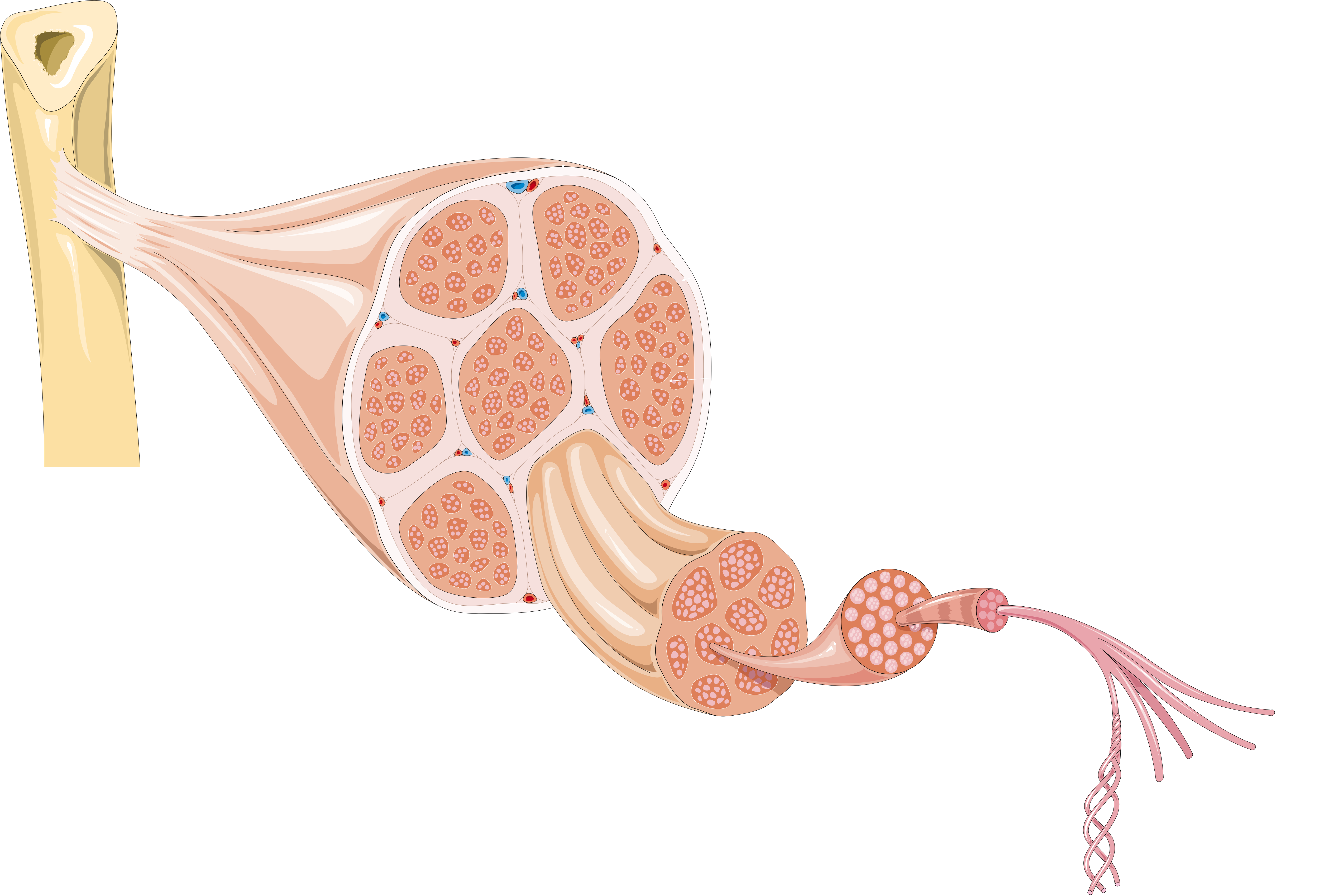
A tendon or sinew is a tough band of fibrous connective tissue, dense fibrous connective tissue that connects skeletal muscle, muscle to bone. It sends the mechanical forces of muscle contraction to the skeletal system, while withstanding tension (physics), tension. Tendons, like ligaments, are made of collagen. The difference is that ligaments connect bone to bone, while tendons connect muscle to bone. There are about 4,000 tendons in the adult human body. Structure A tendon is made of dense regular connective tissue, whose main cellular components are special fibroblasts called tendon cells (tenocytes). Tendon cells synthesize the tendon's extracellular matrix, which abounds with densely-packed collagen fibers. The collagen fibers run parallel to each other and are grouped into fascicles. Each fascicle is bound by an endotendineum, which is a delicate loose connective tissue containing thin collagen fibrils and elastic fibers. A set of fascicles is bound by an epitenon, whi ... [...More Info...] [...Related Items...] OR: [Wikipedia] [Google] [Baidu] |
Tendon Anatomy 1 -- Smart-Servier
A tendon or sinew is a tough band of dense fibrous connective tissue that connects muscle to bone. It sends the mechanical forces of muscle contraction to the skeletal system, while withstanding tension. Tendons, like ligaments, are made of collagen. The difference is that ligaments connect bone to bone, while tendons connect muscle to bone. There are about 4,000 tendons in the adult human body. Structure A tendon is made of dense regular connective tissue, whose main cellular components are special fibroblasts called tendon cells (tenocytes). Tendon cells synthesize the tendon's extracellular matrix, which abounds with densely-packed collagen fibers. The collagen fibers run parallel to each other and are grouped into fascicles. Each fascicle is bound by an endotendineum, which is a delicate loose connective tissue containing thin collagen fibrils and elastic fibers. A set of fascicles is bound by an epitenon, which is a sheath of dense irregular connective tissue. The who ... [...More Info...] [...Related Items...] OR: [Wikipedia] [Google] [Baidu] |
Tendinopathy
Tendinopathy is a type of tendon disorder that results in pain, swelling, and impaired function. The pain is typically worse with movement. It most commonly occurs around the shoulder ( rotator cuff tendinitis, biceps tendinitis), elbow ( tennis elbow, golfer's elbow), wrist, hip, knee ( jumper's knee, popliteus tendinopathy), or ankle ( Achilles tendinitis). Causes may include an injury or repetitive activities. Less common causes include infection, arthritis, gout, thyroid disease, diabetes and the use of quinolone antibiotic medicines. Groups at risk include people who do manual labor, musicians, and athletes. Diagnosis is typically based on symptoms, examination, and occasionally medical imaging. A few weeks following an injury little inflammation remains, with the underlying problem related to weak or disrupted tendon fibrils. Treatment may include rest, NSAIDs, splinting, and physiotherapy. Less commonly steroid injections or surgery may be done. About 80% of over ... [...More Info...] [...Related Items...] OR: [Wikipedia] [Google] [Baidu] |
Tendonitis Tendon Rupture -- Smart-Servier (cropped)
Tendinopathy is a type of tendon disorder that results in pain, swelling, and impaired function. The pain is typically worse with movement. It most commonly occurs around the shoulder (rotator cuff tendinitis, biceps tendinitis), elbow (tennis elbow, golfer's elbow), wrist, hip, knee (jumper's knee, popliteus tendinopathy), or ankle (Achilles tendinitis). Causes may include an injury or repetitive activities. Less common causes include infection, arthritis, gout, thyroid disease, diabetes and the use of quinolone antibiotic medicines. Groups at risk include people who do manual labor, musicians, and athletes. Diagnosis is typically based on symptoms, examination, and occasionally medical imaging. A few weeks following an injury little inflammation remains, with the underlying problem related to weak or disrupted tendon fibrils. Treatment may include rest, NSAIDs, splinting, and physiotherapy. Less commonly steroid injections or surgery may be done. About 80% of overuse te ... [...More Info...] [...Related Items...] OR: [Wikipedia] [Google] [Baidu] |
Hemidesmosomes
Hemidesmosomes are very small stud-like structures found in keratinocytes of the epidermis of skin that attach to the extracellular matrix. They are similar in form to desmosomes when visualized by electron microscopy; however, desmosomes attach to adjacent cells. Hemidesmosomes are also comparable to focal adhesions, as they both attach cells to the extracellular matrix. Instead of desmogleins and desmocollins in the extracellular space, hemidesmosomes utilize integrins. Hemidesmosomes are found in epithelial cells connecting the basal epithelial cells to the lamina lucida, which is part of the basal lamina. Hemidesmosomes are also involved in signaling pathways, such as keratinocyte migration or carcinoma cell intrusion. Structure Hemidesmosomes can be categorized into two types based on their protein constituents. Type 1 hemidesmosomes are found in stratified and pseudo-stratified epithelium. Type 1 hemidesmosomes have five main elements: integrin α6 β4, plectin in its ... [...More Info...] [...Related Items...] OR: [Wikipedia] [Google] [Baidu] |
Human Cells
The list of human cell types provides an enumeration and description of the various specialized cells found within the human body, highlighting their distinct functions, characteristics, and contributions to overall physiological processes. Cells may be classified by their physiological function, histology (microscopic anatomy), lineage, or gene expression. Total number of cells The adult human body is estimated to contain about 30 trillion (3×10¹³) human cells, with the number varying between 20 and 100 trillion depending on factors such as sex, age, and weight. Additionally, there are approximately an equal number of bacterial cells. The exact count of human cells has not yet been empirically measured in its entirety and is estimated using different approaches based on smaller samples of empirical observation. It is generally assumed that these cells share features with each other and thus may be organized as belonging to a smaller number of types. Classification ... [...More Info...] [...Related Items...] OR: [Wikipedia] [Google] [Baidu] |
List Of Human Cell Types Derived From The Germ Layers
This is a list of Cell (biology), cells in humans derived from the three embryonic germ layers – ectoderm, mesoderm, and endoderm. Cells derived from ectoderm Surface ectoderm Skin * Trichocyte (human), Trichocyte * Keratinocyte Anterior pituitary * Gonadotropic cell, Gonadotrope * Corticotropic cell, Corticotrope * Thyrotropic cell, Thyrotrope * Somatotropic cell, Somatotrope * Prolactin cell, Lactotroph Tooth enamel * Ameloblast Neural crest Peripheral nervous system * Neuron * Neuroglia, Glia ** Schwann cell ** Satellite glial cell Neuroendocrine system * Chromaffin cell * Glomus cell Skin * Melanocyte ** Nevus cell * Merkel cell Teeth * Odontoblast * Cementoblast Eyes * Corneal keratocyte Smooth muscle Neural tube Central nervous system * Neuron * Glia ** Astrocyte ** Ependyma, Ependymocytes ** Müller glia (retina) ** Oligodendrocyte ** Oligodendrocyte progenitor cell ** Pituicyte (posterior pituitary) Pineal gland * Pinealocyte Cells derived from mesoderm ... [...More Info...] [...Related Items...] OR: [Wikipedia] [Google] [Baidu] |
CRISPR Gene Editing
CRISPR gene editing (; pronounced like "crisper"; an abbreviation for "clustered regularly interspaced short palindromic repeats") is a genetic engineering technique in molecular biology by which the genomes of living organisms may be modified. It is based on a simplified version of the bacterial CRISPR-Cas9 antiviral defense system. By delivering the Cas9 nuclease complexed with a synthetic guide RNA (gRNA) into a cell, the cell's genome can be cut at a desired location, allowing existing genes to be removed or new ones added ''in vivo''. The technique is considered highly significant in biotechnology and medicine as it enables editing genomes ''in vivo'' and is precise, cost-effective, and efficient. It can be used in the creation of new medicines, agriculture, agricultural products, and genetically modified organisms, or as a means of controlling pathogens and pest control, pests. It also offers potential in the treatment of inherited genetic diseases as well as diseases arisi ... [...More Info...] [...Related Items...] OR: [Wikipedia] [Google] [Baidu] |
Mesenchymal Stem Cell
Mesenchymal stem cells (MSCs), also known as mesenchymal stromal cells or medicinal signaling cells, are multipotent stromal cells that can Cellular differentiation, differentiate into a variety of cell types, including osteoblasts (bone cells), chondrocytes (cartilage cells), myocytes (muscle cells) and adipocytes (fat cells which give rise to Marrow Adipose Tissue, marrow adipose tissue). The primary function of MSCs is to respond to injury and infection by secreting and recruiting a range of biological factors, as well as modulating inflammatory processes to facilitate tissue repair and regeneration. Extensive research interest has led to more than 80,000 peer-reviewed papers on MSCs. Structure Definition Mesenchymal stem cells (MSCs), a term first used (in 1991) by Arnold Caplan at Case Western Reserve University, are characterized morphologically by a small cell body with long, thin cell processes. While the terms ''mesenchymal stem cell'' (MSC) and ''marrow stromal cell ... [...More Info...] [...Related Items...] OR: [Wikipedia] [Google] [Baidu] |
Microfilaments
Microfilaments, also called actin filaments, are protein filaments in the cytoplasm of eukaryotic cells that form part of the cytoskeleton. They are primarily composed of polymers of actin, but are modified by and interact with numerous other proteins in the cell. Microfilaments are usually about 7 nm in diameter and made up of two strands of actin. Microfilament functions include cytokinesis, amoeboid movement, cell motility, changes in cell shape, endocytosis and exocytosis, cell contractility, and mechanical stability. Microfilaments are flexible and relatively strong, resisting buckling by multi-piconewton compressive forces and filament fracture by nanonewton tensile forces. In inducing cell motility, one end of the actin filament elongates while the other end contracts, presumably by myosin II molecular motors. Additionally, they function as part of actomyosin-driven contractile molecular motors, wherein the thin filaments serve as tensile platforms for myosin's ... [...More Info...] [...Related Items...] OR: [Wikipedia] [Google] [Baidu] |


