|
Tail Wagging By Dogs
Tail wagging is the behavior of the dog observed as its tail moves back and forth in the same plane. Within Canidae, specifically ''Canis lupus familiaris'', the tail plays multiple roles, which can include balance, and communication. It is considered a social signal. The behaviour can be categorized by vigorous movement or slight movement of the tip of the tail. Tail wagging can also occur in circular motions, and when the tail is held at maximum height, neutral height, or between the legs. Tail wagging can be used as a social signal within species and convey the emotional state of the dog. The tail wagging behavior of a dog may not always be an indication of its friendliness or happiness, as is commonly believed. Though indeed tail wagging can express positive emotions, tail wagging is also an indication of fear, insecurity, challenging of dominance, establishing social relationships, or a warning that the dog may bite. It is also important to consider the way in which the d ... [...More Info...] [...Related Items...] OR: [Wikipedia] [Google] [Baidu] |
Dog Wagging Tail
The dog (''Canis familiaris'' or ''Canis lupus familiaris'') is a domesticated descendant of the wolf. Also called the domestic dog, it is derived from the extinct Pleistocene wolf, and the modern wolf is the dog's nearest living relative. Dogs were the first species to be domesticated by hunter-gatherers over 15,000 years ago before the development of agriculture. Due to their long association with humans, dogs have expanded to a large number of domestic individuals and gained the ability to thrive on a starch-rich diet that would be inadequate for other canids. The dog has been selectively bred over millennia for various behaviors, sensory capabilities, and physical attributes. Dog breeds vary widely in shape, size, and color. They perform many roles for humans, such as hunting, herding, pulling loads, protection, assisting police and the military, companionship, therapy, and aiding disabled people. Over the millennia, dogs became uniquely adapted to human behavior, and ... [...More Info...] [...Related Items...] OR: [Wikipedia] [Google] [Baidu] |
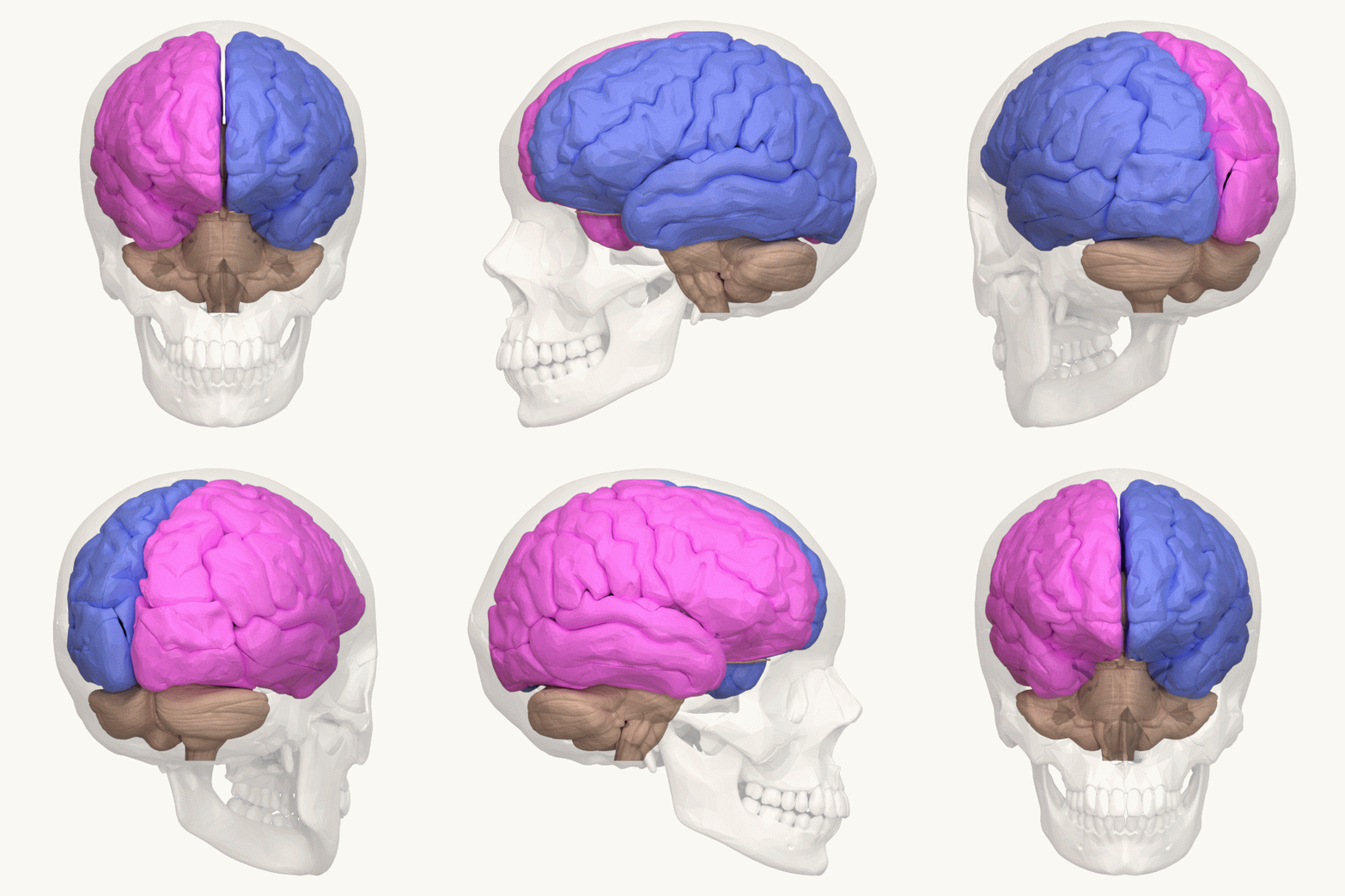
Left Hemisphere
The lateralization of brain function is the tendency for some neural functions or cognitive processes to be specialized to one side of the brain or the other. The median longitudinal fissure separates the human brain into two distinct cerebral hemispheres, connected by the corpus callosum. Although the macrostructure of the two hemispheres appears to be almost identical, different composition of neuronal networks allows for specialized function that is different in each hemisphere. Lateralization of brain structures is based on general trends expressed in healthy patients; however, there are numerous counterexamples to each generalization. Each human's brain develops differently, leading to unique lateralization in individuals. This is different from specialization, as lateralization refers only to the function of one structure divided between two hemispheres. Specialization is much easier to observe as a trend, since it has a stronger anthropological history. The best exam ... [...More Info...] [...Related Items...] OR: [Wikipedia] [Google] [Baidu] |
Dog Communication
Dog communication is the transfer of information between dogs, as well as between dogs and humans. Behaviors associated with dog communication are categorized into visual and vocal. Visual communication includes mouth shape and head position, licking and sniffing, ear and tail positioning, eye gaze, facial expression, and body posture. Dog vocalizations, or auditory communication, can include barks, growls, howls, whines and whimpers, screams, pants and sighs. Dogs also communicate via gustatory communication, utilizing scent and pheromones. Humans can communicate with dogs through a wide variety of methods. Broadly, this includes vocalization, hand signals, body posture and touch. The two species also communicate visually: through domestication, dogs have become particularly adept at "reading" human facial expressions, and they are able to determine human emotional status. When communicating with a human their level of comprehension is generally comparable to a toddler. ... [...More Info...] [...Related Items...] OR: [Wikipedia] [Google] [Baidu] |
Interneuron
Interneurons (also called internuncial neurons, relay neurons, association neurons, connector neurons, intermediate neurons or local circuit neurons) are neurons that connect two brain regions, i.e. not direct motor neurons or sensory neurons. Interneurons are the central nodes of neural circuits, enabling communication between sensory or motor neurons and the central nervous system (CNS). They play vital roles in reflexes, neuronal oscillations, and neurogenesis in the adult mammalian brain. Interneurons can be further broken down into two groups: local interneurons and relay interneurons. Local interneurons have short axons and form circuits with nearby neurons to analyze small pieces of information. Relay interneurons have long axons and connect circuits of neurons in one region of the brain with those in other regions. However, interneurons are generally considered to operate mainly within local brain areas. The interaction between interneurons allow the brain to perform ... [...More Info...] [...Related Items...] OR: [Wikipedia] [Google] [Baidu] |
Lateral Funiculus
The most lateral of the bundles of the anterior nerve roots In anatomy and neurology, the ventral root of spinal nerve, anterior root, or motor root is the efferent motor root of a spinal nerve. At its distal end, the ventral root joins with the dorsal root The dorsal root of spinal nerve (or posterior r ... is generally taken as a dividing line that separates the anterolateral system into two parts. These are the anterior funiculus, between the anterior median fissure and the most lateral of the anterior nerve roots, and the lateral funiculus (or lateral column) between the exit of these roots and the posterolateral sulcus. The lateral funiculus transmits the contralateral corticospinal and spinothalamic tracts. A lateral cutting of the spinal cord results in the transection of both ipsilateral posterior column and lateral funiculus and this produces Brown-Séquard syndrome.Kaplan Qbook - USMLE Step 1 - 5th edition - page References Central nervous system { ... [...More Info...] [...Related Items...] OR: [Wikipedia] [Google] [Baidu] |
Red Nucleus
The red nucleus or nucleus ruber is a structure in the rostral midbrain involved in motor coordination. The red nucleus is pale pink, which is believed to be due to the presence of iron in at least two different forms: hemoglobin and ferritin. The structure is located in the tegmentum of the midbrain next to the substantia nigra and comprises caudal magnocellular and rostral parvocellular components. The red nucleus and substantia nigra are subcortical centers of the extrapyramidal motor system. Function In a vertebrate without a significant corticospinal tract, gait is mainly controlled by the red nucleus. However, in primates, where the corticospinal tract is dominant, the rubrospinal tract may be regarded as vestigial in motor function. Therefore, the red nucleus is less important in primates than in many other mammals. Nevertheless, the crawling of babies is controlled by the red nucleus, as is arm swinging in typical walking. The red nucleus may play an additional ro ... [...More Info...] [...Related Items...] OR: [Wikipedia] [Google] [Baidu] |
Spinal Cord
The spinal cord is a long, thin, tubular structure made up of nervous tissue, which extends from the medulla oblongata in the brainstem to the lumbar region of the vertebral column (backbone). The backbone encloses the central canal of the spinal cord, which contains cerebrospinal fluid. The brain and spinal cord together make up the central nervous system (CNS). In humans, the spinal cord begins at the occipital bone, passing through the foramen magnum and then enters the spinal canal at the beginning of the cervical vertebrae. The spinal cord extends down to between the first and second lumbar vertebrae, where it ends. The enclosing bony vertebral column protects the relatively shorter spinal cord. It is around long in adult men and around long in adult women. The diameter of the spinal cord ranges from in the cervical and lumbar regions to in the thoracic area. The spinal cord functions primarily in the transmission of nerve signals from the motor cortex to the b ... [...More Info...] [...Related Items...] OR: [Wikipedia] [Google] [Baidu] |
Rubrospinal Tract
The rubrospinal tract is a part of the nervous system. It is a part of the lateral indirect extra-pyramidal tract. Structure In the midbrain, it originates in the magnocellular red nucleus, crosses to the other side of the midbrain, and descends in the lateral part of the brainstem tegmentum. In the spinal cord, it travels through the lateral funiculus of the spinal cord, coursing adjacent to the lateral corticospinal tract. Function In humans, the rubrospinal tract is one of several major motor control pathways. It is smaller and has fewer axons than the corticospinal tract, suggesting that it is less important in motor control. It is one of the pathways for the mediation of involuntary movement, along with other extra-pyramidal tracts including the vestibulospinal, tectospinal, and reticulospinal tracts. The tract is responsible for large muscle movement regulation flexor and inhibiting extensor tone as well as fine motor control. It terminates primarily in the cervical and ... [...More Info...] [...Related Items...] OR: [Wikipedia] [Google] [Baidu] |
Right Hemisphere
The lateralization of brain function is the tendency for some neural functions or cognitive processes to be specialized to one side of the brain or the other. The median longitudinal fissure separates the human brain into two distinct cerebral hemispheres, connected by the corpus callosum. Although the macrostructure of the two hemispheres appears to be almost identical, different composition of neuronal networks allows for specialized function that is different in each hemisphere. Lateralization of brain structures is based on general trends expressed in healthy patients; however, there are numerous counterexamples to each generalization. Each human's brain develops differently, leading to unique lateralization in individuals. This is different from specialization, as lateralization refers only to the function of one structure divided between two hemispheres. Specialization is much easier to observe as a trend, since it has a stronger anthropological history. The best exam ... [...More Info...] [...Related Items...] OR: [Wikipedia] [Google] [Baidu] |
Brain
The brain is an organ that serves as the center of the nervous system in all vertebrate and most invertebrate animals. It consists of nervous tissue and is typically located in the head ( cephalization), usually near organs for special senses such as vision, hearing and olfaction. Being the most specialized organ, it is responsible for receiving information from the sensory nervous system, processing those information (thought, cognition, and intelligence) and the coordination of motor control (muscle activity and endocrine system). While invertebrate brains arise from paired segmental ganglia (each of which is only responsible for the respective body segment) of the ventral nerve cord, vertebrate brains develop axially from the midline dorsal nerve cord as a vesicular enlargement at the rostral end of the neural tube, with centralized control over all body segments. All vertebrate brains can be embryonically divided into three parts: the forebrain (prosencep ... [...More Info...] [...Related Items...] OR: [Wikipedia] [Google] [Baidu] |


