|
T1R1
Taste receptor type 1 member 1 is a protein that in humans is encoded by the ''TAS1R1'' gene. Structure The protein encoded by the ''TAS1R1'' gene is a G protein-coupled receptor with seven trans-membrane domains and is a component of the heterodimeric amino acid taste receptor T1R1+3. This receptor is formed as a dimer of the TAS1R1 and TAS1R3 proteins. Moreover, the TAS1R1 protein is not functional outside of formation of the 1+3 heterodimer. The TAS1R1+3 receptor has been shown to respond to L-amino acids but not to their D-enantiomers or other compounds. This ability to bind L- amino acids, specifically L- glutamine, enables the body to sense the umami, or savory, taste. Multiple transcript variants encoding several different isoforms have been found for this gene, which may account for differing taste thresholds among individuals for the umami taste. Another interesting quality of the TAS1R1 and TAS1R2 proteins is their spontaneous activity in the absence of the extra ... [...More Info...] [...Related Items...] OR: [Wikipedia] [Google] [Baidu] |
TAS1R2
Taste receptor type 1 member 2 is a protein that in humans is encoded by the ''TAS1R2'' gene. The sweet taste receptor is predominantly formed as a dimer of T1R2 and T1R3 by which different organisms sense this taste. In songbirds, however, the T1R2 monomer does not exist, and they sense the sweet taste through the umami taste receptor (T1R1 and T1R3) as a result of an evolutionary change that it has undergone. Structure The protein encoded by the ''TAS1R2'' gene is a G protein-coupled receptor with seven trans-membrane domains and is a component of the heterodimeric amino acid taste receptor T1R2+3. This receptor is formed as a dimer of the TAS1R2 and TAS1R3 proteins. Moreover, the TAS1R2 protein is not functional without formation of the 2+3 heterodimer. Another interesting quality of these receptors expressed by ''TAS1R2'' and '' TAS1R1'' genes, is their spontaneous activity in the absence of the extracellular domains and binding ligands. This may mean that the extracellula ... [...More Info...] [...Related Items...] OR: [Wikipedia] [Google] [Baidu] |
Taste Receptor
A taste receptor or tastant is a type of cellular receptor which facilitates the sensation of taste. When food or other substances enter the mouth, molecules interact with saliva and are bound to taste receptors in the oral cavity and other locations. Molecules which give a sensation of taste are considered "sapid". Vertebrate taste receptors are divided into two families: * Type 1, sweet, first characterized in 2001: – * Type 2, bitter, first characterized in 2000: In humans there are 25 known different bitter receptors, in cats there are 12, in chickens there are three, and in mice there are 35 known different bitter receptors. Visual, olfactive, "sapictive" (the perception of tastes), trigeminal (hot, cool), mechanical, all contribute to the perception of ''taste''. Of these, transient receptor potential cation channel subfamily V member 1 ( TRPV1) vanilloid receptors are responsible for the perception of heat from some molecules such as capsaicin, and a CMR1 receptor i ... [...More Info...] [...Related Items...] OR: [Wikipedia] [Google] [Baidu] |
Gustducin
Gustducin is a G protein associated with taste and the gustatory system, found in some taste receptor cells. Research on the discovery and isolation of gustducin is recent. It is known to play a large role in the transduction of bitter, sweet and umami stimuli. Its pathways (especially for detecting bitter stimuli) are many and diverse. An intriguing feature of gustducin is its similarity to transducin. These two G proteins have been shown to be structurally and functionally similar, leading researchers to believe that the sense of taste evolved in a similar fashion to the sense of sight. Gustducin is a heterotrimeric protein composed of the products of the GNAT3 (α-subunit), GNB1 (β-subunit) and GNG13 (γ-subunit). Discovery Gustducin was discovered in 1992 when degenerate oligonucleotide primers were synthesized and mixed with a taste tissue cDNA library. The DNA products were amplified by the polymerase chain reaction method, and eight positive clones were show ... [...More Info...] [...Related Items...] OR: [Wikipedia] [Google] [Baidu] |
Glossopharyngeal Nerve
The glossopharyngeal nerve (), also known as the ninth cranial nerve, cranial nerve IX, or simply CN IX, is a cranial nerve that exits the brainstem from the sides of the upper medulla, just anterior (closer to the nose) to the vagus nerve. Being a mixed nerve (sensorimotor), it carries afferent sensory and efferent motor information. The motor division of the glossopharyngeal nerve is derived from the basal plate of the embryonic medulla oblongata, whereas the sensory division originates from the cranial neural crest. Structure From the anterior portion of the medulla oblongata, the glossopharyngeal nerve passes laterally across or below the flocculus, and leaves the skull through the central part of the jugular foramen. From the superior and inferior ganglia in jugular foramen, it has its own sheath of dura mater. The inferior ganglion on the inferior surface of petrous part of temporal is related with a triangular depression into which the aqueduct of cochlea opens. On the ... [...More Info...] [...Related Items...] OR: [Wikipedia] [Google] [Baidu] |
Chorda Tympani
The chorda tympani is a branch of the facial nerve that originates from the taste buds in the front of the tongue, runs through the middle ear, and carries taste messages to the brain. It joins the facial nerve (cranial nerve VII) inside the facial canal, at the level where the facial nerve exits the skull via the stylomastoid foramen, but exits through the petrotympanic fissure and descends in the infratemporal fossa. The chorda tympani is part of one of three cranial nerves that are involved in taste. The taste system involves a complicated feedback loop, with each nerve acting to inhibit the signals of other nerves. Structure The chorda tympani exits the cranial cavity through the internal acoustic meatus along with the facial nerve, then it travels through the middle ear, where it runs from posterior to anterior across the tympanic membrane. It passes between the malleus and the incus, on the medial surface of the neck of the malleus. The nerve continues through the pet ... [...More Info...] [...Related Items...] OR: [Wikipedia] [Google] [Baidu] |
Fungiform Papilla
Lingual papillae (singular papilla) are small structures on the upper surface of the tongue that give it its characteristic rough texture. The four types of papillae on the human tongue have different structures and are accordingly classified as circumvallate (or vallate), fungiform, filiform, and foliate. All except the filiform papillae are associated with taste buds. Structure In living subjects, lingual papillae are more readily seen when the tongue is dry. There are four types of papillae present on the tongue: Filiform papillae Filiform papillae are the most numerous of the lingual papillae. They are fine, small, cone-shaped papillae covering most of the dorsum of the tongue. They are responsible for giving the tongue its texture and are responsible for the sensation of touch. Unlike the other kinds of papillae, filiform papillae do not contain taste buds. They cover most of the front two-thirds of the tongue's surface. They appear as very small, conical or cylindrical ... [...More Info...] [...Related Items...] OR: [Wikipedia] [Google] [Baidu] |
Phospholipase
A phospholipase is an enzyme that hydrolyzes phospholipids into fatty acids and other lipophilic substances. Acids trigger the release of bound calcium from cellular stores and the consequent increase in free cytosolic Ca2+, an essential step in calcium signaling to regulate intracellular processes. There are four major classes, termed A, B, C, and D, which are distinguished by the type of reaction which they catalyze: *Phospholipase A ** Phospholipase A1 – cleaves the ''sn''-1 acyl chain (where ''sn'' refers to stereospecific numbering). **Phospholipase A2 – cleaves the ''sn''-2 acyl chain, releasing arachidonic acid. *Phospholipase B – cleaves both ''sn''-1 and ''sn''-2 acyl chains; this enzyme is also known as a lysophospholipase. *Phospholipase C – cleaves before the phosphate, releasing diacylglycerol and a phosphate-containing head group. PLCs play a central role in signal transduction, releasing the second messenger inositol triphosphate. * Phospholipase D – ... [...More Info...] [...Related Items...] OR: [Wikipedia] [Google] [Baidu] |
Transient Receptor Potential
Transient receptor potential channels (TRP channels) are a group of ion channels located mostly on the plasma membrane of numerous animal cell types. Most of these are grouped into two broad groups: Group 1 includes TRPC ( "C" for canonical), TRPV ("V" for vanilloid), TRPVL ("VL" for vanilloid-like), TRPM ("M" for melastatin), TRPS ("S" for soromelastatin), TRPN ("N" for no mechanoreceptor potential C), and TRPA ("A" for ankyrin). Group 2 consists of TRPP ("P" for polycystic) and TRPML ("ML" for mucolipin). Other less-well categorized TRP channels exist, including yeast channels and a number of Group 1 and Group 2 channels present in non-animals. Many of these channels mediate a variety of sensations such as pain, temperature, different kinds of tastes, pressure, and vision. In the body, some TRP channels are thought to behave like microscopic thermometers and used in animals to sense hot or cold. Some TRP channels are activated by molecules found in spices like garlic (a ... [...More Info...] [...Related Items...] OR: [Wikipedia] [Google] [Baidu] |
Phosphatidylinositol
Phosphatidylinositol (or Inositol Phospholipid) consists of a family of lipids as illustrated on the right, where red is x, blue is y, and black is z, in the context of independent variation, a class of the phosphatidylglycerides. In such molecules the isomer of the inositol group is assumed to be the myo- conformer unless otherwise stated. Typically phosphatidylinositols form a minor component on the cytosolic side of eukaryotic cell membranes. The phosphate group gives the molecules a negative charge at physiological pH. The form of phosphatidylinositol comprising the isomer ''muco''-inositol acts as a sensory receptor in the taste function of the sensory system. In this context it is often referred to as PtdIns, but that does not imply any molecular difference from phosphatidylinositols comprising the myo- conformers of inositol. The phosphatidylinositol can be phosphorylated to form phosphatidylinositol phosphate (PI-4-P, referred to as PIP in close context or informa ... [...More Info...] [...Related Items...] OR: [Wikipedia] [Google] [Baidu] |
Second Messenger Systems
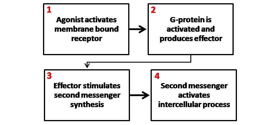
Second messengers are intracellular signaling molecules released by the cell in response to exposure to extracellular signaling molecules—the first messengers. (Intercellular signals, a non-local form or cell signaling, encompassing both first messengers and second messengers, are classified as autocrine, juxtacrine, paracrine, and endocrine depending on the range of the signal.) Second messengers trigger physiological changes at cellular level such as proliferation, differentiation, migration, survival, apoptosis and depolarization. They are one of the triggers of intracellular signal transduction cascades. Examples of second messenger molecules include cyclic AMP, cyclic GMP, inositol triphosphate, diacylglycerol, and calcium. First messengers are extracellular factors, often hormones or neurotransmitters, such as epinephrine, growth hormone, and serotonin. Because peptide hormones and neurotransmitters typically are biochemically hydrophilic molecules, these first messengers ... [...More Info...] [...Related Items...] OR: [Wikipedia] [Google] [Baidu] |
Cyclic Guanosine Monophosphate
Cyclic guanosine monophosphate (cGMP) is a cyclic nucleotide derived from guanosine triphosphate (GTP). cGMP acts as a second messenger much like cyclic AMP. Its most likely mechanism of action is activation of intracellular protein kinases in response to the binding of membrane-impermeable peptide hormones to the external cell surface. Synthesis Guanylate cyclase (GC) catalyzes cGMP synthesis. This enzyme converts GTP to cGMP. Peptide hormones such as the atrial natriuretic factor activate membrane-bound GC, while soluble GC (sGC) is typically activated by nitric oxide to stimulate cGMP synthesis. sGC can be inhibited by ODQ (1H- ,2,4xadiazolo ,3-auinoxalin-1-one). Functions cGMP is a common regulator of ion channel conductance, glycogenolysis, and cellular apoptosis. It also relaxes smooth muscle tissues. In blood vessels, relaxation of vascular smooth muscles leads to vasodilation and increased blood flow. At presynaptic terminals in the striatum, cGMP controls the e ... [...More Info...] [...Related Items...] OR: [Wikipedia] [Google] [Baidu] |


