|
T-shaped Uterus
A t-shaped uterus is a type of uterine malformation wherein the uterus is shaped resembling the letter T. This is typically observed in DES-exposed women. It is recognised in the ESHRE/ESGE classification, and is associated with failed implantation, increased risk of ectopic pregnancy, miscarriage and preterm delivery. There is a surgical procedure to correct the malformation. Causes The T-shaped malformation is commonly associated with in-utero exposure to diethylstilbestrol (the so-called " DES daughters"). It is also presented congenitally. Diagnosis Women are often diagnosed with this condition after several failed pregnancies, proceeded by exploratory diagnostic procedures, such as magnetic resonance, sonography, and particularly hysterosalpingography. In such studies, a widening of the interstitial and isthmus of uterine tube is observed, as well as constrictions or narrowing of the uterus as a whole, especially the lower and lateral portions, hence the "t" denomination. ... [...More Info...] [...Related Items...] OR: [Wikipedia] [Google] [Baidu] |
Hysterosalpingography
Hysterosalpingography (HSG), also known as uterosalpingography, is a radiologic procedure to investigate the shape of the uterine cavity and the shape and patency of the Fallopian tubes. It is a special x-ray using dye to look at the womb (uterus) and Fallopian tubes. It injects a radio-opaque material into the cervical canal, and usually fluoroscopy with image intensification. A normal result shows the filling of the uterine cavity and the bilateral filling of the Fallopian tube with the injection material. To demonstrate tubal rupture, spillage of the material into the peritoneal cavity needs to be observed. Hysterosalpingography has vital role in treatment of infertility, especially in the case of fallopian tube blockage. Uses HSG is considered a diagnostic procedure. It is used in the workup of infertile females to assess the patency of fallopian tubes, assess the competency of the cervix or congenital abnormality of the uterus in multiple miscarriages, assess the patenc ... [...More Info...] [...Related Items...] OR: [Wikipedia] [Google] [Baidu] |
Isthmus Of Uterine Tube
The fallopian tubes, also known as uterine tubes, oviducts or salpinges (singular salpinx), are paired tubes in the human female that stretch from the uterus to the ovaries. The fallopian tubes are part of the female reproductive system. In other mammals they are only called oviducts. Each tube is a muscular hollow organ that is on average between 10 and 14 cm in length, with an external diameter of 1 cm. It has four described parts: the intramural part, isthmus, ampulla, and infundibulum with associated fimbriae. Each tube has two openings a proximal opening nearest and opening to the uterus, and a distal opening furthest and opening to the abdomen. The fallopian tubes are held in place by the mesosalpinx, a part of the broad ligament mesentery that wraps around the tubes. Another part of the broad ligament, the mesovarium suspends the ovaries in place. An egg cell is transported from an ovary to a fallopian tube where it may be fertilized in the ampulla of the t ... [...More Info...] [...Related Items...] OR: [Wikipedia] [Google] [Baidu] |
Mammal Reproductive System
The reproductive system of an organism, also known as the genital system, is the biological system made up of all the anatomical organs involved in sexual reproduction. Many non-living substances such as fluids, hormones, and pheromones are also important accessories to the reproductive system. Unlike most organ systems, the sexes of differentiated species often have significant differences. These differences allow for a combination of genetic material between two individuals, which allows for the possibility of greater genetic fitness of the offspring. Reproductive System 2001 Body Guide powered by Adam Animals In mammals, the major organs of the reproductive system include the external |
Vaginal Adenosis
Vaginal adenosis is a benign abnormality in the vagina, commonly thought to be caused by intrauterine and neonatal exposure of diethylstilbestrol and other progestogens and nonsteroidal estrogens, however it has also been observed in otherwise healthy women and has been considered at times idiopathic or congenital. Postpubertal lesions have also been observed to grow ''de novo''. It has a rather common incidence, of about 10% of adult women. Causes Vaginal adenosis is characterised by the presence of metaplastic cervical or endometrial epithelium within the vaginal wall, considered as derived from Müllerian epithelium islets in later life. In women who were exposed to certain chemicals, vaginal adenosis may arise in up to 90%. Since these contraceptives were discontinued, incidence has dropped dramatically. Risk is however still present in subsequent generations due to recent exposure. It is thought steroid hormones play a stimulatory growth in adenosis formation. Vaginal ... [...More Info...] [...Related Items...] OR: [Wikipedia] [Google] [Baidu] |
Haemorrhage
Bleeding, hemorrhage, haemorrhage or blood loss, is blood escaping from the circulatory system from damaged blood vessels. Bleeding can occur internally, or externally either through a natural opening such as the mouth, nose, ear, urethra, vagina or anus, or through a puncture in the skin. Hypovolemia is a massive decrease in blood volume, and death by excessive loss of blood is referred to as exsanguination. Typically, a healthy person can endure a loss of 10–15% of the total blood volume without serious medical difficulties (by comparison, blood donation typically takes 8–10% of the donor's blood volume). The stopping or controlling of bleeding is called hemostasis and is an important part of both first aid and surgery. Types * Upper head ** Intracranial hemorrhage – bleeding in the skull. ** Cerebral hemorrhage – a type of intracranial hemorrhage, bleeding within the brain tissue itself. ** Intracerebral hemorrhage – bleeding in the brain caused by the ruptur ... [...More Info...] [...Related Items...] OR: [Wikipedia] [Google] [Baidu] |
Asherman's Syndrome
Asherman's syndrome (AS) is an acquired uterine condition that occurs when scar tissue (adhesion (medicine), adhesions) forms inside the uterus and/or the cervix. It is characterized by variable scarring inside the uterine cavity, where in many cases the front and back walls of the uterus stick to one another. AS can be the cause of menstrual disturbances, infertility, and placental abnormalities. Although the first case of intrauterine adhesion was published in 1894 by Heinrich Fritsch, it was only after 54 years that a full description of Asherman syndrome was carried out by Joseph Asherman. A number of other terms have been used to describe the condition and related conditions including: uterine/cervical atresia, traumatic uterine atrophy, sclerotic endometrium, and endometrial sclerosis. There is not any one cause of AS. Risk factors can include myomectomy, cesarean section, infections, age, genital tuberculosis, and obesity. Genetic predisposition to AS is being investigated. ... [...More Info...] [...Related Items...] OR: [Wikipedia] [Google] [Baidu] |
Placenta Accreta
Placenta accreta occurs when all or part of the placenta attaches abnormally to the ''myometrium'' (the muscular layer of the uterine wall). Three grades of abnormal placental attachment are defined according to the depth of attachment and invasion into the muscular layers of the uterus: # ''Accreta – chorionic villi'' attached to the myometrium, rather than being restricted within the '' decidua basalis''. # ''Increta – chorionic villi'' invaded into the ''myometrium''. # ''Percreta – chorionic villi'' invaded through the ''perimetrium'' (uterine serosa). Because of abnormal attachment to the myometrium, placenta accreta is associated with an increased risk of heavy bleeding at the time of attempted vaginal delivery. The need for transfusion of blood products is frequent, and surgical removal of the uterus (hysterectomy) is sometimes required to control life-threatening bleeding. Rates of placenta accreta are increasing. As of 2016, placenta accreta affects an estimated 1 ... [...More Info...] [...Related Items...] OR: [Wikipedia] [Google] [Baidu] |
Endometrium
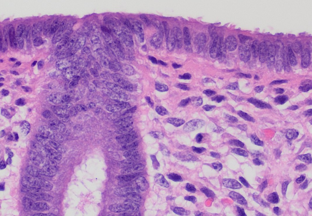
The endometrium is the inner epithelial layer, along with its mucous membrane, of the mammalian uterus. It has a basal layer and a functional layer: the basal layer contains stem cells which regenerate the functional layer. The functional layer thickens and then is shed during menstruation in humans and some other mammals, including apes, Old World monkeys, some species of bat, the elephant shrew and the Cairo spiny mouse. In most other mammals, the endometrium is reabsorbed in the estrous cycle. During pregnancy, the glands and blood vessels in the endometrium further increase in size and number. Vascular spaces fuse and become interconnected, forming the placenta, which supplies oxygen and nutrition to the embryo and fetus.Blue Histology - Female Reproductive System . ... [...More Info...] [...Related Items...] OR: [Wikipedia] [Google] [Baidu] |
Preterm Birth
Preterm birth, also known as premature birth, is the birth of a baby at fewer than 37 weeks gestational age, as opposed to full-term delivery at approximately 40 weeks. Extreme preterm is less than 28 weeks, very early preterm birth is between 28 and 32 weeks, early preterm birth occurs between 32 and 36 weeks, late preterm birth is between 34 and 36 weeks' gestation. These babies are also known as premature babies or colloquially preemies (American English) or premmies (Australian English). Symptoms of preterm labor include uterine contractions which occur more often than every ten minutes and/or the leaking of fluid from the vagina before 37 weeks. Premature infants are at greater risk for cerebral palsy, delays in development, hearing problems and problems with their vision. The earlier a baby is born, the greater these risks will be. The cause of spontaneous preterm birth is often not known. Risk factors include diabetes, high blood pressure, multiple gestation (bei ... [...More Info...] [...Related Items...] OR: [Wikipedia] [Google] [Baidu] |
Surgical Incision
In surgery, a surgical incision is a cut made through the skin and soft tissue to facilitate an operation or procedure. Often, multiple incisions are possible for an operation. In general, a surgical incision is made as small and unobtrusive as possible to facilitate safe and timely operating conditions. Anatomy Surgical incisions are planned based on the expected extent of exposure needed for the specific operation planned. Within each region of the body, several incisions are common. Head and neck * Wilde's incision – This post-aural incision is used for a variant mastoiditis drainage, and was named after Sir William Wilde, an ENT surgeon in Dublin who first described it at the end of the nineteenth century. His son, Oscar Wilde's, death was stated by his doctors to be due to meningitis stemming from an ear infection. He had recently had an operation, believed by some to be a mastoidectomy. Chest * Median sternotomy – This is the primary incision used for cardiac p ... [...More Info...] [...Related Items...] OR: [Wikipedia] [Google] [Baidu] |
Metroplasty
Metroplasty (also called Strassman metroplasty, uteroplasty or hysteroplasty) is a reconstructive surgery used to repair congenital anomalies of the uterus, including septate uterus A uterine septum is a form of a Congenital disorder, congenital uterine malformation, malformation where the uterus, uterine cavity is partitioned by a longitudinal septum; the outside of the uterus has a normal typical shape. The wedge-like parti ... and bicornuate uterus. The surgery entails removing the abnormal tissue that separates the cornua of the uterus, then using several layers of stitches to create a normal shape. References {{Medicine Gynecological surgery ... [...More Info...] [...Related Items...] OR: [Wikipedia] [Google] [Baidu] |
Hysteroscopy
Hysteroscopy is the inspection of the uterine cavity by endoscopy with access through the cervix. It allows for the diagnosis of intrauterine pathology and serves as a method for surgical intervention (operative hysteroscopy). Hysteroscope A hysteroscope is an endoscope that carries optical and light channels or fibers. It is introduced in a sheath that provides an inflow and outflow channel for insufflation of the uterine cavity. In addition, an operative channel may be present to introduce scissors, graspers or biopsy instruments. A hysteroscopic resectoscope is similar to a transurethral resectoscope and allows entry of an electric loop to shave off tissue, for instance to eliminate a fibroid. A ''contact hysteroscope'' is a hysteroscope that does not use distention media. Procedure Hysteroscopy has been carried out in hospitals, surgical centers and doctors' offices. It is best carried out when the endometrium is relatively thin, that is after a menstruation. Both diagnost ... [...More Info...] [...Related Items...] OR: [Wikipedia] [Google] [Baidu] |
.jpg)


