|
Systole
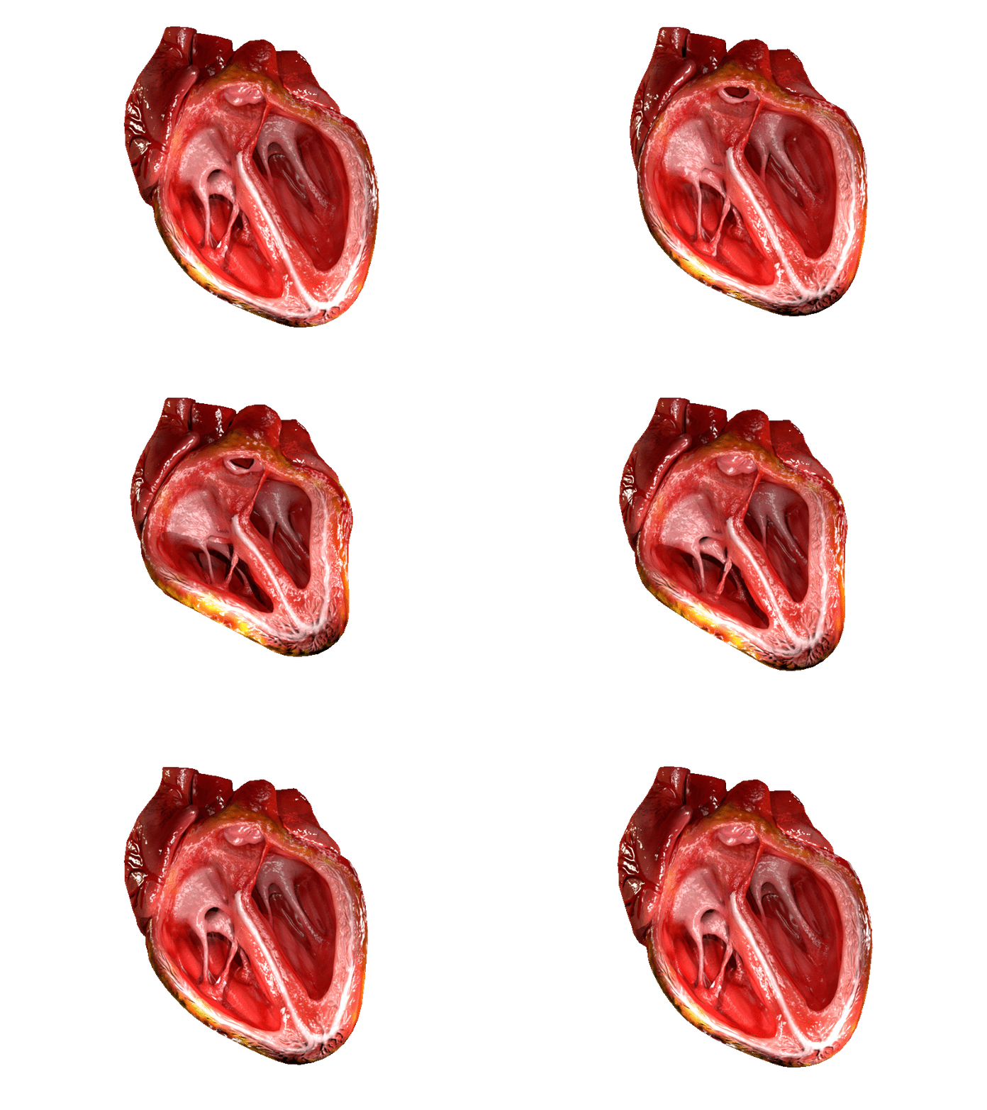
Systole ( ) is the part of the cardiac cycle during which some chambers of the heart contract after refilling with blood. Its contrasting phase is diastole, the relaxed phase of the cardiac cycle when the chambers of the heart are refilling with blood. Etymology The term originates, via Neo-Latin, from Ancient Greek (''sustolē''), from (''sustéllein'' 'to contract'; from ''sun'' 'together' + ''stéllein'' 'to send'), and is similar to the use of the English term ''to squeeze''. Terminology, general explanation The mammalian heart has four chambers: the left atrium above the left ventricle (lighter pink, see graphic), which two are connected through the mitral (or bicuspid) valve; and the right atrium above the right ventricle (lighter blue), connected through the tricuspid valve. The atria are the receiving blood chambers for the circulation of blood and the ventricles are the discharging chambers. In late ventricular diastole, the atrial chambers contract and send ... [...More Info...] [...Related Items...] OR: [Wikipedia] [Google] [Baidu] |
Heart Systole
The heart is a muscular Organ (biology), organ found in humans and other animals. This organ pumps blood through the blood vessels. The heart and blood vessels together make the circulatory system. The pumped blood carries oxygen and nutrients to the tissue, while carrying metabolic waste such as carbon dioxide to the lungs. In humans, the heart is approximately the size of a closed fist and is located between the lungs, in the middle compartment of the thorax, chest, called the mediastinum. In humans, the heart is divided into four chambers: upper left and right Atrium (heart), atria and lower left and right Ventricle (heart), ventricles. Commonly, the right atrium and ventricle are referred together as the right heart and their left counterparts as the left heart. In a healthy heart, blood flows one way through the heart due to heart valves, which prevent cardiac regurgitation, backflow. The heart is enclosed in a protective sac, the pericardium, which also contains a sma ... [...More Info...] [...Related Items...] OR: [Wikipedia] [Google] [Baidu] |
Cardiac Muscle
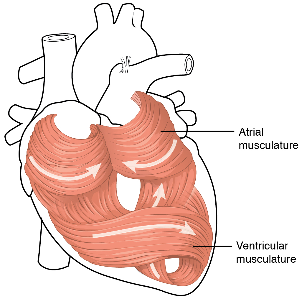
Cardiac muscle (also called heart muscle or myocardium) is one of three types of vertebrate muscle tissues, the others being skeletal muscle and smooth muscle. It is an involuntary, striated muscle that constitutes the main tissue of the wall of the heart. The cardiac muscle (myocardium) forms a thick middle layer between the outer layer of the heart wall (the pericardium) and the inner layer (the endocardium), with blood supplied via the coronary circulation. It is composed of individual cardiac muscle cells joined by intercalated discs, and encased by collagen fibers and other substances that form the extracellular matrix. Cardiac muscle contracts in a similar manner to skeletal muscle, although with some important differences. Electrical stimulation in the form of a cardiac action potential triggers the release of calcium from the cell's internal calcium store, the sarcoplasmic reticulum. The rise in calcium causes the cell's myofilaments to slide past each other i ... [...More Info...] [...Related Items...] OR: [Wikipedia] [Google] [Baidu] |
Histological
Histology, also known as microscopic anatomy or microanatomy, is the branch of biology that studies the microscopic anatomy of biological tissue (biology), tissues. Histology is the microscopic counterpart to gross anatomy, which looks at larger structures visible without a microscope. Although one may divide microscopic anatomy into ''organology'', the study of organs, ''histology'', the study of tissues, and ''cytology'', the study of cell (biology), cells, modern usage places all of these topics under the field of histology. In medicine, histopathology is the branch of histology that includes the microscopic identification and study of diseased tissue. In the field of paleontology, the term paleohistology refers to the histology of fossil organisms. Biological tissues Animal tissue classification There are four basic types of animal tissues: muscle tissue, nervous tissue, connective tissue, and epithelial tissue. All animal tissues are considered to be subtypes of these ... [...More Info...] [...Related Items...] OR: [Wikipedia] [Google] [Baidu] |
P Wave (electrocardiography)
In cardiology, the P wave on an Electrocardiography, electrocardiogram (ECG) represents atrial depolarization, which results in atrial contraction, or systole#Atrial systole, atrial systole. Physiology The P wave is a summation wave generated by the depolarization front as it transits the atria. Normally the right atrium depolarizes slightly earlier than left atrium since the depolarization wave originates in the sinoatrial node, in the high right atrium and then travels to and through the left atrium. The depolarization front is carried through the atria along semi-specialized conduction pathways including Bachmann's bundle resulting in uniform shaped waves. Depolarization originating elsewhere in the atria (atrial ectopics) result in P waves with a different morphology from normal. Pathology Peaked P waves (> 0.25 mV) suggest right atrial enlargement, cor pulmonale, (''P pulmonale'' rhythm), but have a low predictive value (~20%). A P wave with increased amplitude can in ... [...More Info...] [...Related Items...] OR: [Wikipedia] [Google] [Baidu] |
Third-degree Heart Block
Third-degree atrioventricular block (AV block) is a medical condition in which the electrical impulse generated in the sinoatrial node (SA node) in the atrium of the heart can not propagate to the ventricles. Because the impulse is blocked, an accessory pacemaker in the lower chambers will typically activate the ventricles. This is known as an '' escape rhythm''. Since this accessory pacemaker also activates independently of the impulse generated at the SA node, two independent rhythms can be noted on the electrocardiogram (ECG). * The P waves with a regular P-to-P interval (in other words, a sinus rhythm) represent the first rhythm. * The QRS complexes with a regular R-to-R interval represent the second rhythm. The PR interval will be variable, as the hallmark of complete heart block is the lack of any apparent relationship between P waves and QRS complexes. Presentation People with third-degree AV block typically experience severe bradycardia (an abnormally low measured hea ... [...More Info...] [...Related Items...] OR: [Wikipedia] [Google] [Baidu] |
Atrial Flutter
Atrial flutter (AFL) is a common abnormal heart rhythm that starts in the atrial chambers of the heart. When it first occurs, it is usually associated with a fast heart rate and is classified as a type of supraventricular tachycardia (SVT). Atrial flutter is characterized by a sudden-onset (usually) regular abnormal heart rhythm on an electrocardiogram (ECG) in which the heart rate is fast. Symptoms may include a feeling of the heart beating too fast, too hard, or skipping beats, chest discomfort, difficulty breathing, a feeling as if one's stomach has dropped, a feeling of being light-headed, or loss of consciousness. Although this abnormal heart rhythm typically occurs in individuals with cardiovascular disease (e.g., high blood pressure, coronary artery disease, and cardiomyopathy) and diabetes mellitus, it may occur spontaneously in people with otherwise normal hearts. It is typically not a stable rhythm and often degenerates into atrial fibrillation (AF). But rar ... [...More Info...] [...Related Items...] OR: [Wikipedia] [Google] [Baidu] |
Atrial Fibrillation
Atrial fibrillation (AF, AFib or A-fib) is an Heart arrhythmia, abnormal heart rhythm (arrhythmia) characterized by fibrillation, rapid and irregular beating of the Atrium (heart), atrial chambers of the heart. It often begins as short periods of abnormal cardiac cycle, beating, which become longer or continuous over time. It may also start as other forms of arrhythmia such as atrial flutter that then transform into AF. Episodes can be asymptomatic. Symptomatic episodes may involve heart palpitations, syncope (medicine), fainting, Presyncope, lightheadedness, Unconsciousness, loss of consciousness, or shortness of breath. Atrial fibrillation is associated with an increased risk of heart failure, dementia, and stroke. It is a type of supraventricular tachycardia. Atrial fibrillation frequently results from bursts of tachycardia that originate in muscle bundles extending from the Atrium (heart), atrium to the pulmonary veins. Pulmonary vein isolation by catheter ablation, trans ... [...More Info...] [...Related Items...] OR: [Wikipedia] [Google] [Baidu] |
Left Ventricular Hypertrophy
Left ventricular hypertrophy (LVH) is thickening of the heart muscle of the left ventricle of the heart, that is, left-sided ventricular hypertrophy and resulting increased left ventricular mass. Causes While ventricular hypertrophy occurs naturally as a reaction to aerobic exercise and strength training, it is most frequently referred to as a pathological reaction to cardiovascular disease, or high blood pressure. It is one aspect of ventricular remodeling. While LVH itself is not a disease, it is usually a marker for disease involving the heart. Disease processes that can cause LVH include any disease that increases the afterload that the heart has to contract against, and some primary diseases of the muscle of the heart. Causes of increased afterload that can cause LVH include aortic stenosis, aortic insufficiency and hypertension. Primary disease of the muscle of the heart that cause LVH are known as hypertrophic cardiomyopathies, which can lead into heart failure. ... [...More Info...] [...Related Items...] OR: [Wikipedia] [Google] [Baidu] |
Tricuspid
The tricuspid valve, or right atrioventricular valve, is on the right dorsal side of the mammalian heart, at the superior portion of the right ventricle. The function of the valve is to allow blood to flow from the right atrium to the right ventricle during diastole, and to close to prevent backflow ( regurgitation) from the right ventricle into the right atrium during right ventricular contraction (systole). Structure The tricuspid valve usually has three cusps or leaflets, named the anterior, posterior, and septal cusps. Each leaflet is connected via chordae tendineae to the anterior, posterior, and septal papillary muscles of the right ventricle, respectively. Tricuspid valves may also occur with two or four leaflets; the number may change over a lifetime. Function The tricuspid valve functions as a one-way valve that closes during ventricular systole to prevent regurgitation of blood from the right ventricle back into the right atrium. It opens during ventricular dia ... [...More Info...] [...Related Items...] OR: [Wikipedia] [Google] [Baidu] |
Atrioventricular Valve
A heart valve is a biological one-way valve that allows blood to flow in one direction through the chambers of the heart. A mammalian heart usually has four valves. Together, the valves determine the direction of blood flow through the heart. Heart valves are opened or closed by a difference in blood pressure on each side. The mammalian heart has two atrioventricular valves separating the upper atria from the lower ventricles: the mitral valve in the left heart, and the tricuspid valve in the right heart. The two semilunar valves are at the entrance of the arteries leaving the heart. These are the aortic valve at the aorta, and the pulmonary valve at the pulmonary artery. The heart also has a coronary sinus valve and an inferior vena cava valve, not discussed here. Structure The heart valves and the chambers are lined with endocardium. Heart valves separate the atria from the ventricles, or the ventricles from a blood vessel. Heart valves are situated around the fibro ... [...More Info...] [...Related Items...] OR: [Wikipedia] [Google] [Baidu] |
Myocardium
Cardiac muscle (also called heart muscle or myocardium) is one of three types of vertebrate muscle tissues, the others being skeletal muscle and smooth muscle. It is an involuntary, striated muscle that constitutes the main tissue of the wall of the heart. The cardiac muscle (myocardium) forms a thick middle layer between the outer layer of the heart wall (the pericardium) and the inner layer (the endocardium), with blood supplied via the coronary circulation. It is composed of individual cardiac muscle cells joined by intercalated discs, and encased by collagen fibers and other substances that form the extracellular matrix. Cardiac muscle contracts in a similar manner to skeletal muscle, although with some important differences. Electrical stimulation in the form of a cardiac action potential triggers the release of calcium from the cell's internal calcium store, the sarcoplasmic reticulum. The rise in calcium causes the cell's myofilaments to slide past each other ... [...More Info...] [...Related Items...] OR: [Wikipedia] [Google] [Baidu] |
Heart Diasystole
The heart is a muscular Organ (biology), organ found in humans and other animals. This organ pumps blood through the blood vessels. The heart and blood vessels together make the circulatory system. The pumped blood carries oxygen and nutrients to the tissue, while carrying metabolic waste such as carbon dioxide to the lungs. In humans, the heart is approximately the size of a closed fist and is located between the lungs, in the middle compartment of the thorax, chest, called the mediastinum. In humans, the heart is divided into four chambers: upper left and right Atrium (heart), atria and lower left and right Ventricle (heart), ventricles. Commonly, the right atrium and ventricle are referred together as the right heart and their left counterparts as the left heart. In a healthy heart, blood flows one way through the heart due to heart valves, which prevent cardiac regurgitation, backflow. The heart is enclosed in a protective sac, the pericardium, which also contains a sma ... [...More Info...] [...Related Items...] OR: [Wikipedia] [Google] [Baidu] |


