|
Supratrochlear
The supratrochlear nerve is a branch of the frontal nerve, itself a branch of the ophthalmic nerve (CN V1) from the trigeminal nerve (CN V). It provides sensory innervation to the skin of the forehead and the upper eyelid. Structure Origin The supratrochlear nerve is the smaller of the two terminal branches of the frontal nerve (the other being the supraorbital nerve). It arises midway between the base and apex of the orbit where the frontal nerve splits into said terminal branches. Course The supratrochlear nerve passes medially above the trochlea of the superior oblique muscle. It then travels anteriorly above the levator palpebrae superioris muscle. It exits the orbit through the supratrochlear notch or foramen. It then ascends onto the forehead beneath the corrugator supercilii muscle and frontalis muscle. It finally divides into sensory branches. The supratrochlear nerve travels with the supratrochlear artery, a branch of the ophthalmic artery. Branches Before ... [...More Info...] [...Related Items...] OR: [Wikipedia] [Google] [Baidu] |
Supratrochlear Artery
The supratrochlear artery (or frontal artery) is one of the terminal branches of the ophthalmic artery. It arises within the orbit. It exits the orbit alongside the supratrochlear nerve. It contributes arterial supply to the skin, muscles and pericranium of the forehead. Anatomy It branches from the ophthalmic artery near the trochlea of the superior oblique muscle in the orbit. Origin The supratrochlear artery branches from the ophthalmic artery in the orbit near the trochlea of the superior oblique muscle. Course After branching from the ophthalmic artery, it passes anteriorly through the superomedial orbit. It travels medial to the trochlear nerve. With the supratrochlear nerve, the supratrochlear artery exits the orbit through the supratrochlear notch (variably present), medial to the supraorbital foramen. It then ascends on the forehead. Anastomoses The supratrochlear artery anastomoses with the contralateral supratrochlear artery, and the ipsilateral supraorbit ... [...More Info...] [...Related Items...] OR: [Wikipedia] [Google] [Baidu] |
Supratrochlear Notch
The supratrochlear nerve is a branch of the frontal nerve, itself a branch of the ophthalmic nerve (CN V1) from the trigeminal nerve (CN V). It provides sensory innervation to the skin of the forehead and the upper eyelid. Structure Origin The supratrochlear nerve is the smaller of the two terminal branches of the frontal nerve (the other being the supraorbital nerve). It arises midway between the base and apex of the orbit where the frontal nerve splits into said terminal branches. Course The supratrochlear nerve passes medially above the trochlea of the superior oblique muscle. It then travels anteriorly above the levator palpebrae superioris muscle. It exits the orbit through the supratrochlear notch or foramen. It then ascends onto the forehead beneath the corrugator supercilii muscle and frontalis muscle. It finally divides into sensory branches. The supratrochlear nerve travels with the supratrochlear artery, a branch of the ophthalmic artery. Branches Before ... [...More Info...] [...Related Items...] OR: [Wikipedia] [Google] [Baidu] |
Scalp
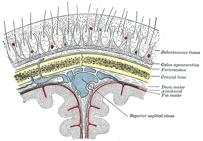
The scalp is the area of the head where head hair grows. It is made up of skin, layers of connective and fibrous tissues, and the membrane of the skull. Anatomically, the scalp is part of the epicranium, a collection of structures covering the cranium. The scalp is bordered by the face at the front, and by the neck at the sides and back. The scientific study of hair and scalp is called trichology. Structure Layers The scalp is usually described as having five layers, which can be remembered using the mnemonic 'SCALP': * S: Skin. The skin of the scalp contains numerous hair follicles and sebaceous glands. * C: Connective tissue. A dense subcutaneous layer of fat and fibrous tissue that lies beneath the skin, containing the nerves and vessels of the scalp. * A: Aponeurosis. The epicranial aponeurosis or galea aponeurotica is a tough layer of dense fibrous tissue which anchors the above layers in place. It runs from the frontalis muscle anteriorly to the occipitalis ... [...More Info...] [...Related Items...] OR: [Wikipedia] [Google] [Baidu] |
Frontalis Muscle
The frontalis muscle () is a muscle which covers parts of the forehead of the skull. Some sources consider the frontalis muscle to be a distinct muscle. However, Terminologia Anatomica currently classifies it as part of the occipitofrontalis muscle along with the occipitalis muscle. In humans, the frontalis muscle only serves for facial expressions. The frontalis muscle is supplied by the facial nerve and receives blood from the supraorbital and supratrochlear arteries. Structure The frontalis muscle is thin, of a quadrilateral form, and intimately adherent to the superficial fascia. It is broader than the occipitalis and its fibers are longer and paler in color. It is located on the front of the head. The muscle has no bony attachments. Its medial fibers are continuous with those of the procerus; its intermediate fibers blend with the corrugator and orbicularis oculi muscles, thus attached to the skin of the eyebrows; and its lateral fibers are also blended with the latte ... [...More Info...] [...Related Items...] OR: [Wikipedia] [Google] [Baidu] |
Supraorbital Nerve
The supraorbital nerve is one of two terminal branches - the other being the supratrochlear nerve - of the frontal nerve (itself a branch of the ophthalmic nerve (CN V1)). It exits the orbit via the supraorbital foramen/notch before splitting into a medial branch and a lateral branch. It innervates the skin of the forehead, upper eyelid, and the root of the nose. Structure Origin The supraorbital nerve branches from the frontal nerve midway between the base and apex of the orbit. Course It travels anteriorly superior to the levator palpebrae superioris muscle. It exits the orbit through the supraorbital foramen/notch in the superior margin orbit, exiting it lateral to the supratrochlear nerve. It then ascends onto the forehead deep to the corrugator supercilii muscle and frontalis muscles. Fate It divides into a medial branch and lateral branch - usually after emerging from the orbit, but sometimes already within the orbit. Distribution The supraorbital nerv ... [...More Info...] [...Related Items...] OR: [Wikipedia] [Google] [Baidu] |
Infratrochlear Nerve
The infratrochlear nerve is a branch of the nasociliary nerve (itself a branch of the ophthalmic nerve (CN V1)) in the orbit In celestial mechanics, an orbit (also known as orbital revolution) is the curved trajectory of an object such as the trajectory of a planet around a star, or of a natural satellite around a planet, or of an artificial satellite around an .... It exits the orbit inferior to the trochlea of superior oblique. It provides sensory innervation to structures of the orbit and skin of adjacent structures. Structure The nasociliary nerve terminates by bifurcating into the infratrochlear and the anterior ethmoidal nerves. The infratrochlear nerve travels anteriorly in the orbit along the upper border of the medial rectus muscle and underneath the trochlea of the superior oblique muscle. It exits the orbit medially and divides into small sensory branches. Distribution The infratrochlear nerve provides sensory innervation to the skin of the eyelids ... [...More Info...] [...Related Items...] OR: [Wikipedia] [Google] [Baidu] |
Ophthalmic Artery
The ophthalmic artery (OA) is an artery of the head. It is the first branch of the internal carotid artery distal to the cavernous sinus. Branches of the ophthalmic artery supply all the structures in the orbit around the eye, as well as some structures in the nose, face, and meninges. Occlusion of the ophthalmic artery or its branches can produce sight-threatening conditions. Structure The ophthalmic artery emerges from the internal carotid artery. This is usually just after the internal carotid artery emerges from the cavernous sinus. In some cases, the ophthalmic artery branches just before the internal carotid exits the carotid sinus. The ophthalmic artery emerges along the medial side of the anterior clinoid process. It runs anteriorly, passing through the optic canal inferolaterally to the optic nerve. It can also pass superiorly to the optic nerve in a minority of cases. In the posterior third of the cone of the orbit, the ophthalmic artery turns sharply and med ... [...More Info...] [...Related Items...] OR: [Wikipedia] [Google] [Baidu] |
Frontal Nerve
The frontal nerve is the largest branch of the ophthalmic nerve (V1), itself a branch of the trigeminal nerve (CN V). It supplies sensation to the skin of the forehead, the mucosa of the frontal sinus, and the skin of the upper eyelid. It may be affected by schwannoma. Structure The frontal nerve is a branch of the ophthalmic nerve (V1), itself a branch of the trigeminal nerve (CN V). The frontal nerve branches immediately before entering the superior orbital fissure. In then travels superolateral to the annulus of Zinn between the lacrimal nerve and inferior ophthalmic vein. After entering the orbit it travels anteriorly between the roof periosteum and the levator palpebrae superioris. Midway between the apex and base of the orbit it divides into two branches, the supratrochlear nerve and supraorbital nerve. Functions The two branches of the frontal nerve provide sensory innervation to the skin of the forehead, mucosa of the frontal sinus (an air sinus), and the skin of th ... [...More Info...] [...Related Items...] OR: [Wikipedia] [Google] [Baidu] |
Forehead
In human anatomy, the forehead is an area of the head bounded by three features, two of the skull and one of the scalp. The top of the forehead is marked by the hairline, the edge of the area where hair on the scalp grows. The bottom of the forehead is marked by the supraorbital ridge, the bone feature of the skull above the eyes. The two sides of the forehead are marked by the temporal ridge, a bone feature that links the supraorbital ridge to the coronal suture line and beyond. However, the eyebrows do not form part of the forehead. In '' Terminologia Anatomica'', ''sinciput'' is given as the Latin equivalent to "forehead" (etymology of ''sinciput'': from ''semi-'' "half" and ''caput'' "head".). Structure The bone of the forehead is the squamous part of the frontal bone. The overlying muscles are the occipitofrontalis, procerus, and corrugator supercilii muscles, all of which are controlled by the temporal branch of the facial nerve. The sensory nerves of the forehea ... [...More Info...] [...Related Items...] OR: [Wikipedia] [Google] [Baidu] |
Supraorbital Artery
The supraorbital artery is a branch of the ophthalmic artery. It passes anteriorly within the orbit to exit the orbit through the supraorbital foramen or notch alongside the supraorbital nerve, splitting into two terminal branches which go on to form anastomoses with arteries of the head. Structure Origin The supraorbital artery arises from the ophthalmic artery. Course and relations It travels anteriorly in the orbit by passing superior to the eye and medial to the superior rectus and levator palpebrae superioris. It then joins the supraorbital nerve to jointly pass between the periosteum of the roof of the orbit and the levator palpebrae superioris towards the supraorbital foramen or notch. After passing through the supraorbital foramen or notch, it often splits into a superficial branch and a deep branch. Distribution The supraorbital artery contributes arterial supply to: the superior rectus muscle, superior oblique muscle, levator palpebrae muscles, periorbita, t ... [...More Info...] [...Related Items...] OR: [Wikipedia] [Google] [Baidu] |
Upper Eyelid
An eyelid ( ) is a thin fold of skin that covers and protects an eye. The levator palpebrae superioris muscle retracts the eyelid, exposing the cornea to the outside, giving vision. This can be either voluntarily or involuntarily. "Palpebral" (and "blepharal") means relating to the eyelids. Its key function is to regularly spread the tears and other secretions on the eye surface to keep it moist, since the cornea must be continuously moist. They keep the eyes from drying out when asleep. Moreover, the blink reflex protects the eye from foreign bodies. A set of specialized hairs known as lashes grow from the upper and lower eyelid margins to further protect the eye from dust and debris. The appearance of the human upper eyelid often varies between different populations. The prevalence of an epicanthic fold covering the inner corner of the eye account for the majority of East Asian and Southeast Asian populations, and is also found in varying degrees among other population ... [...More Info...] [...Related Items...] OR: [Wikipedia] [Google] [Baidu] |

