|
Supersecondary Structure
A supersecondary structure is a compact three-dimensional protein structure of several adjacent elements of a secondary structure that is smaller than a protein domain or a subunit. Supersecondary structures can act as nucleations in the process of protein folding. Examples Helix supersecondary structures Helix hairpin A helix hairpin, also known as an alpha-alpha hairpin, is composed of two antiparallel alpha helices connected by a loop of two or more residues. True to its name, it resembles a hairpin. A longer loop has a greater number of possible conformations. If short strands connect the helices, then the individual helices will pack together through their hydrophobic residues. The function of a helix hairpin is unknown; however, a four helix bundle is composed of two helix hairpins, which have important ligand binding sites. Helix corner A helix corner, also called an alpha-alpha corner, has two alpha helices almost at right angles to each other connected by a ... [...More Info...] [...Related Items...] OR: [Wikipedia] [Google] [Baidu] |
Protein Structure
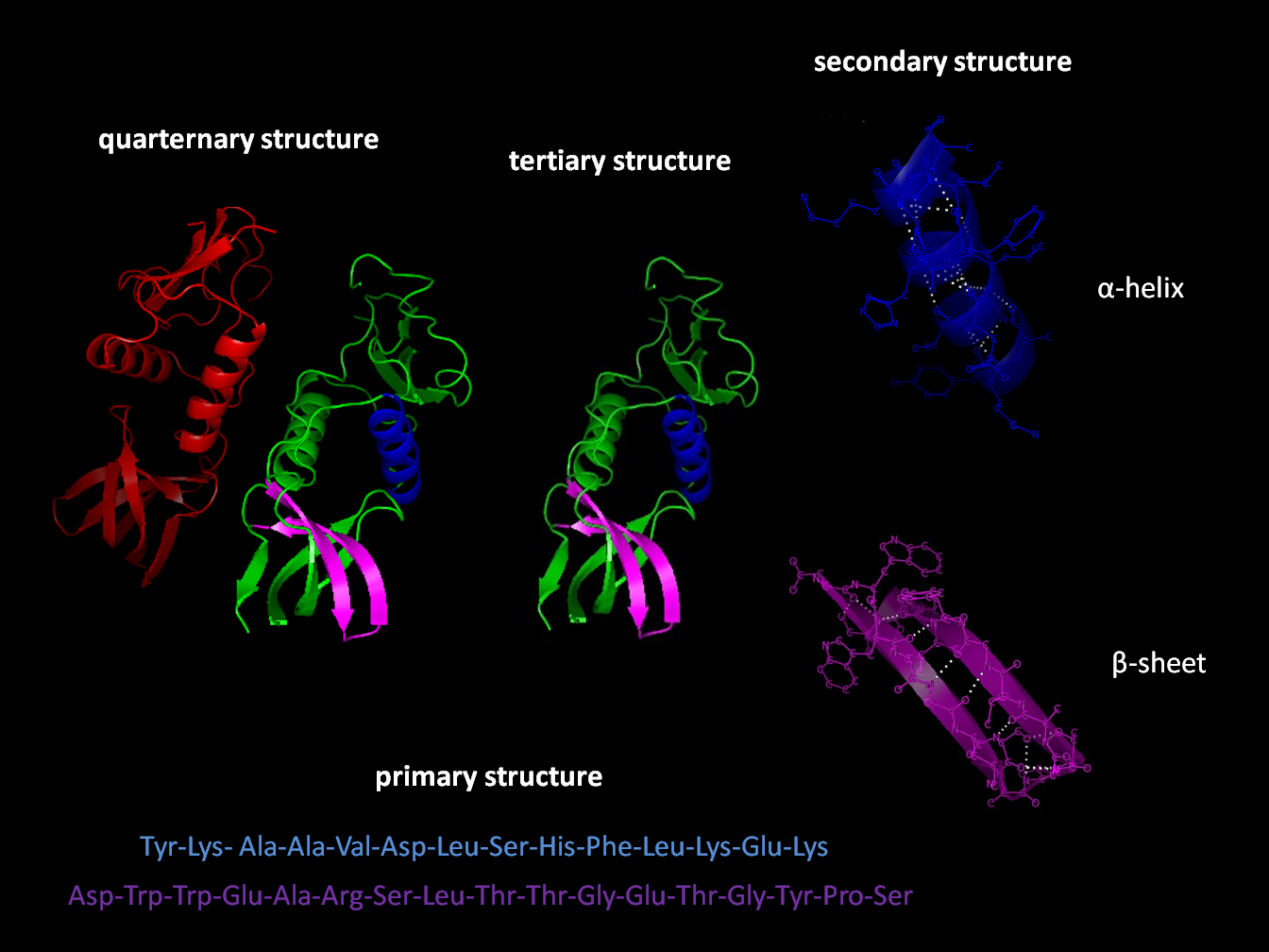
Protein structure is the three-dimensional arrangement of atoms in an amino acid-chain molecule. Proteins are polymers specifically polypeptides formed from sequences of amino acids, which are the monomers of the polymer. A single amino acid monomer may also be called a ''residue'', which indicates a repeating unit of a polymer. Proteins form by amino acids undergoing condensation reactions, in which the amino acids lose one water molecule per reaction in order to attach to one another with a peptide bond. By convention, a chain under 30 amino acids is often identified as a peptide, rather than a protein. To be able to perform their biological function, proteins fold into one or more specific spatial conformations driven by a number of non-covalent interactions, such as hydrogen bonding, ionic interactions, Van der Waals forces, and hydrophobic packing. To understand the functions of proteins at a molecular level, it is often necessary to determine their three-dimensiona ... [...More Info...] [...Related Items...] OR: [Wikipedia] [Google] [Baidu] |
PDB 1j4m EBI
PDB or pdb may refer to: Organizations * Party of German-speaking Belgians (German: '), a former Belgian political party * Promised Day Brigade, a former Iraqi organization Science and technology * Protein Data Bank, a biological molecule database ** Protein Data Bank (file format) * Potato dextrose broth, a microbiological growth medium * Pee Dee Belemnite, a reference standard for isotopes; see ''δ''13C Computing * PDB (Palm OS), a record database format * Pluggable database, in Oracle Database * Program database, a debugging information format * Python Debugger (pdb), of the Python programming language; see Stepping Other uses * Chess Problem Database Server (PDB Server), a repository for chess problems * Pousette-Dart Band, an American band * President's Daily Brief, a US intelligence document See also * 1,4-Dichlorobenzene or ''para''-dichlorobenzene (PDCB), a chemical * Bangladesh Power Development Board The Bangladesh Power Development Board (BPDB) is a governmen ... [...More Info...] [...Related Items...] OR: [Wikipedia] [Google] [Baidu] |
Protein Folding
Protein folding is the physical process by which a protein, after Protein biosynthesis, synthesis by a ribosome as a linear chain of Amino acid, amino acids, changes from an unstable random coil into a more ordered protein tertiary structure, three-dimensional structure. This structure permits the protein to become biologically functional or active. The folding of many proteins begins even during the translation of the polypeptide chain. The amino acids interact with each other to produce a well-defined three-dimensional structure, known as the protein's native state. This structure is determined by the amino-acid sequence or primary structure. The correct three-dimensional structure is essential to function, although some parts of functional proteins Intrinsically unstructured proteins, may remain unfolded, indicating that protein dynamics are important. Failure to fold into a native structure generally produces inactive proteins, but in some instances, misfolded proteins have ... [...More Info...] [...Related Items...] OR: [Wikipedia] [Google] [Baidu] |
Nicotinamide Adenine Dinucleotide
Nicotinamide adenine dinucleotide (NAD) is a Cofactor (biochemistry), coenzyme central to metabolism. Found in all living cell (biology), cells, NAD is called a dinucleotide because it consists of two nucleotides joined through their phosphate groups. One nucleotide contains an adenine nucleobase and the other, nicotinamide. NAD exists in two forms: an Redox, oxidized and reduced form, abbreviated as NAD and NADH (H for hydrogen), respectively. In cellular metabolism, NAD is involved in redox reactions, carrying electrons from one reaction to another, so it is found in two forms: NAD is an oxidizing agent, accepting electrons from other molecules and becoming reduced; with H+, this reaction forms NADH, which can be used as a reducing agent to donate electrons. These electron transfer reactions are the main function of NAD. It is also used in other cellular processes, most notably as a substrate (biochemistry), substrate of enzymes in adding or removing chemical groups to or fr ... [...More Info...] [...Related Items...] OR: [Wikipedia] [Google] [Baidu] |
Michael Rossmann
Michael G. Rossmann (30 July 1930 – 14 May 2019) was a German-American physicist, microbiologist, and Hanley Distinguished Professor of Biological Sciences at Purdue University who led a team of researchers to be the first to map the structure of a human common cold virus to an atomic level. He also discovered the Rossmann fold protein motif. His most well recognised contribution to structural biology is the development of a phasing technique named molecular replacement, which has led to about three quarters of depositions in the Protein Data Bank. Education Born in Frankfurt, Germany, Rossmann studied physics and mathematics at the University of London, where he received BSc and MSc degrees. He moved to Glasgow in 1953 where he taught physics in the technical college and received his Ph.D. in chemical crystallography in 1956. He attributes his initial interest in crystallography to Kathleen Lonsdale, whom he heard speak as a schoolboy. Rossmann began his career as a crys ... [...More Info...] [...Related Items...] OR: [Wikipedia] [Google] [Baidu] |
Rossman Fold
Rossmann, Roßmann or Rossman may refer to: Surname * Amy Y. Rossman (born 1946), American mycologist * Benjamin Rossman (born 1980), American-Canadian mathematician * Bubby Rossman (born 1992), American Major League Baseball player * Claude Rossman (1881–1928), American Major League Baseball player * Dirk Rossmann (born 1946), German billionaire businessman and founder of Rossmann * Douglas A. Rossman (1936–2015), U.S. herpetologist * Edmund Roßmann (1918–2005), German fighter pilot during World War II * Ernst Dieter Rossmann (born 1951), German politician * George Rossman (1885–1967), American lawyer and judge * George R. Rossman (born 1944), American mineralogist and professor * Henryk Rossman (1896–1937), Polish lawyer and political activist * Karl Roßmann (1916–2002), German officer in the Luftwaffe during World War II * Louis Rossmann (born 1988), American right to repair activist * Michael Rossmann (1930–2019), German-American physicist * Mike Rossman ... [...More Info...] [...Related Items...] OR: [Wikipedia] [Google] [Baidu] |
β-sheets
The beta sheet (β-sheet, also β-pleated sheet) is a common motif of the regular protein secondary structure. Beta sheets consist of beta strands (β-strands) connected laterally by at least two or three backbone hydrogen bonds, forming a generally twisted, pleated sheet. A β-strand is a stretch of polypeptide chain typically 3 to 10 amino acids long with backbone in an extended conformation. The supramolecular association of β-sheets has been implicated in the formation of the fibrils and protein aggregates observed in amyloidosis, Alzheimer's disease and other proteinopathies. History The first β-sheet structure was proposed by William Astbury in the 1930s. He proposed the idea of hydrogen bonding between the peptide bonds of parallel or antiparallel extended β-strands. However, Astbury did not have the necessary data on the bond geometry of the amino acids in order to build accurate models, especially since he did not then know that the peptide bond was planar. A ref ... [...More Info...] [...Related Items...] OR: [Wikipedia] [Google] [Baidu] |
Anthrax Toxin Protein Key Motif
Anthrax is an infection caused by the bacterium ''Bacillus anthracis'' or ''Bacillus cereus'' biovar ''anthracis''. Infection typically occurs by contact with the skin, inhalation, or intestinal absorption. Symptom onset occurs between one day and more than two months after the infection is contracted. The skin form presents with a small blister with surrounding swelling that often turns into a painless ulcer with a black center. The inhalation form presents with fever, chest pain, and shortness of breath. The intestinal form presents with diarrhea (which may contain blood), abdominal pains, nausea, and vomiting. According to the U.S. Centers for Disease Control and Prevention, the first clinical descriptions of cutaneous anthrax were given by Maret in 1752 and Fournier in 1769. Before that, anthrax had been described only in historical accounts. The German scientist Robert Koch was the first to identify ''Bacillus anthracis'' as the bacterium that causes anthrax. Anthrax i ... [...More Info...] [...Related Items...] OR: [Wikipedia] [Google] [Baidu] |
Beta Bulge
A beta bulge can be described as a localized disruption of the regular hydrogen bonding of beta sheet by inserting extra residues into one or both hydrogen bonded β-strands. Types β-bulges can be grouped according to their length of the disruption, the number of residues inserted into each strand, whether the disrupted β-strands are parallel or antiparallel and by their dihedral angles (which controls the placement of their side chains). Two types occur commonly. One, the ''classic beta bulge'', occurs within, or at the edge of, antiparallel beta-sheet; the first residue at the outwards bulge typically has the αR, rather than the normal β, conformation. The other type is the G1 ''beta bulge'', of which there are two common sorts, both mainly occurring in association with antiparallel sheet; one residue has the αL conformation and is usually a glycine. In one sort, the beta bulge loop, one of the hydrogen bonds of the beta-bulge also forms a beta turn or alpha turn, such that ... [...More Info...] [...Related Items...] OR: [Wikipedia] [Google] [Baidu] |
Glycine
Glycine (symbol Gly or G; ) is an amino acid that has a single hydrogen atom as its side chain. It is the simplest stable amino acid. Glycine is one of the proteinogenic amino acids. It is encoded by all the codons starting with GG (GGU, GGC, GGA, GGG). Glycine disrupts the formation of alpha-helices in secondary protein structure. Its small side chain causes it to favor random coils instead. Glycine is also an inhibitory neurotransmitter – interference with its release within the spinal cord (such as during a '' Clostridium tetani'' infection) can cause spastic paralysis due to uninhibited muscle contraction. It is the only achiral proteinogenic amino acid. It can fit into both hydrophilic and hydrophobic environments, due to its minimal side chain of only one hydrogen atom. History and etymology Glycine was discovered in 1820 by French chemist Henri Braconnot when he hydrolyzed gelatin by boiling it with sulfuric acid. He originally called it "sugar of ... [...More Info...] [...Related Items...] OR: [Wikipedia] [Google] [Baidu] |
Beta Sheet
The beta sheet (β-sheet, also β-pleated sheet) is a common motif of the regular protein secondary structure. Beta sheets consist of beta strands (β-strands) connected laterally by at least two or three backbone hydrogen bonds, forming a generally twisted, pleated sheet. A β-strand is a stretch of polypeptide chain typically 3 to 10 amino acids long with backbone in an extended conformation. The supramolecular association of β-sheets has been implicated in the formation of the fibrils and protein aggregates observed in amyloidosis, Alzheimer's disease and other proteinopathies. History The first β-sheet structure was proposed by William Astbury in the 1930s. He proposed the idea of hydrogen bonding between the peptide bonds of parallel or antiparallel extended β-strands. However, Astbury did not have the necessary data on the bond geometry of the amino acids in order to build accurate models, especially since he did not then know that the peptide bond was planar. ... [...More Info...] [...Related Items...] OR: [Wikipedia] [Google] [Baidu] |
Structural Motif
In a chain-like biological molecule, such as a protein or nucleic acid, a structural motif is a common three-dimensional structure that appears in a variety of different, evolutionarily unrelated molecules. A structural motif does not have to be associated with a sequence motif; it can be represented by different and completely unrelated sequences in different proteins or RNA. In nucleic acids Depending upon the sequence and other conditions, nucleic acids can form a variety of structural motifs which is thought to have biological significance. ;Stem-loop: Stem-loop intramolecular base pairing is a pattern that can occur in single-stranded DNA or, more commonly, in RNA. The structure is also known as a hairpin or hairpin loop. It occurs when two regions of the same strand, usually complementary in nucleotide sequence when read in opposite directions, base-pair to form a double helix that ends in an unpaired loop. The resulting structure is a key building block of many ... [...More Info...] [...Related Items...] OR: [Wikipedia] [Google] [Baidu] |


