|
Superior Salivatory Nucleus
The salivatory nuclei are two general visceral efferent fiber, general visceral efferent Nucleus (anatomy), nuclei located in the caudal pons, dorsal and lateral to the facial nucleus. Their neurons give rise to preganglionic nerve fibers, preganglionic Parasympathetic nervous system, parasympathetic nerve fibers in the control of Salivary gland, salivation.Digital version The superior salivatory nucleus supplies fibers to the intermediate nerve (part of the facial nerve (CN VII). The inferior salivatory nucleus supplies fibers to the glossopharyngeal nerve (CN IX). The nuclei may also be involved in parasympathetic control of (extracranial and intracranial) head vasculature. Superior salivatory nucleus The superior salivatory nucleus (or nucleus salivatorius superior) is a visceral motor cranial nerve nucleus of the facial nerve, facial nerve (CN VII). It is located in the pontine tegmentum. It projects pre-ganglionic visceral motor parasympathetic efferents (via Facial nerve, C ... [...More Info...] [...Related Items...] OR: [Wikipedia] [Google] [Baidu] |
General Visceral Efferent Fiber
General visceral efferent fibers (GVE), visceral efferents or autonomic efferents are the efferent nerve fibers of the autonomic nervous system (also known as the ''visceral efferent nervous system'') that provide motor innervation to smooth muscle, cardiac muscle, and glands (contrast with special visceral efferent (SVE) fibers) through postganglionic varicosities. GVE fibers may be either sympathetic or parasympathetic. Cranial and sacral spinal fibers are parasympathetic GVE fibers, while thoracic and lumbar spinal cord give rise to sympathetic GVE fibers. The cranial nerves containing GVE fibers include the oculomotor nerve (CN III), the facial nerve (CN VII), the glossopharyngeal nerve (CN IX) and the vagus nerve (CN X).Mehta, Samir et al. Step-Up: A High-Yield, Systems-Based Review for the USMLE Step 1. Baltimore, MD: LWW, 2003. Additional images File:Gray840.png, Sympathetic connections of the ciliary and superior cervical ganglia. File:Gray839.png, Autonomic nervous ... [...More Info...] [...Related Items...] OR: [Wikipedia] [Google] [Baidu] |
Preganglionic
In the autonomic nervous system, nerve fibers from the central nervous system to the autonomic ganglion, ganglion are known as preganglionic nerve fibers. All preganglionic fibers, whether they are in the sympathetic nervous system, sympathetic division or in the parasympathetic nervous system, parasympathetic division, are cholinergic (that is, these fibers use acetylcholine as their neurotransmitter) and they are myelinated. Sympathetic nervous system, Sympathetic preganglionic fibers tend to be shorter than parasympathetic preganglionic fibers because sympathetic ganglia are often closer to the spinal cord than are the Parasympathetic nervous system, parasympathetic ganglia. Another major difference between the two ANS (autonomic nervous systems) is divergence. Whereas in the parasympathetic division there is a divergence factor of roughly 1:4, in the sympathetic division there can be a divergence of up to 1:20. This is due to the number of synapses formed by the preganglio ... [...More Info...] [...Related Items...] OR: [Wikipedia] [Google] [Baidu] |
Saliva
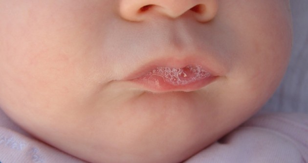
Saliva (commonly referred as spit or drool) is an extracellular fluid produced and secreted by salivary glands in the mouth. In humans, saliva is around 99% water, plus electrolytes, mucus, white blood cells, epithelial cells (from which DNA can be extracted), enzymes (such as lingual lipase and amylase), and antimicrobial agents (such as secretory IgA, and lysozymes). The enzymes found in saliva are essential in beginning the process of digestion of dietary starches and fats. These enzymes also play a role in breaking down food particles entrapped within dental crevices, thus protecting teeth from bacterial decay. Saliva also performs a lubricating function, wetting food and permitting the initiation of swallowing, and protecting the oral mucosa from drying out. Saliva has specialized purposes for a variety of animal species beyond predigestion. Certain swifts construct nests with their sticky saliva. The foundation of bird's nest soup is an aerodramus nest. Venom ... [...More Info...] [...Related Items...] OR: [Wikipedia] [Google] [Baidu] |
Parotid Gland
The parotid gland is a major salivary gland in many animals. In humans, the two parotid glands are present on either side of the mouth and in front of both ears. They are the largest of the salivary glands. Each parotid is wrapped around the mandibular ramus, and secretes serous saliva through the parotid duct into the mouth, to facilitate mastication and swallowing and to begin the digestion of starches. There are also two other types of salivary glands; they are submandibular and sublingual glands. Sometimes accessory parotid glands are found close to the main parotid glands. The venom glands of snakes are a modification of the parotid salivary glands. Etymology The word ''parotid'' literally means "beside the ear". From Greek παρωτίς (stem παρωτιδ-) : (gland) behind the ear < παρά - pará : in front, and οὖς - ous (stem ὠτ-, ōt-) : ear. Structure The parotid glands are a pair of mainly[...More Info...] [...Related Items...] OR: [Wikipedia] [Google] [Baidu] |
Parasympathetic Nervous System
The parasympathetic nervous system (PSNS) is one of the three divisions of the autonomic nervous system, the others being the sympathetic nervous system and the enteric nervous system. The autonomic nervous system is responsible for regulating the body's unconscious actions. The parasympathetic system is responsible for stimulation of "rest-and-digest" or "feed-and-breed" activities that occur when the body is at rest, especially after eating, including sexual arousal, salivation, lacrimation (tears), urination, digestion, and defecation. Its action is described as being complementary to that of the sympathetic nervous system, which is responsible for stimulating activities associated with the fight-or-flight response. Nerve fibres of the parasympathetic nervous system arise from the central nervous system. Specific nerves include several cranial nerves, specifically the oculomotor nerve, facial nerve, glossopharyngeal nerve, and vagus nerve. Three spinal nerves ... [...More Info...] [...Related Items...] OR: [Wikipedia] [Google] [Baidu] |
General Visceral Efferent Fibers
General visceral efferent fibers (GVE), visceral efferents or autonomic efferents are the efferent nerve fibers of the autonomic nervous system (also known as the ''visceral efferent nervous system'') that provide motor innervation to smooth muscle, cardiac muscle, and glands (contrast with special visceral efferent (SVE) fibers) through postganglionic varicosities. GVE fibers may be either sympathetic or parasympathetic. Cranial and sacral spinal fibers are parasympathetic GVE fibers, while thoracic and lumbar spinal cord give rise to sympathetic GVE fibers. The cranial nerves containing GVE fibers include the oculomotor nerve (CN III), the facial nerve (CN VII), the glossopharyngeal nerve (CN IX) and the vagus nerve (CN X).Mehta, Samir et al. Step-Up: A High-Yield, Systems-Based Review for the USMLE Step 1. Baltimore, MD: LWW, 2003. Additional images File:Gray840.png, Sympathetic connections of the ciliary and superior cervical ganglia. File:Gray839.png, Autonomic nervou ... [...More Info...] [...Related Items...] OR: [Wikipedia] [Google] [Baidu] |
Medulla Oblongata
The medulla oblongata or simply medulla is a long stem-like structure which makes up the lower part of the brainstem. It is anterior and partially inferior to the cerebellum. It is a cone-shaped neuronal mass responsible for autonomic (involuntary) functions, ranging from vomiting to sneezing. The medulla contains the cardiovascular center, the respiratory center, vomiting and vasomotor centers, responsible for the autonomic functions of breathing, heart rate and blood pressure as well as the sleep–wake cycle. "Medulla" is from Latin, ‘pith or marrow’. And "oblongata" is from Latin, ‘lengthened or longish or elongated'. During embryonic development, the medulla oblongata develops from the myelencephalon. The myelencephalon is a secondary brain vesicle which forms during the maturation of the rhombencephalon, also referred to as the hindbrain. The bulb is an archaic term for the medulla oblongata. In modern clinical usage, the word bulbar (as in bulbar palsy) is r ... [...More Info...] [...Related Items...] OR: [Wikipedia] [Google] [Baidu] |
Neuron
A neuron (American English), neurone (British English), or nerve cell, is an membrane potential#Cell excitability, excitable cell (biology), cell that fires electric signals called action potentials across a neural network (biology), neural network in the nervous system. They are located in the nervous system and help to receive and conduct impulses. Neurons communicate with other cells via synapses, which are specialized connections that commonly use minute amounts of chemical neurotransmitters to pass the electric signal from the presynaptic neuron to the target cell through the synaptic gap. Neurons are the main components of nervous tissue in all Animalia, animals except sponges and placozoans. Plants and fungi do not have nerve cells. Molecular evidence suggests that the ability to generate electric signals first appeared in evolution some 700 to 800 million years ago, during the Tonian period. Predecessors of neurons were the peptidergic secretory cells. They eventually ga ... [...More Info...] [...Related Items...] OR: [Wikipedia] [Google] [Baidu] |
Dorsal Longitudinal Fasciculus
The dorsal longitudinal fasciculus (DLF) is a distinctive nerve tract in the midbrain. It extends from the hypothalamus rostrally to the spinal cord caudally, and contains both descending and ascending fibers. Descending fibers arise in the hypothalamus to project directly or indirectly onto autonomic nuclei and lower motor neurons of the brainstem and spinal cord; the descending component is involved in controlling chewing, swallowing, salivation and gastrointestinal secretory function, and shivering. Among the ascending fibers is a serotonin pathway arising in the raphe nuclei. Anatomy Ascending fibers Fibres arising from the nuclei of the reticular formation ascend in the DLF to terminate in the hypothalamus. It conveys visceral information to the brain. Fibers arising from the parabrachial area pass in the DLF to convey taste and general visceral sensation from the nucleus tractus solitarii to the posterior nucleus and periventricular nuclei of the hypothalamus. A ... [...More Info...] [...Related Items...] OR: [Wikipedia] [Google] [Baidu] |
Nucleus Of Solitary Tract
The solitary nucleus (SN) (nucleus of the solitary tract, nucleus solitarius, or nucleus tractus solitarii) is a series of neurons whose cell bodies form a roughly vertical column of grey matter in the medulla oblongata of the brainstem. Their axons form the bulk of the enclosed solitary tract. The solitary nucleus can be divided into different parts including dorsomedial, dorsolateral, and ventrolateral subnuclei. The solitary nucleus receives general visceral and special visceral inputs from the facial nerve (CN VII), glossopharyngeal nerve (CN IX) and vagus nerve (CN X); it receives and relays stimuli related to taste and visceral sensation. It sends outputs to various parts of the brain, such as the hypothalamus, thalamus, and reticular formation, forming circuits that contribute to autonomic regulation. Cells along the length of the SN are arranged roughly in accordance with function; for instance, cells involved in taste are located in the rostral part, while those recei ... [...More Info...] [...Related Items...] OR: [Wikipedia] [Google] [Baidu] |
Chorda Tympani
Chorda tympani is a branch of the facial nerve that carries gustatory (taste) sensory innervation from the front of the tongue and parasympathetic ( secretomotor) innervation to the submandibular and sublingual salivary glands. Chorda tympani has a complex course from the brainstem, through the temporal bone and middle ear, into the infratemporal fossa, and ending in the oral cavity. Structure Chorda tympani fibers emerge from the pons of the brainstem as part of the intermediate nerve of the facial nerve. The facial nerve exits the cranial cavity through the internal acoustic meatus and enters the facial canal. In the facial canal, the chorda tympani branches off the facial nerve and enters the lateral wall of the tympanic cavity inside the middle ear where it runs across the tympanic membrane (from posterior to anterior) and medial to the neck of the malleus. The chorda then exits the skull by descending through the petrotympanic fissure into the infrate ... [...More Info...] [...Related Items...] OR: [Wikipedia] [Google] [Baidu] |
Salivary Glands
The salivary glands in many vertebrates including mammals are exocrine glands that produce saliva through a system of Duct (anatomy), ducts. Humans have three paired major salivary glands (Parotid gland, parotid, Submandibular gland, submandibular, and sublingual gland, sublingual), as well as hundreds of minor salivary glands. Salivary glands can be classified as Serous gland, serous, Mucous gland, mucous, or seromucous gland, seromucous (mixed). In Serous fluid, serous secretions, the main type of protein secreted is alpha-amylase, an enzyme that breaks down starch into maltose and glucose, whereas in Mucus, mucous secretions, the main protein secreted is mucin, which acts as a lubricant. In humans, 1200 to 1500 ml of saliva are produced every day. The secretion of saliva (salivation) is mediated by Parasympathetic nervous system, parasympathetic stimulation; acetylcholine is the active neurotransmitter and binds to Muscarinic acetylcholine receptor M1, muscarinic receptors in ... [...More Info...] [...Related Items...] OR: [Wikipedia] [Google] [Baidu] |


