|
Superior Cluneal Nerves
The superior cluneal nerves are pure sensory nerves that innervate the skin of the upper part of the buttocks. They are the terminal ends of the L1-L3 spinal nerve dorsal rami lateral branches.Waldman SD. Atlas of Uncommon Pain Syndromes. 3rd ed. Philadelphia, PA: Saunders/Elsevier; 2014. They are one of three different types of cluneal nerves (middle and inferior cluneal nerves being the other two). Dysfunction of the superior cluneal nerves is often due to entrapment as the nerves cross the iliac crest – this can result in numbness, tingling or pain in the low back and upper buttocks region. Superior cluneal nerve dysfunction is a clinical diagnosis that can be supported by diagnostic nerve blocks. Anatomy The superior cluneal nerves are a group of nerves that were first described by Maigne et al. in 1989 as a source of low back pain. These nerves are grouped as the superior cluneal nerves due to their trajectory over the iliac spine, as opposed to the lateral, medial and in ... [...More Info...] [...Related Items...] OR: [Wikipedia] [Google] [Baidu] |
Dorsal Rami
The dorsal ramus of spinal nerve (or posterior ramus of spinal nerve, or posterior primary division) is the posterior division of a spinal nerve. The dorsal ramus (Latin for branch, plural ''rami'' ) is the dorsal branch of a spinal nerve that forms from the dorsal root of the nerve after it emerges from the spinal cord. The spinal nerve is formed from the dorsal and ventral rami. The dorsal ramus carries information that supplies muscles and skin sensation to the human back. Structure Ventral root axons join with dorsal root ganglia to form mixed spinal nerves (below). These then merge to form peripheral nerves. Shortly after this spinal nerve forms, it then branches into the dorsal ramus and ventral ramus. Spinal nerves are mixed nerves that carry both sensory and motor information. It also branches to form the grey and the white rami communicantes which make connections with the sympathetic ganglia. After it is formed, the dorsal ramus of each spinal nerve travels backward ... [...More Info...] [...Related Items...] OR: [Wikipedia] [Google] [Baidu] |
Buttocks
The buttocks (singular: buttock) are two rounded portions of the exterior anatomy of most mammals, located on the posterior of the pelvic region. In humans, the buttocks are located between the lower back and the perineum. They are composed of a layer of exterior skin and underlying subcutaneous fat superimposed on a left and right gluteus maximus and gluteus medius muscles. The two gluteus maximus muscles are the largest muscles in the human body. They are responsible for movements such as straightening the body into the upright (standing) posture when it is bent at the waist; maintaining the body in the upright posture by keeping the hip joints extended; and propelling the body forward via further leg (hip) extension when walking or running. In the seated position, the buttocks bear the weight of the upper body and take that weight off the feet. In many cultures, the buttocks play a role in sexual attraction. Many cultures have also used the buttocks as a primary targe ... [...More Info...] [...Related Items...] OR: [Wikipedia] [Google] [Baidu] |
Spinal Nerve
A spinal nerve is a mixed nerve, which carries motor, sensory, and autonomic signals between the spinal cord and the body. In the human body there are 31 pairs of spinal nerves, one on each side of the vertebral column. These are grouped into the corresponding cervical, thoracic, lumbar, sacral and coccygeal regions of the spine. There are eight pairs of cervical nerves, twelve pairs of thoracic nerves, five pairs of lumbar nerves, five pairs of sacral nerves, and one pair of coccygeal nerves. The spinal nerves are part of the peripheral nervous system. Structure Each spinal nerve is a mixed nerve, formed from the combination of nerve fibers from its dorsal and ventral roots. The dorsal root is the afferent sensory root and carries sensory information to the brain. The ventral root is the efferent motor root and carries motor information from the brain. The spinal nerve emerges from the spinal column through an opening ( intervertebral foramen) between adjacent vert ... [...More Info...] [...Related Items...] OR: [Wikipedia] [Google] [Baidu] |
Latissimus Dorsi
The latissimus dorsi () is a large, flat muscle on the back that stretches to the sides, behind the arm, and is partly covered by the trapezius on the back near the midline. The word latissimus dorsi (plural: ''latissimi dorsorum'') comes from Latin and means "broadest uscleof the back", from "latissimus" ( la, broadest)' and "dorsum" ( la, back). The pair of muscles are commonly known as "lats", especially among bodybuilders. The latissimus dorsi is the largest muscle in the upper body. The latissimus dorsi is responsible for extension, adduction, transverse extension also known as horizontal abduction (or horizontal extension), flexion from an extended position, and (medial) internal rotation of the shoulder joint. It also has a synergistic role in extension and lateral flexion of the lumbar spine. Due to bypassing the scapulothoracic joints and attaching directly to the spine, the actions the latissimi dorsi have on moving the arms can also influence the movement of the s ... [...More Info...] [...Related Items...] OR: [Wikipedia] [Google] [Baidu] |
Iliac Crest
The crest of the ilium (or iliac crest) is the superior border of the wing of ilium and the superiolateral margin of the greater pelvis. Structure The iliac crest stretches posteriorly from the anterior superior iliac spine (ASIS) to the posterior superior iliac spine (PSIS). Behind the ASIS, it divides into an outer and inner lip separated by the intermediate zone. The outer lip bulges laterally into the iliac tubercle. Platzer (2004), p 186 Palpable in its entire length, the crest is convex superiorly but is sinuously curved, being concave inward in front, concave outward behind. Palastanga (2006), p 243 It is thinner at the center than at the extremities. Development The iliac crest is derived from endochondral bone. Function To the external lip are attached the '' Tensor fasciae latae'', ''Obliquus externus abdominis'', and ''Latissimus dorsi'', and along its whole length the ''fascia lata''; to the intermediate line, the ''Obliquus internus abdominis''. To the i ... [...More Info...] [...Related Items...] OR: [Wikipedia] [Google] [Baidu] |
Facet Joint
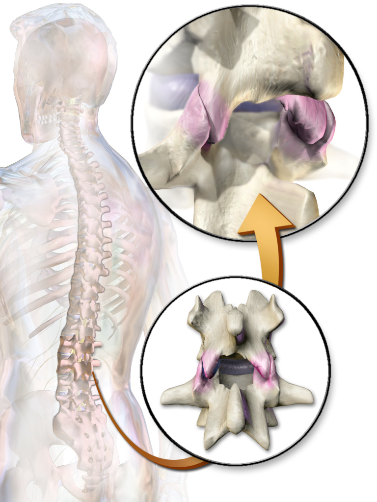
The facet joints (or zygapophysial joints, zygapophyseal, apophyseal, or Z-joints) are a set of synovial, plane joints between the articular processes of two adjacent vertebrae. There are two facet joints in each spinal motion segment and each facet joint is innervated by the recurrent meningeal nerves. Innervation Innervation to the facet joints vary between segments of the spinal, but they are generally innervated by medial branch nerves that come off the dorsal rami. It is thought that these nerves are for primary sensory input, though there is some evidence that they have some motor input local musculature. Within the cervical spine, most joints are innervated by the medial branch nerve (a branch of the dorsal rami) from the same levels. In other words, the facet joint between C4 and C5 vertebral segments is innervated by the C4 and C5 medial branch nerves. However, there are two exceptions: # The facet joint between C2 and C3 is innervated by the third occipital ner ... [...More Info...] [...Related Items...] OR: [Wikipedia] [Google] [Baidu] |
Sacroiliac Joint Dysfunction
The term sacroiliac joint dysfunction refers to abnormal motion in the sacroiliac joint, either too much motion or too little motion, that causes pain in this region. Signs and symptoms Common symptoms include lower back pain, buttocks pain, sciatic leg pain, groin pain, hip pain (for explanation of leg, groin, and hip pain, see referred pain), urinary frequency, and "transient numbness, prickling, or tingling". Pain can range from dull aching to sharp and stabbing and increases with physical activity. Symptoms also worsen with prolonged or sustained positions (i.e., sitting, standing, lying). Bending forward, stair climbing, hill climbing, and rising from a seated position can also provoke pain. Pain can increase during menstruation in women. People with severe and disabling sacroiliac joint dysfunction can develop insomnia and depression. Sacral torsion that is untreated over a long period of time can cause severe Achilles tendinosis. Causes Hypermobility Sacroiliac joint ... [...More Info...] [...Related Items...] OR: [Wikipedia] [Google] [Baidu] |
Lumbosacral Radiculopathy
Sciatica is pain going down the leg from the lower back. This pain may go down the back, outside, or front of the leg. Onset is often sudden following activities like heavy lifting, though gradual onset may also occur. The pain is often described as shooting. Typically, symptoms are only on one side of the body. Certain causes, however, may result in pain on both sides. Lower back pain is sometimes present. Weakness or numbness may occur in various parts of the affected leg and foot. About 90% of sciatica is due to a spinal disc herniation pressing on one of the lumbar or sacral nerve roots. Spondylolisthesis, spinal stenosis, piriformis syndrome, pelvic tumors, and pregnancy are other possible causes of sciatica. The straight-leg-raising test is often helpful in diagnosis. The test is positive if, when the leg is raised while a person is lying on their back, pain shoots below the knee. In most cases medical imaging is not needed. However, imaging may be obtained if bowel or blad ... [...More Info...] [...Related Items...] OR: [Wikipedia] [Google] [Baidu] |
COX-2 Inhibitors
COX-2 inhibitors are a type of nonsteroidal anti-inflammatory drug (NSAID) that directly targets cyclooxygenase-2, COX-2, an enzyme responsible for inflammation and pain. Targeting selectivity for COX-2 reduces the risk of peptic ulceration and is the main feature of celecoxib, rofecoxib, and other members of this drug class. After several COX-2-inhibiting drugs were approved for marketing, data from clinical trials revealed that COX-2 inhibitors caused a significant increase in heart attacks and strokes, with some drugs in the class having worse risks than others. Rofecoxib (sold under the brand name Vioxx) was taken off the market in 2004 because of these concerns, while celecoxib (sold under the brand name Celebrex) and traditional NSAIDs received boxed warnings on their labels. Many COX-2-specific inhibitors have been removed from the US market. As of December 2011, only Celebrex (generic name of celecoxib) is still available for purchase in the United States. In the European ... [...More Info...] [...Related Items...] OR: [Wikipedia] [Google] [Baidu] |
Spinal Nerves
A spinal nerve is a mixed nerve, which carries motor, sensory, and autonomic signals between the spinal cord and the body. In the human body there are 31 pairs of spinal nerves, one on each side of the vertebral column. These are grouped into the corresponding cervical, thoracic, lumbar, sacral and coccygeal regions of the spine. There are eight pairs of cervical nerves, twelve pairs of thoracic nerves, five pairs of lumbar nerves, five pairs of sacral nerves, and one pair of coccygeal nerves. The spinal nerves are part of the peripheral nervous system. Structure Each spinal nerve is a mixed nerve, formed from the combination of nerve fibers from its dorsal and ventral roots. The dorsal root is the afferent sensory root and carries sensory information to the brain. The ventral root is the efferent motor root and carries motor information from the brain. The spinal nerve emerges from the spinal column through an opening ( intervertebral foramen) between adjacent ve ... [...More Info...] [...Related Items...] OR: [Wikipedia] [Google] [Baidu] |


