|
Startle
In animals, including humans, the startle response is a largely unconscious defensive response to sudden or threatening stimuli, such as sudden noise or sharp movement, and is associated with negative affect.Rammirez-Moreno, David. "A computational model for the modulation of the prepulse inhibition of the acoustic startle reflex". ''Biological Cybernetics'', 2012, p. 169 Usually the onset of the startle response is a startle reflex reaction. The startle reflex is a brainstem reflectory reaction (reflex) that serves to protect vulnerable parts, such as the back of the neck (whole-body startle) and the eyes (eyeblink) and facilitates escape from sudden stimuli. It is found across many different species, throughout all stages of life. A variety of responses may occur depending on the affected individual's emotional state, body posture, preparation for execution of a motor task, or other activities. The startle response is implicated in the formation of specific phobias. Startle ... [...More Info...] [...Related Items...] OR: [Wikipedia] [Google] [Baidu] |
Moro Reflex
The Moro reflex is an infantile reflex that develops between 28 and 32 weeks of gestation and disappears at 3–6 months of age. It is a response to a sudden loss of support and involves three distinct components: # spreading out the arms (abduction) # pulling the arms in ( adduction) # crying (usually) It is distinct from the startle reflex. Unlike the startle reflex, the Moro reflex does not decrease with repeated stimulation. The primary significance of the Moro reflex is in evaluating integration of the central nervous system. Eliciting the Moro reflex Ernst Moro elicited the Moro reflex by slapping the pillow on both sides of the infant's head. Other methods have been used since then, including rapidly lowering the infant (while supported) to a sudden stop and pinching the skin of the abdomen. Today, the most common method is the head drop, where the infant is supported in both hands and tilted suddenly so the head is a few centimeters lower than the level of the body ... [...More Info...] [...Related Items...] OR: [Wikipedia] [Google] [Baidu] |
Reflex
In biology, a reflex, or reflex action, is an involuntary, unplanned sequence or action and nearly instantaneous response to a stimulus. Reflexes are found with varying levels of complexity in organisms with a nervous system. A reflex occurs via neural pathways in the nervous system called reflex arcs. A stimulus initiates a neural signal, which is carried to a synapse. The signal is then transferred across the synapse to a motor neuron which evokes a target response. These neural signals do not always travel to the brain, so many reflexes are an automatic response to a stimulus that does not receive or need conscious thought. Many reflexes are fine-tuned to increase organism survival and self-defense. This is observed in reflexes such as the startle reflex, which provides an automatic response to an unexpected stimuli, and the feline righting reflex, which reorients a cat's body when falling to ensure safe landing. The simplest type of reflex, a short-latency reflex, has ... [...More Info...] [...Related Items...] OR: [Wikipedia] [Google] [Baidu] |
Nucleus Reticularis Pontis Caudalis
The caudal pontine reticular nucleus or nucleus reticularis pontis caudalis is a portion of the reticular formation, composed of gigantocellular neurons. In rabbits and cats it is exclusively giant cells, however in humans there are normally sized cells as well. In rodents, it has been shown to play a role in the acoustic startle response. The caudal pontine reticular nucleus is rostral to the gigantocellular reticular nucleus and is located in the caudal pons. The caudal pontine reticular nucleus has been known to mediate head movement, in concert with the gigantocellular nucleus and the superior colliculus. The neurons in the dorsal half of this nucleus fire rhythmically during mastication, and in an anesthetized animal it is possible to induce mastication via electrical stimulation of the nucleus reticularis pontis caudalis or adjacent areas of the gigantocellular nucleus.Scott G, Effect of lidocaine and NMDA injections into the medial pontobulbar reticular formation on ... [...More Info...] [...Related Items...] OR: [Wikipedia] [Google] [Baidu] |
Stria Terminalis
The stria terminalis (or terminal stria) is a structure in the brain consisting of a band of fibers running along the lateral margin of the ventricular surface of the thalamus. Serving as a major output pathway of the amygdala, the stria terminalis runs from its centromedial division to the ventromedial nucleus of the hypothalamus. Anatomy The stria terminalis covers the superior thalamostriate vein, marking a line of separation between the thalamus and the caudate nucleus as seen upon gross dissection of the ventricles of the brain, viewed from the superior aspect. The stria terminalis extends from the region of the interventricular foramina to the temporal horn of the lateral ventricle, carrying fibers from the amygdala to the septal nuclei, hypothalamic, and thalamic areas of the brain. It also carries fibers projecting from these areas back to the amygdala. Bed nucleus of the stria terminalis (BNST) The activity of the bed nucleus of the stria terminalis correlate ... [...More Info...] [...Related Items...] OR: [Wikipedia] [Google] [Baidu] |
The European Journal Of Neuroscience
The ''European Journal of Neuroscience'' is a biweekly peer-reviewed scientific journal covering all aspects of neuroscience. It was established in 1989 with Ray Guillery (then at the University of Oxford) as the founding editor-in-chief. The editor-in-chief is John J. Foxe ( University of Rochester) The journal is published by Wiley-Blackwell on behalf of the Federation of European Neuroscience Societies. According to the '' Journal Citation Reports'', the journal has a 2020 impact factor of 3.386. Authors can elect to have accepted articles published as open access. EJN adopted transparent peer-review in January 2017. Sections Articles will appear within one of the five sections of the journal: * Molecular and Synaptic Mechanisms * Systems Neuroscience * Behavioural Neuroscience * Cognitive Neuroscience * Clinical and Translational Neuroscience Features EJN also publishespecialanissues related to topical issue in neuroscience. Editors-in-chief The following pers ... [...More Info...] [...Related Items...] OR: [Wikipedia] [Google] [Baidu] |
Masseter Muscle
In human anatomy, the masseter is one of the muscles of mastication. Found only in mammals, it is particularly powerful in herbivores to facilitate chewing of plant matter. The most obvious muscle of mastication is the masseter muscle, since it is the most superficial and one of the strongest. Structure The masseter is a thick, somewhat quadrilateral muscle, consisting of three heads, superficial, deep and coronoid. The fibers of superficial and deep heads are continuous at their insertion. Superficial head The superficial head, the larger, arises by a thick, tendinous aponeurosis from the temporal process of the zygomatic bone, and from the anterior two-thirds of the inferior border of the zygomatic arch. Its fibers pass inferior and posterior, to be inserted into the angle of the mandible and inferior half of the lateral surface of the ramus of the mandible. Deep head The deep head is much smaller, and more muscular in texture. It arises from the posterior third of the l ... [...More Info...] [...Related Items...] OR: [Wikipedia] [Google] [Baidu] |
Electromyography
Electromyography (EMG) is a technique for evaluating and recording the electrical activity produced by skeletal muscles. EMG is performed using an instrument called an electromyograph to produce a record called an electromyogram. An electromyograph detects the electric potential generated by muscle cells when these cells are electrically or neurologically activated. The signals can be analyzed to detect abnormalities, activation level, or recruitment order, or to analyze the biomechanics of human or animal movement. Needle EMG is an electrodiagnostic medicine technique commonly used by neurologists. Surface EMG is a non-medical procedure used to assess muscle activation by several professionals, including physiotherapists, kinesiologists and biomedical engineers. In Computer Science, EMG is also used as middleware in gesture recognition towards allowing the input of physical action to a computer as a form of human-computer interaction. Clinical uses EMG testing has a variety ... [...More Info...] [...Related Items...] OR: [Wikipedia] [Google] [Baidu] |
Orbicularis Oculi Muscle
The orbicularis oculi is a muscle in the face that closes the eyelids. It arises from the nasal part of the frontal bone, from the frontal process of the maxilla in front of the lacrimal groove, and from the anterior surface and borders of a short fibrous band, the medial palpebral ligament. From this origin, the fibers are directed laterally, forming a broad and thin layer, which occupies the eyelids or palpebræ, surrounds the circumference of the orbit, and spreads over the temple, and downward on the cheek. Structure There are at least 3 clearly defined sections of the orbicularis muscle. However, it is not clear whether the lacrimal section is a separate section, or whether it is just an extension of the preseptal and pretarsal sections. Orbital orbicularis The orbital portion is thicker and of a reddish color; its fibers form a complete ellipse without interruption at the lateral palpebral commissure; the upper fibers of this portion blend with the frontalis and corrug ... [...More Info...] [...Related Items...] OR: [Wikipedia] [Google] [Baidu] |
PET Scan
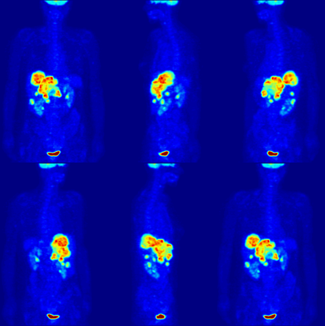
Positron emission tomography (PET) is a functional imaging technique that uses radioactive substances known as radiotracers to visualize and measure changes in metabolic processes, and in other physiological activities including blood flow, regional chemical composition, and absorption. Different tracers are used for various imaging purposes, depending on the target process within the body. For example: * Fluorodeoxyglucose ( 18F">sup>18FDG or FDG) is commonly used to detect cancer; * 18Fodium fluoride">sup>18Fodium fluoride (Na18F) is widely used for detecting bone formation; * Oxygen-15 (15O) is sometimes used to measure blood flow. PET is a common imaging technique, a medical scintillography technique used in nuclear medicine. A radiopharmaceutical – a radioisotope attached to a drug – is injected into the body as a tracer. When the radiopharmaceutical undergoes beta plus decay, a positron is emitted, and when the positron interacts with an ordinary electron, ... [...More Info...] [...Related Items...] OR: [Wikipedia] [Google] [Baidu] |
Affect (psychology)
Affect, in psychology, refers to the underlying experience of feeling, emotion or mood. History The modern conception of affect developed in the 19th century with Wilhelm Wundt. The word comes from the German ''Gefühl'', meaning "feeling." A number of experiments have been conducted in the study of social and psychological affective preferences (i.e., what people like or dislike). Specific research has been done on preferences, attitudes, impression formation, and decision-making. This research contrasts findings with recognition memory (old-new judgments), allowing researchers to demonstrate reliable distinctions between the two. Affect-based judgments and cognitive processes have been examined with noted differences indicated, and some argue affect and cognition are under the control of separate and partially independent systems that can influence each other in a variety of ways ( Zajonc, 1980). Both affect and cognition may constitute independent sources of effects ... [...More Info...] [...Related Items...] OR: [Wikipedia] [Google] [Baidu] |
Hippocampus
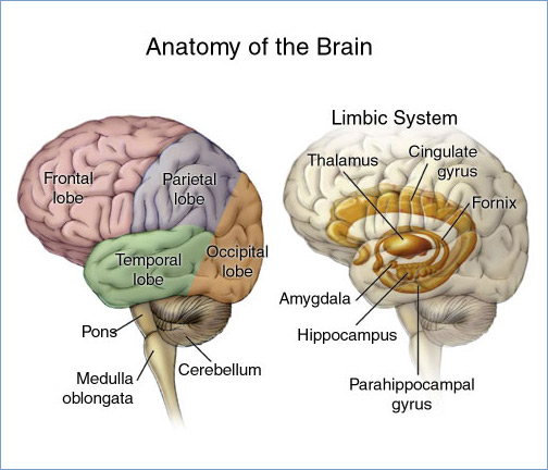
The hippocampus (via Latin from Greek , ' seahorse') is a major component of the brain of humans and other vertebrates. Humans and other mammals have two hippocampi, one in each side of the brain. The hippocampus is part of the limbic system, and plays important roles in the consolidation of information from short-term memory to long-term memory, and in spatial memory that enables navigation. The hippocampus is located in the allocortex, with neural projections into the neocortex in humans, as well as primates. The hippocampus, as the medial pallium, is a structure found in all vertebrates. In humans, it contains two main interlocking parts: the hippocampus proper (also called ''Ammon's horn''), and the dentate gyrus. In Alzheimer's disease (and other forms of dementia), the hippocampus is one of the first regions of the brain to suffer damage; short-term memory loss and disorientation are included among the early symptoms. Damage to the hippocampus can also result ... [...More Info...] [...Related Items...] OR: [Wikipedia] [Google] [Baidu] |


