|
Spi-1
Transcription factor PU.1 is a protein that in humans is encoded by the ''SPI1'' gene. Function This gene encodes an ETS-domain transcription factor that activates gene expression during myeloid and B-lymphoid cell development. The nuclear protein binds to a purine-rich sequence known as the PU-box found on enhancers of target genes, and regulates their expression in coordination with other transcription factors and cofactors. The protein can also regulate alternative splicing of target genes. Multiple transcript variants encoding different isoforms have been found for this gene. The PU.1 transcription factor is essential for hematopoiesis and cell fate decisions. PU.1 can physically interact with a variety of regulatory factors like SWI/SNF, TFIID, GATA-2, GATA-1 and c-Jun. The protein-protein interactions between these factors can regulate PU.1-dependent cell fate decisions. PU.1 can modulate the expression of 3000 genes in hematopoietic cells including cytokines. It is exp ... [...More Info...] [...Related Items...] OR: [Wikipedia] [Google] [Baidu] |
FUS (gene)
RNA-binding protein fused in sarcoma/translocated in liposarcoma (FUS/TLS), also known as heterogeneous nuclear ribonucleoprotein P2 is a protein that in humans is encoded by the ''FUS'' gene. Discovery FUS/TLS was initially identified as a fusion protein (FUS-CHOP) produced as a result of chromosomal translocations in human cancers, especially liposarcomas. In these instances, the promoter and N-terminal part of FUS/TLS is translocated to the C-terminal domain of various DNA-binding transcription factors (e.g. C/EBP homologous protein, CHOP) conferring a strong transcriptional activation domain onto the fusion proteins. FUS/TLS was independently identified as the hnRNP P2 protein, a subunit of a complex involved in the maturation of pre-mRNA. Structure FUS/TLS is a member of the FET protein family that also includes the Ewing sarcoma breakpoint region 1, EWS protein, the TATA-binding protein TBP-associated factor TAFII68/TAF15, and the Drosophila cabeza/SARF protein. FUS/T ... [...More Info...] [...Related Items...] OR: [Wikipedia] [Google] [Baidu] |
Protein
Proteins are large biomolecules and macromolecules that comprise one or more long chains of amino acid residue (biochemistry), residues. Proteins perform a vast array of functions within organisms, including Enzyme catalysis, catalysing metabolic reactions, DNA replication, Cell signaling, responding to stimuli, providing Cytoskeleton, structure to cells and Fibrous protein, organisms, and Intracellular transport, transporting molecules from one location to another. Proteins differ from one another primarily in their sequence of amino acids, which is dictated by the Nucleic acid sequence, nucleotide sequence of their genes, and which usually results in protein folding into a specific Protein structure, 3D structure that determines its activity. A linear chain of amino acid residues is called a polypeptide. A protein contains at least one long polypeptide. Short polypeptides, containing less than 20–30 residues, are rarely considered to be proteins and are commonly called pep ... [...More Info...] [...Related Items...] OR: [Wikipedia] [Google] [Baidu] |
T Cell
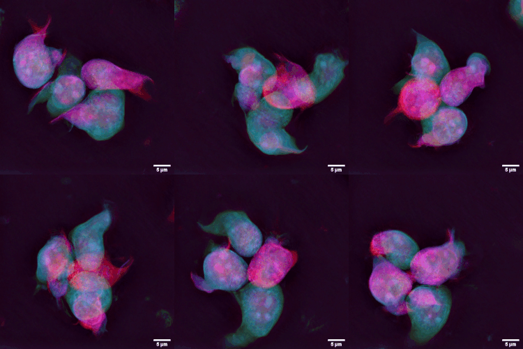
T cells (also known as T lymphocytes) are an important part of the immune system and play a central role in the adaptive immune response. T cells can be distinguished from other lymphocytes by the presence of a T-cell receptor (TCR) on their cell surface receptor, cell surface. T cells are born from hematopoietic stem cells, found in the bone marrow. Developing T cells then migrate to the thymus gland to develop (or mature). T cells derive their name from the thymus. After migration to the thymus, the precursor cells mature into several distinct types of T cells. T cell differentiation also continues after they have left the thymus. Groups of specific, differentiated T cell subtypes have a variety of important functions in controlling and shaping the immune response. One of these functions is immune-mediated cell death, and it is carried out by two major subtypes: Cytotoxic T cell, CD8+ "killer" (cytotoxic) and T helper cell, CD4+ "helper" T cells. (These are named for the presen ... [...More Info...] [...Related Items...] OR: [Wikipedia] [Google] [Baidu] |
IRF4
Interferon regulatory factor 4 (IRF4) also known as MUM1 is a protein that in humans is encoded by the ''IRF4'' gene. IRF4 functions as a key regulatory transcription factor in the development of human immune cells.Nam S, Lim J-S (2016). "Essential role of interferon regulatory factor 4 (IRF4) in immune cell development." ''Arch. Pharm. Res''. 39: 1548–1555doi:10.1007/s12272-016-0854-1Shaffer AL, Tolga Emre NC, Romesser PB, Staudt LM (2009). "IRF4: Immunity. Malignancy! Therapy?" ''Clinical Cancer Research''. 15 (9): 2954-2961doi:10.1158/1078-0432.CCR-08-1845/ref> The expression of IRF4 is essential for the differentiation of T lymphocytes and B lymphocytes as well as certain myeloid cells. Dysregulation of the ''IRF4'' gene can result in ''IRF4'' functioning either as an oncogene or a tumor-suppressor, depending on the context of the modification. The ''MUM1'' symbol is also the current HGNC official symbol for melanoma associated antigen (mutated) 1 (HGNC:29641). Immune ce ... [...More Info...] [...Related Items...] OR: [Wikipedia] [Google] [Baidu] |
Lymphocyte
A lymphocyte is a type of white blood cell (leukocyte) in the immune system of most vertebrates. Lymphocytes include T cells (for cell-mediated and cytotoxic adaptive immunity), B cells (for humoral, antibody-driven adaptive immunity), and innate lymphoid cells (ILCs; "innate T cell-like" cells involved in mucosal immunity and homeostasis), of which natural killer cells are an important subtype (which functions in cell-mediated, cytotoxic innate immunity). They are the main type of cell found in lymph, which prompted the name "lymphocyte" (with ''cyte'' meaning cell). Lymphocytes make up between 18% and 42% of circulating white blood cells. Types The three major types of lymphocyte are T cells, B cells and natural killer (NK) cells. They can also be classified as small lymphocytes and large lymphocytes based on their size and appearance. Lymphocytes can be identified by their large nucleus. T cells and B cells T cells (thymus cells) and B cells ( bone marrow- ... [...More Info...] [...Related Items...] OR: [Wikipedia] [Google] [Baidu] |
Inflammation
Inflammation (from ) is part of the biological response of body tissues to harmful stimuli, such as pathogens, damaged cells, or irritants. The five cardinal signs are heat, pain, redness, swelling, and loss of function (Latin ''calor'', ''dolor'', ''rubor'', ''tumor'', and ''functio laesa''). Inflammation is a generic response, and therefore is considered a mechanism of innate immunity, whereas adaptive immunity is specific to each pathogen. Inflammation is a protective response involving immune cells, blood vessels, and molecular mediators. The function of inflammation is to eliminate the initial cause of cell injury, clear out damaged cells and tissues, and initiate tissue repair. Too little inflammation could lead to progressive tissue destruction by the harmful stimulus (e.g. bacteria) and compromise the survival of the organism. However inflammation can also have negative effects. Too much inflammation, in the form of chronic inflammation, is associated with variou ... [...More Info...] [...Related Items...] OR: [Wikipedia] [Google] [Baidu] |
Extracellular Matrix
In biology, the extracellular matrix (ECM), also called intercellular matrix (ICM), is a network consisting of extracellular macromolecules and minerals, such as collagen, enzymes, glycoproteins and hydroxyapatite that provide structural and biochemical support to surrounding cells. Because multicellularity evolved independently in different multicellular lineages, the composition of ECM varies between multicellular structures; however, cell adhesion, cell-to-cell communication and differentiation are common functions of the ECM. The animal extracellular matrix includes the interstitial matrix and the basement membrane. Interstitial matrix is present between various animal cells (i.e., in the intercellular spaces). Gels of polysaccharides and fibrous proteins fill the interstitial space and act as a compression buffer against the stress placed on the ECM. Basement membranes are sheet-like depositions of ECM on which various epithelial cells rest. Each type of connective tissue ... [...More Info...] [...Related Items...] OR: [Wikipedia] [Google] [Baidu] |
Fibroblast
A fibroblast is a type of cell (biology), biological cell typically with a spindle shape that synthesizes the extracellular matrix and collagen, produces the structural framework (Stroma (tissue), stroma) for animal Tissue (biology), tissues, and plays a critical role in wound healing. Fibroblasts are the most common cells of connective tissue in animals. Structure Fibroblasts have a branched cytoplasm surrounding an elliptical, speckled cell nucleus, nucleus having two or more nucleoli. Active fibroblasts can be recognized by their abundant rough endoplasmic reticulum (RER). Inactive fibroblasts, called 'fibrocytes', are smaller, spindle-shaped, and have less RER. Although disjointed and scattered when covering large spaces, fibroblasts often locally align in parallel clusters when crowded together. Unlike the epithelial cells lining the body structures, fibroblasts do not form flat monolayers and are not restricted by a polarizing attachment to a basal lamina on one side, a ... [...More Info...] [...Related Items...] OR: [Wikipedia] [Google] [Baidu] |
Fibrosis
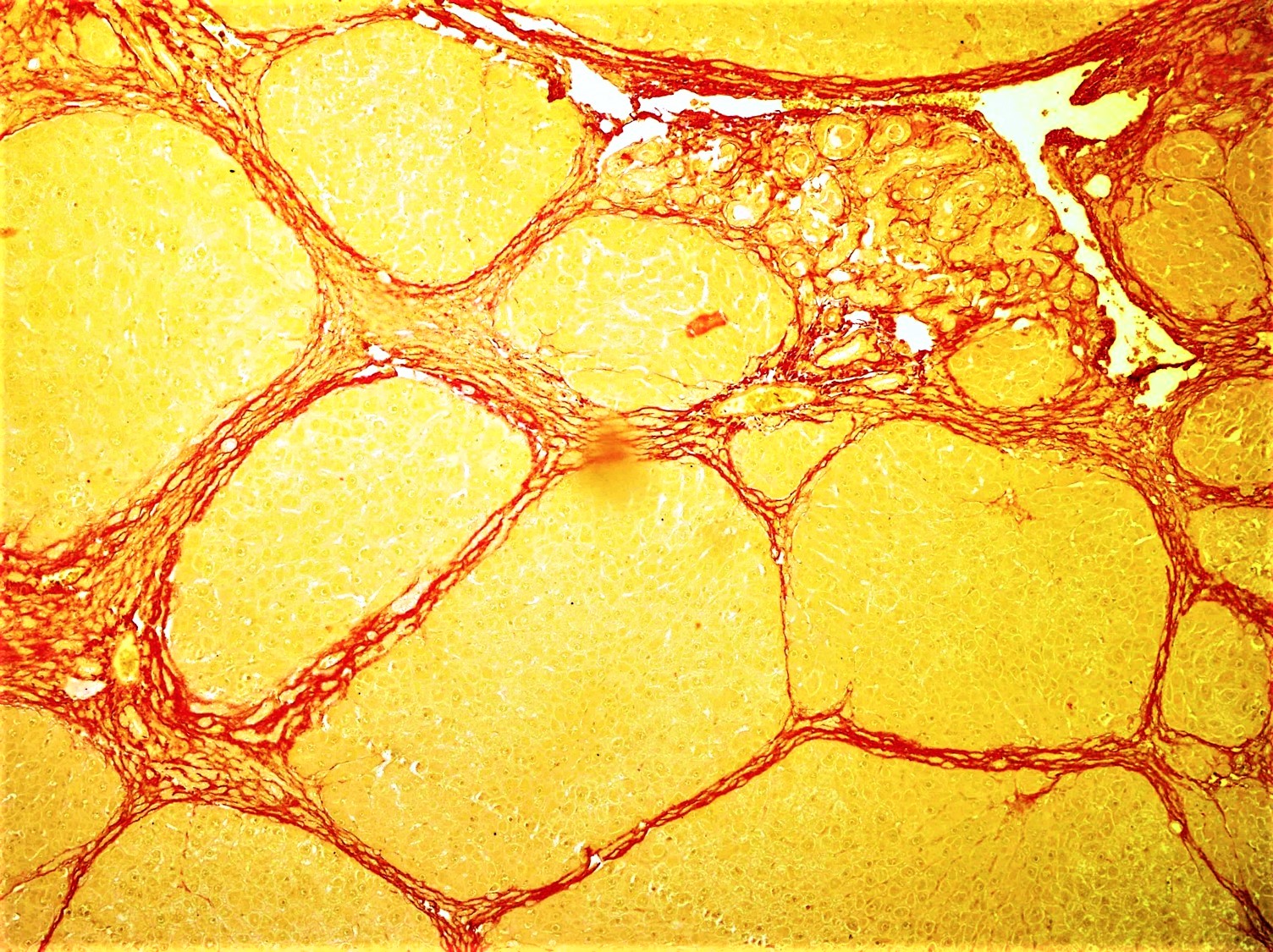
Fibrosis, also known as fibrotic scarring, is the development of fibrous connective tissue in response to an injury. Fibrosis can be a normal connective tissue deposition or excessive tissue deposition caused by a disease. Repeated injuries, chronic inflammation and repair are susceptible to fibrosis, where an accidental excessive accumulation of extracellular matrix components, such as the collagen, is produced by fibroblasts, leading to the formation of a permanent fibrotic scar. In response to injury, this is called scarring, and if fibrosis arises from a single cell line, this is called a fibroma. Physiologically, fibrosis acts to deposit connective tissue, which can interfere with or totally inhibit the normal architecture and function of the underlying organ or tissue. Fibrosis can be used to describe the pathological state of excess deposition of fibrous tissue, as well as the process of connective tissue deposition in healing. Defined by the pathological accumulation of ... [...More Info...] [...Related Items...] OR: [Wikipedia] [Google] [Baidu] |
Megakaryocyte
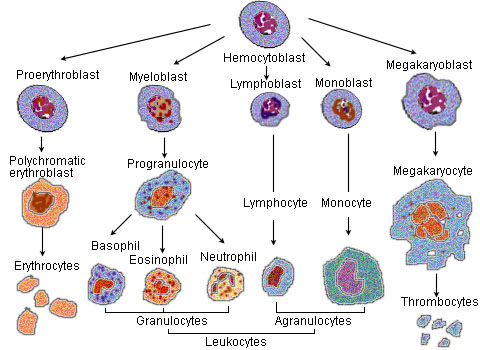
A megakaryocyte () is a large bone marrow cell with a lobation, lobated nucleus that produces blood platelets (thrombocytes), which are necessary for normal blood coagulation, clotting. In humans, megakaryocytes usually account for 1 out of 10,000 bone marrow cells, but can increase in number nearly 10-fold during the course of certain diseases. Owing to variations in neoclassical compound, combining forms and spelling, synonyms include megalokaryocyte and megacaryocyte. Structure In general, megakaryocytes are 10 to 15 times larger than a typical red blood cell, averaging 50–100 μm in diameter. During its maturation, the megakaryocyte grows in size and replicates its DNA without cytokinesis in a process called mitosis#Errors and other variations, endomitosis. As a result, the nucleus of the megakaryocyte can become very large and lobulated, which, under a light microscope, can give the false impression that there are several nuclei. In some cases, the nucleus may contain up to ... [...More Info...] [...Related Items...] OR: [Wikipedia] [Google] [Baidu] |
Reticulocyte
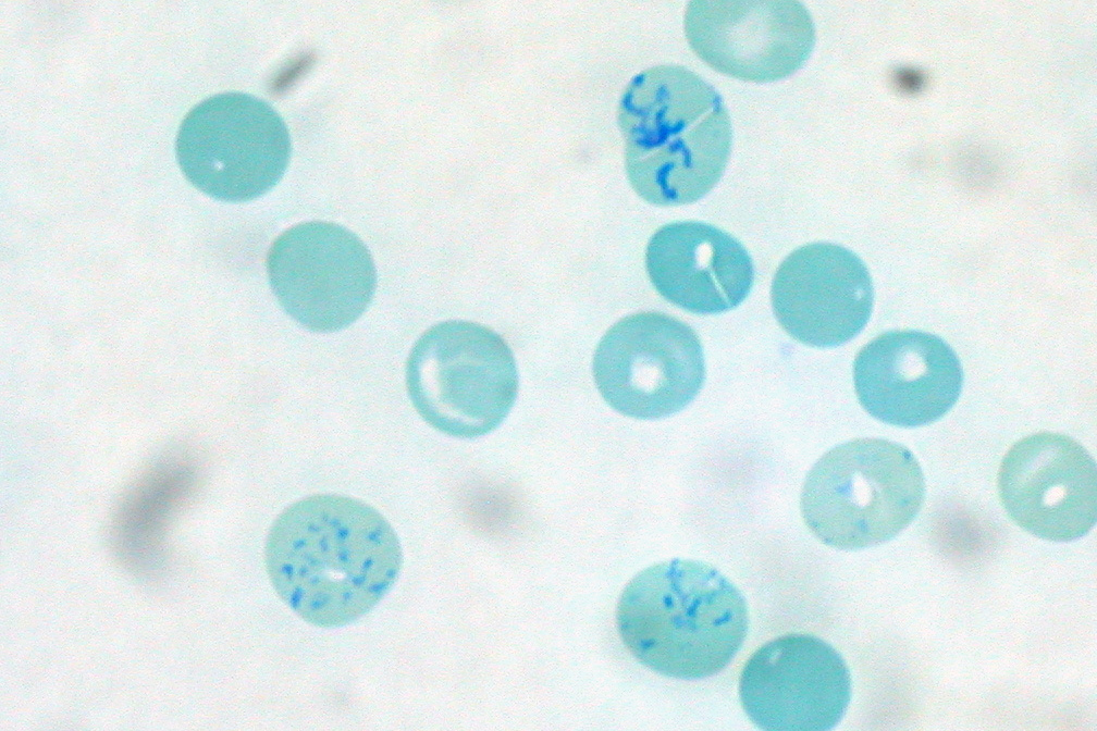
In hematology, reticulocytes are immature red blood cells (RBCs). In the process of erythropoiesis (red blood cell formation), reticulocytes develop and mature in the bone marrow and then circulate for about a day in the blood stream before developing into mature red blood cells. Like mature red blood cells, in mammals, reticulocytes do not have a cell nucleus. They are called reticulocytes because of a reticular (mesh-like) network of ribosomal RNA that becomes visible under a microscope with certain stains such as new methylene blue and Romanowsky stain. Clinical significance To accurately measure reticulocyte counts, automated counters use a combination of laser excitation, detectors and a fluorescent dye that marks RNA and DNA (such as titan yellow or polymethine). Reticulocytes appear slightly bluer than other red cells when looked at with the normal Romanowsky stain. Reticulocytes are also relatively large, a characteristic that is described by the mean corpuscular ... [...More Info...] [...Related Items...] OR: [Wikipedia] [Google] [Baidu] |


