|
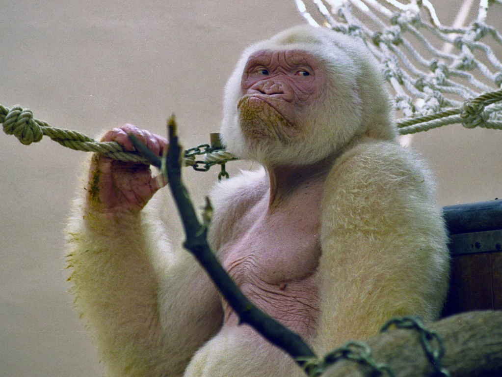
Snowflake (albino Gorilla)
Snowflake (, , ; – 24 November 2003) was a western lowland gorilla who is the world's only known albino gorilla to date. He was kept at Barcelona Zoo in Barcelona, Catalonia, Spain, from 1966 until his death in 2003. History Snowflake was captured in the Río Muni region in Spanish Guinea on 1 October 1966 by Benito Mañé, a farmer of the Fang people. Mañé had killed the rest of Snowflake's gorilla group, who were all typically colored gorillas. Mañé then kept Snowflake at his home for four days before transporting him to Bata, where he was purchased by primatologist Jordi Sabater Pi. Originally named ''Nfumu Ngui'' in Fang language ("white gorilla") by his captor, he was then nicknamed ''Floquet de Neu'' ( Catalan for "little snowflake") by his keeper, Jordi Sabater Pi. Characteristics Snowflake was a western lowland gorilla with non-syndromic oculocutaneous albinism. He had poor vision, though tests to determine whether he had a central blind spot did not find one. ... [...More Info...] [...Related Items...] OR: [Wikipedia] [Google] [Baidu] |
Western Lowland Gorilla
The western lowland gorilla (''Gorilla gorilla gorilla'') is one of two Critically Endangered subspecies of the western gorilla (''Gorilla gorilla'') that lives in Montane ecosystems#Montane forests, montane, Old-growth forest, primary and secondary forest, secondary forest and lowland swampland in central Africa in Angola (Cabinda Province), Cameroon, Central African Republic, Republic of the Congo, Democratic Republic of the Congo, Equatorial Guinea and Gabon. It is the nominate subspecies of the western gorilla, and the smallest of the four gorilla subspecies. The western lowland gorilla is the only subspecies kept in zoos with the exception of Amahoro (gorilla), Amahoro, a female eastern lowland gorilla at Antwerp Zoo, and a few mountain gorillas kept captive in the Democratic Republic of the Congo. Description The western lowland gorilla is the smallest subspecies of gorilla but still has exceptional size and strength. This species of gorillas exhibits pronounced sexual ... [...More Info...] [...Related Items...] OR: [Wikipedia] [Google] [Baidu] |
Sclera
The sclera, also known as the white of the eye or, in older literature, as the tunica albuginea oculi, is the opaque, fibrous, protective outer layer of the eye containing mainly collagen and some crucial elastic fiber. In the development of the embryo, the sclera is derived from the neural crest. In children, it is thinner and shows some of the underlying pigment, appearing slightly blue. In the elderly, fatty deposits on the sclera can make it appear slightly yellow. People with dark skin can have naturally darkened sclerae, the result of melanin pigmentation. In humans, and some other vertebrates, the whole sclera is white or pale, contrasting with the coloured iris (anatomy), iris. The cooperative eye hypothesis suggests that the pale sclera evolved as a method of nonverbal communication that makes it easier for one individual to identify where another individual is looking. Other mammals with white or pale sclera include chimpanzees, many orangutans, some gorillas, and bon ... [...More Info...] [...Related Items...] OR: [Wikipedia] [Google] [Baidu] |
Recessive Gene
In genetics, dominance is the phenomenon of one variant (allele) of a gene on a chromosome masking or overriding the effect of a different variant of the same gene on the other copy of the chromosome. The first variant is termed dominant and the second is called recessive. This state of having two different variants of the same gene on each chromosome is originally caused by a mutation in one of the genes, either new (''de novo'') or inherited. The terms autosomal dominant or autosomal recessive are used to describe gene variants on non-sex chromosomes ( autosomes) and their associated traits, while those on sex chromosomes (allosomes) are termed X-linked dominant, X-linked recessive or Y-linked; these have an inheritance and presentation pattern that depends on the sex of both the parent and the child (see Sex linkage). Since there is only one Y chromosome, Y-linked traits cannot be dominant or recessive. Additionally, there are other forms of dominance, such as incomplete ... [...More Info...] [...Related Items...] OR: [Wikipedia] [Google] [Baidu] |
Biotope
A biotope is an area of uniform environmental conditions providing a living place for a specific assemblage of flora (plants), plants and fauna (animals), animals. ''Biotope'' is almost synonymous with the term habitat (ecology), "habitat", which is more commonly used in English-speaking countries. However, in some countries these two terms are distinguished: the subject of a habitat is a population, the subject of a biotope is a ''biocoenosis'' or "biological community". It is an English loanword derived from the German '':de:Biotop, Biotop'', which in turn came from the Greek ''bios'' (meaning 'life') and ''topos'' ('place'). (The related word ''geotope'' has made its way into the English language by the same route, from the German '':de:Geotop, Geotop''.) Ecology The concept of a biotope was first advocated by Ernst Haeckel (1834–1919), a German zoologist famous for the recapitulation theory. In his book ''General Morphology'' (1866), which defines the term "ecology", he st ... [...More Info...] [...Related Items...] OR: [Wikipedia] [Google] [Baidu] |
Photophobia
Photophobia is a medical symptom of abnormal intolerance to visual perception of light. As a medical symptom, photophobia is not a morbid fear or phobia, but an experience of discomfort or pain to the eyes due to light exposure or by presence of actual physical sensitivity of the eyes, though the term is sometimes additionally applied to abnormal or irrational fear of light, such as heliophobia. The term ''photophobia'' comes . Causes Patients may develop photophobia as a result of several different medical conditions, related to the human eye, eye, the nervous system, genetic, or other causes. Photophobia may manifest itself in an increased response to light starting at any step in the visual system, such as: * Too much light entering the eye. Too much light can enter the eye if it is damaged, such as with corneal abrasion and retinal damage, or if its pupil is unable to normally constrict (seen with damage to the oculomotor nerve). * Due to albinism, the lack of pigment in th ... [...More Info...] [...Related Items...] OR: [Wikipedia] [Google] [Baidu] |
Pupil
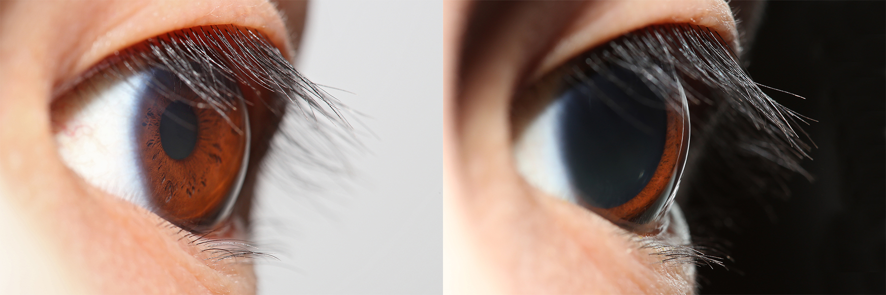
The pupil is a hole located in the center of the iris of the eye that allows light to strike the retina.Cassin, B. and Solomon, S. (1990) ''Dictionary of Eye Terminology''. Gainesville, Florida: Triad Publishing Company. It appears black because light rays entering the pupil are either absorbed by the tissues inside the eye directly, or absorbed after diffuse reflections within the eye that mostly miss exiting the narrow pupil. The size of the pupil is controlled by the iris, and varies depending on many factors, the most significant being the amount of light in the environment. The term "pupil" was coined by Gerard of Cremona. In humans, the pupil is circular, but its shape varies between species; some cats, reptiles, and foxes have vertical slit pupils, goats and sheep have horizontally oriented pupils, and some catfish have annular types. In optical terms, the anatomical pupil is the eye's aperture and the iris is the aperture stop. The image of the pupil as seen from o ... [...More Info...] [...Related Items...] OR: [Wikipedia] [Google] [Baidu] |
Blood Vessel
Blood vessels are the tubular structures of a circulatory system that transport blood throughout many Animal, animals’ bodies. Blood vessels transport blood cells, nutrients, and oxygen to most of the Tissue (biology), tissues of a Body (biology), body. They also take waste and carbon dioxide away from the tissues. Some tissues such as cartilage, epithelium, and the lens (anatomy), lens and cornea of the eye are not supplied with blood vessels and are termed ''avascular''. There are five types of blood vessels: the arteries, which carry the blood away from the heart; the arterioles; the capillaries, where the exchange of water and chemicals between the blood and the tissues occurs; the venules; and the veins, which carry blood from the capillaries back towards the heart. The word ''vascular'', is derived from the Latin ''vas'', meaning ''vessel'', and is mostly used in relation to blood vessels. Etymology * artery – late Middle English; from Latin ''arteria'', from Gree ... [...More Info...] [...Related Items...] OR: [Wikipedia] [Google] [Baidu] |
Choroid
The choroid, also known as the choroidea or choroid coat, is a part of the uvea, the vascular layer of the eye. It contains connective tissues, and lies between the retina and the sclera. The human choroid is thickest at the far extreme rear of the eye (at 0.2 mm), while in the outlying areas it narrows to 0.1 mm. The choroid provides oxygen and nourishment to the outer layers of the retina. Along with the ciliary body and iris, the choroid forms the uveal tract. The structure of the choroid is generally divided into four layers (classified in order of furthest away from the retina to closest): *Haller's layer – outermost layer of the choroid consisting of larger diameter blood vessels; * Sattler's layer – layer of medium diameter blood vessels; * Choriocapillaris – layer of capillaries; and * Bruch's membrane (synonyms: Lamina basalis, Complexus basalis, Lamina vitra) – innermost layer of the choroid. Blood supply There are two circulations of the eye: ... [...More Info...] [...Related Items...] OR: [Wikipedia] [Google] [Baidu] |
Fundus (eye)
The fundus of the eye is the interior surface of the eye opposite the lens and includes the retina, optic disc, macula, fovea, and posterior pole.Cassin, B. and Solomon, S. ''Dictionary of Eye Terminology''. Gainesville, Florida: Triad Publishing Company, 1990. The fundus can be examined by ophthalmoscopy and/or fundus photography. Variation The color of the fundus varies both between and within species. In one study of primates the retina is blue, green, yellow, orange, and red; only the human fundus (from a lightly pigmented blond person) is red. The major differences noted among the "higher" primate species were size and regularity of the border of macular area, size and shape of the optic disc, apparent 'texturing' of retina, and pigmentation of retina. Clinical significance Medical signs that can be detected from observation of eye fundus (generally by funduscopy) include hemorrhages, exudates, cotton wool spots, blood vessel abnormalities ( tortuosity, puls ... [...More Info...] [...Related Items...] OR: [Wikipedia] [Google] [Baidu] |


