|
Skull
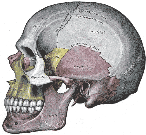
The skull, or cranium, is typically a bony enclosure around the brain of a vertebrate. In some fish, and amphibians, the skull is of cartilage. The skull is at the head end of the vertebrate. In the human, the skull comprises two prominent parts: the neurocranium and the facial skeleton, which evolved from the first pharyngeal arch. The skull forms the frontmost portion of the axial skeleton and is a product of cephalization and vesicular enlargement of the brain, with several special senses structures such as the eyes, ears, nose, tongue and, in fish, specialized tactile organs such as barbels near the mouth. The skull is composed of three types of bone: cranial bones, facial bones and ossicles, which is made up of a number of fused flat and irregular bones. The cranial bones are joined at firm fibrous junctions called sutures and contains many foramina, fossae, processes, and sinuses. In zoology, the openings in the skull are called fenestrae, the most ... [...More Info...] [...Related Items...] OR: [Wikipedia] [Google] [Baidu] |
Axial Skeleton
The axial skeleton is the core part of the endoskeleton made of the bones of the head and trunk of vertebrates. In the human skeleton, it consists of 80 bones and is composed of the skull (28 bones, including the cranium, mandible and the middle ear ossicles), the vertebral column (26 bones, including vertebrae, sacrum and coccyx), the rib cage (25 bones, including ribs and sternum), and the hyoid bone. The axial skeleton is joined to the appendicular skeleton (which support the limbs) via the shoulder girdles and the pelvis. Structure Flat bones house the brain and other vital organs. This article mainly deals with the axial skeletons of humans; however, it is important to understand its evolutionary lineage. The human axial skeleton consists of 81 different bones forming the medial core of the body and connects the pelvis to the body, where the appendicular skeleton attaches. As the skeleton grows older the bones get weaker with the exception of the skull. The skull ... [...More Info...] [...Related Items...] OR: [Wikipedia] [Google] [Baidu] |
Neurocranium
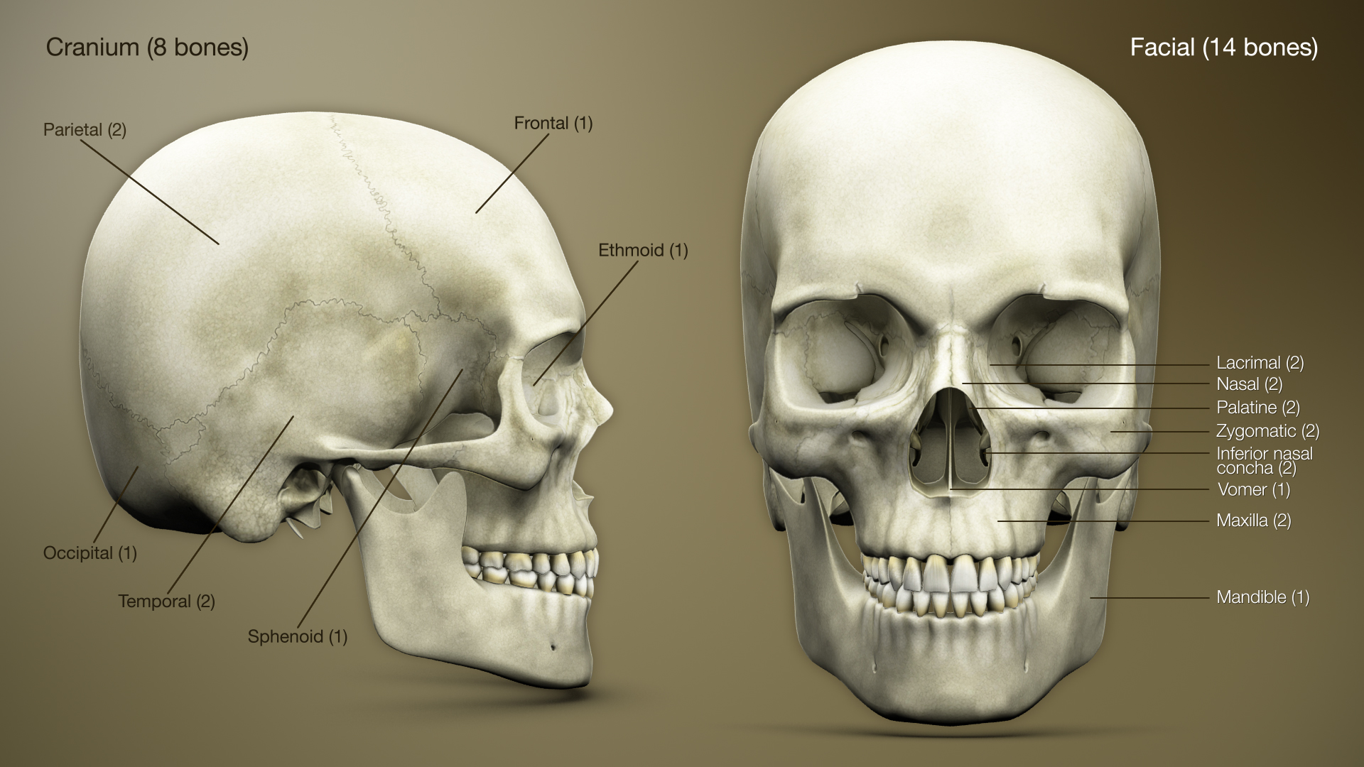
In human anatomy, the neurocranium, also known as the braincase, brainpan, brain-pan, or brainbox, is the upper and back part of the skull, which forms a protective case around the brain. In the human skull, the neurocranium includes the calvaria or skullcap. The remainder of the skull is the facial skeleton. In comparative anatomy, neurocranium is sometimes used synonymously with endocranium or chondrocranium. Structure The neurocranium is divided into two portions: * the membranous part, consisting of flat bones, which surround the brain; and * the cartilaginous part, or chondrocranium, which forms bones of the base of the skull. In humans, the neurocranium is usually considered to include the following eight bones: * 1 ethmoid bone * 1 frontal bone * 1 occipital bone * 2 parietal bones * 1 sphenoid bone * 2 temporal bones The ossicles (three on each side) are usually not included as bones of the neurocranium. There may variably also be extra sutural bones present. ... [...More Info...] [...Related Items...] OR: [Wikipedia] [Google] [Baidu] |
Nose
A nose is a sensory organ and respiratory structure in vertebrates. It consists of a nasal cavity inside the head, and an external nose on the face. The external nose houses the nostrils, or nares, a pair of tubes providing airflow through the nose for Respiration (physiology), respiration. Where the nostrils pass through the nasal cavity they widen, are known as nasal fossae, and contain nasal concha, turbinates and olfactory mucosa. The nasal cavity also connects to the paranasal sinuses (dead-end air cavities for pressure buffering and humidification). From the nasal cavity, the nostrils continue into the pharynx, a switch track valve connecting the respiratory system, respiratory and digestive systems. In humans, the nose is located centrally on the face and serves as an alternative respiratory passage especially during suckling for infants. The protruding nose that is completely separate from the mouth part is a characteristic found only in theria, therian mammals. It has b ... [...More Info...] [...Related Items...] OR: [Wikipedia] [Google] [Baidu] |
Foramina
In anatomy and osteology, a foramen (; : foramina, or foramens ; ) is an opening or enclosed gap within the dense connective tissue (bones and deep fasciae) of extant and extinct amniote animals, typically to allow passage of nerves, arteries, veins or other soft tissue structures (e.g. muscle tendon) from one body compartment to another. Skull The skulls of vertebrates have foramina through which nerves, arteries, veins, and other structures pass. The human skull has many foramina, collectively referred to as the cranial foramina. Spine Within the vertebral column (spine) of vertebrates, including the human spine, each bone has an opening at both its top and bottom to allow nerves, arteries, veins, etc. to pass through. Other * Apical foramen, the hole at the tip of the root of a tooth * Foramen ovale (heart), a hole between the venous and arterial sides of the fetal heart * Transverse foramen, one of a pair of openings in each cervical vertebra, in which ... [...More Info...] [...Related Items...] OR: [Wikipedia] [Google] [Baidu] |
Facial Bone
The facial skeleton comprises the ''facial bones'' that may attach to build a portion of the skull. The remainder of the skull is the neurocranium. In human anatomy and development, the facial skeleton is sometimes called the ''membranous viscerocranium'', which comprises the mandible and dermatocranial elements that are not part of the braincase. Structure In the human skull, the facial skeleton consists of fourteen bones in the face: * Inferior turbinal (2) * Lacrimal bones (2) * Mandible * Maxilla (2) * Nasal bones (2) * Palatine bones (2) * Vomer * Zygomatic bones (2) Variations Elements of the ''cartilaginous viscerocranium'' (i.e., splanchnocranial elements), such as the hyoid bone, are sometimes considered part of the facial skeleton. The ethmoid bone (or a part of it) and also the sphenoid bone are sometimes included, but otherwise considered part of the neurocranium. Because the maxillary bones are fused, they are often collectively listed as only one bone. The mandi ... [...More Info...] [...Related Items...] OR: [Wikipedia] [Google] [Baidu] |
Facial Skeleton
The facial skeleton comprises the ''facial bones'' that may attach to build a portion of the skull. The remainder of the skull is the neurocranium. In human anatomy and development, the facial skeleton is sometimes called the ''membranous viscerocranium'', which comprises the mandible and dermatocranial elements that are not part of the braincase. Structure In the human skull, the facial skeleton consists of fourteen bones in the face: * Inferior turbinal (2) * Lacrimal bones (2) * Mandible * Maxilla (2) * Nasal bones (2) * Palatine bones (2) * Vomer * Zygomatic bones (2) Variations Elements of the ''cartilaginous viscerocranium'' (i.e., splanchnocranial elements), such as the hyoid bone, are sometimes considered part of the facial skeleton. The ethmoid bone (or a part of it) and also the sphenoid bone are sometimes included, but otherwise considered part of the neurocranium. Because the maxillary bones are fused, they are often collectively listed as only one bone. ... [...More Info...] [...Related Items...] OR: [Wikipedia] [Google] [Baidu] |
Volume Rendering
In scientific visualization and computer graphics, volume rendering is a set of techniques used to display a 2D projection of a 3D discretely sampled data set, typically a 3D scalar field. A typical 3D data set is a group of 2D slice images acquired by a CT, MRI, or MicroCT scanner. Usually these are acquired in a regular pattern (e.g., one slice for each millimeter of depth) and usually have a regular number of image pixels in a regular pattern. This is an example of a regular volumetric grid, with each volume element, or voxel represented by a single value that is obtained by sampling the immediate area surrounding the voxel. To render a 2D projection of the 3D data set, one first needs to define a camera in space relative to the volume. Also, one needs to define the opacity and color of every voxel. This is usually defined using an RGBA (for red, green, blue, alpha) transfer function that defines the RGBA value for every possible voxel value. For example, a volu ... [...More Info...] [...Related Items...] OR: [Wikipedia] [Google] [Baidu] |
Head
A head is the part of an organism which usually includes the ears, brain, forehead, cheeks, chin, eyes, nose, and mouth, each of which aid in various sensory functions such as sight, hearing, smell, and taste. Some very simple animals may not have a head, but many bilaterally symmetric forms do, regardless of size. Heads develop in animals by an evolutionary trend known as cephalization. In bilaterally symmetrical animals, nervous tissue concentrate at the anterior region, forming structures responsible for information processing. Through biological evolution, sense organs and feeding structures also concentrate into the anterior region; these collectively form the head. Human head The human head is an anatomical unit that consists of the skull, hyoid bone and cervical vertebrae. The skull consists of the brain case which encloses the cranial cavity, and the facial skeleton, which includes the mandible. There are eight bones in the brain case ... [...More Info...] [...Related Items...] OR: [Wikipedia] [Google] [Baidu] |
Suture (joint)
In anatomy, fibrous joints are joints connected by fibrous tissue, consisting mainly of collagen. These are fixed joints where bones are united by a layer of white fibrous tissue of varying thickness. In the skull, the joints between the bones are called sutures. Such immovable joints are also referred to as synarthroses. Types Most fibrous joints are also called "fixed" or "immovable". These joints have no joint cavity and are connected via fibrous connective tissue. * Sutures: The skull bones are connected by fibrous joints called '' sutures''. In fetal skulls, the sutures are wide to allow slight movement during birth. They later become rigid ( synarthrodial). * Syndesmosis: Some of the long bones in the body such as the radius and ulna in the forearm are joined by a '' syndesmosis'' (along the interosseous membrane). Syndemoses are slightly moveable ( amphiarthrodial). The distal tibiofibular joint is another example. * A '' gomphosis'' is a joint between the roo ... [...More Info...] [...Related Items...] OR: [Wikipedia] [Google] [Baidu] |
Amphibian
Amphibians are ectothermic, anamniote, anamniotic, tetrapod, four-limbed vertebrate animals that constitute the class (biology), class Amphibia. In its broadest sense, it is a paraphyletic group encompassing all Tetrapod, tetrapods, but excluding the amniotes (tetrapods with an amniotic membrane, such as modern reptiles, birds and mammals). All extant taxon, extant (living) amphibians belong to the monophyletic subclass (biology), subclass Lissamphibia, with three living order (biology), orders: Anura (frogs and toads), Urodela (salamanders), and Gymnophiona (caecilians). Evolved to be mostly semiaquatic, amphibians have adapted to inhabit a wide variety of habitats, with most species living in freshwater ecosystem, freshwater, wetland or terrestrial ecosystems (such as riparian woodland, fossorial and even arboreal habitats). Their biological life cycle, life cycle typically starts out as aquatic animal, aquatic larvae with gills known as tadpoles, but some species have devel ... [...More Info...] [...Related Items...] OR: [Wikipedia] [Google] [Baidu] |
Ossicle
The ossicles (also called auditory ossicles) are three irregular bones in the middle ear of humans and other mammals, and are among the smallest bones in the human body. Although the term "ossicle" literally means "tiny bone" (from Latin ''ossiculum'') and may refer to any small bone throughout the body, it typically refers specifically to the malleus, incus and stapes ("hammer, anvil, and stirrup") of the middle ear. The auditory ossicles serve as a kinematic chain to transmit and amplify (intensity (physics), intensify) sound vibrations collected from the air by the ear drum to the fluid-filled labyrinth (inner ear), labyrinth (cochlea). The absence or pathology of the auditory ossicles would constitute a moderate-to-severe conductive hearing loss. Structure The ossicles are, in order from the eardrum to the inner ear (from superficial to deep): the malleus, incus, and stapes, terms that in Latin are translated as "the hammer, anvil, and stirrup". * The malleus () articulat ... [...More Info...] [...Related Items...] OR: [Wikipedia] [Google] [Baidu] |
Touch
The somatosensory system, or somatic sensory system is a subset of the sensory nervous system. The main functions of the somatosensory system are the perception of external stimuli, the perception of internal stimuli, and the regulation of body position and balance ( proprioception). It is believed to act as a pathway between the different sensory modalities within the body. As of 2024 debate continued on the underlying mechanisms, correctness and validity of the somatosensory system model, and whether it impacts emotions in the body. The somatosensory system has been thought of as having two subdivisions; *one for the detection of mechanosensory information related to touch. Mechanosensory information includes that of light touch, vibration, pressure and tension in the skin. Much of this information belongs to the sense of touch which is a general somatic sense in contrast to the special senses of sight, smell, taste, hearing, and balance. * one for the nociception ... [...More Info...] [...Related Items...] OR: [Wikipedia] [Google] [Baidu] |


