|
Serratus Posterior Inferior Muscle
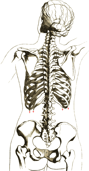
The serratus posterior inferior muscle, also known as the posterior serratus muscle, is a muscle of the human body. Structure The muscle is situated at the junction of the thoracic and lumbar regions. It has an irregularly quadrilateral form, broader than the serratus posterior superior muscle, and separated from it by a wide interval. It arises by a thin aponeurosis from the spinous processes of the lower two thoracic and upper two or three lumbar vertebrae. Passing obliquely upward and lateralward, it becomes fleshy, and divides into four flat digitations. These are inserted into the inferior borders of the lower four ribs, a little beyond their angles. The thin aponeurosis of origin is intimately blended with the thoracolumbar fascia, and aponeurosis of the latissimus dorsi muscle. Function The serratus posterior inferior draws the lower ribs backward and downward to assist in rotation and extension of the trunk. This movement of the ribs may also contribute to i ... [...More Info...] [...Related Items...] OR: [Wikipedia] [Google] [Baidu] |
Vertebrae
Each vertebra (: vertebrae) is an irregular bone with a complex structure composed of bone and some hyaline cartilage, that make up the vertebral column or spine, of vertebrates. The proportions of the vertebrae differ according to their spinal segment and the particular species. The basic configuration of a vertebra varies; the vertebral body (also ''centrum'') is of bone and bears the load of the vertebral column. The upper and lower surfaces of the vertebra body give attachment to the intervertebral discs. The posterior part of a vertebra forms a vertebral arch, in eleven parts, consisting of two pedicles (pedicle of vertebral arch), two laminae, and seven process (anatomy), processes. The laminae give attachment to the ligamenta flava (ligaments of the spine). There are vertebral notches formed from the shape of the pedicles, which form the intervertebral foramina when the vertebrae articulation (anatomy), articulate. These foramina are the entry and exit conduits for the spi ... [...More Info...] [...Related Items...] OR: [Wikipedia] [Google] [Baidu] |
Aponeurosis
An aponeurosis (; : aponeuroses) is a flattened tendon by which muscle attaches to bone or fascia. Aponeuroses exhibit an ordered arrangement of collagen fibres, thus attaining high tensile strength in a particular direction while being vulnerable to tensional or shear forces in other directions. They have a shiny, whitish-silvery color, are histologically similar to tendons, and are very sparingly supplied with blood vessels and nerves. When dissected, aponeuroses are papery and peel off by sections. The primary regions with thick aponeuroses are in the ventral abdominal region, the dorsal lumbar region, the ventriculus in birds, and the palmar (palms) and plantar (soles) regions. Anatomy Anterior abdominal aponeuroses The anterior abdominal aponeuroses are located just superficial to the rectus abdominis muscle. It has for its borders the external oblique, pectoralis muscles, and the latissimus dorsi. Posterior lumbar aponeuroses The posterior lumbar aponeuroses are sit ... [...More Info...] [...Related Items...] OR: [Wikipedia] [Google] [Baidu] |
Serratus Anterior Muscle
The serratus anterior is a muscle of the chest. It originates at the side of the chest from the upper 8 or 9 ribs; it inserts along the entire length of the anterior aspect of the medial border of the scapula. It is innervated by the long thoracic nerve from the brachial plexus. The serratus anterior acts to pull the scapula forward around the thorax. The muscle is named from Latin: ''serrare'' = to saw (referring to the shape); and ''anterior'' = on the front side of the body. Structure Origin Serratus anterior normally originates by nine or ten muscle slips – arising from either the 1st to 8th ribs, or the 1st to 9th ribs; because two slips usually arise from the 2nd rib, the number of slips is greater than the number of ribs from which they originate. Insertion The muscle is inserted along the medial border of the scapula between the superior and inferior angle of the scapula. The muscle is divided into three parts according to the points of insertion: * the s ... [...More Info...] [...Related Items...] OR: [Wikipedia] [Google] [Baidu] |
Twelfth Rib
The rib cage or thoracic cage is an endoskeletal enclosure in the thorax of most vertebrates that comprises the ribs, vertebral column and sternum, which protect the vital organs of the thoracic cavity, such as the heart, lungs and great vessels and support the shoulder girdle to form the core part of the axial skeleton. A typical human thoracic cage consists of 12 pairs of ribs and the adjoining costal cartilages, the sternum (along with the manubrium and xiphoid process), and the 12 thoracic vertebrae articulating with the ribs. The thoracic cage also provides attachments for extrinsic skeletal muscles of the neck, upper limbs, upper abdomen and back, and together with the overlying skin and associated fascia and muscles, makes up the thoracic wall. In tetrapods, the rib cage intrinsically holds the muscles of respiration ( diaphragm, intercostal muscles, etc.) that are crucial for active inhalation and forced exhalation, and therefore has a major ventilatory function in th ... [...More Info...] [...Related Items...] OR: [Wikipedia] [Google] [Baidu] |
Lung
The lungs are the primary Organ (biology), organs of the respiratory system in many animals, including humans. In mammals and most other tetrapods, two lungs are located near the Vertebral column, backbone on either side of the heart. Their function in the respiratory system is to extract oxygen from the atmosphere and transfer it into the bloodstream, and to release carbon dioxide from the bloodstream into the atmosphere, in a process of gas exchange. Respiration is driven by different muscular systems in different species. Mammals, reptiles and birds use their musculoskeletal systems to support and foster breathing. In early tetrapods, air was driven into the lungs by the pharyngeal muscles via buccal pumping, a mechanism still seen in amphibians. In humans, the primary muscle that drives breathing is the Thoracic diaphragm, diaphragm. The lungs also provide airflow that makes Animal communication#Auditory, vocalisation including speech possible. Humans have two lungs, a ri ... [...More Info...] [...Related Items...] OR: [Wikipedia] [Google] [Baidu] |
Exhalation
Exhalation (or expiration) is the flow of the breathing, breath out of an organism. In animals, it is the movement of air from the lungs out of the airways, to the external environment during breathing. This happens due to elastic properties of the lungs, as well as the internal intercostal muscles which lower the rib cage and decrease thoracic volume. As the thoracic diaphragm relaxes during exhalation it causes the tissue it has depressed to rise superiorly and put pressure on the lungs to expel the air. During Hyperpnea, forced exhalation, as when blowing out a candle, expiratory muscles including the abdominal muscles and internal intercostal muscles generate abdominal and thoracic pressure, which forces air out of the lungs. Exhaled air is 4% carbon dioxide, a waste product of cellular respiration during the production of energy, which is stored as Adenosine triphosphate, ATP. Exhalation has a complementary relationship to inhalation which together make up the respirator ... [...More Info...] [...Related Items...] OR: [Wikipedia] [Google] [Baidu] |
Inhalation
Inhalation (or inspiration) happens when air or other gases enter the lungs. Inhalation of air Inhalation of air, as part of the cycle of breathing, is a vital process for all human life. The process is autonomic (though there are exceptions in some disease states) and does not need conscious control or effort. However, breathing can be consciously controlled or interrupted (within limits). Breathing allows oxygen (which humans and a lot of other species need for survival) to enter the lungs, from where it can be absorbed into the bloodstream. Other substances – accidental Examples of accidental inhalation includes inhalation of water (e.g. in drowning), smoke, food, vomitus and less common foreign substances (e.g. tooth fragments, coins, batteries, small toy parts, needles). Other substances – deliberate Recreational use Nitrous oxide ("laughing gas") has been used recreationally since 1899 for its ability to induce euphoria, hallucinogenic states and relaxa ... [...More Info...] [...Related Items...] OR: [Wikipedia] [Google] [Baidu] |
Trunk (anatomy)
The torso or trunk is an anatomical term for the central part, or the core, of the body of many animals (including human beings), from which the head, neck, limbs, tail and other appendages extend. The tetrapod torso — including that of a human — is usually divided into the ''thoracic'' segment (also known as the upper torso, where the forelimbs extend), the '' abdominal'' segment (also known as the "mid-section" or "midriff"), and the ''pelvic'' and '' perineal'' segments (sometimes known together with the abdomen as the lower torso, where the hindlimbs extend). Anatomy Major organs In humans, most critical organs, with the notable exception of the brain, are housed within the torso. In the upper chest, the heart and lungs are protected by the rib cage, and the abdomen contains most of the organs responsible for digestion: the stomach, which breaks down partially digested food via gastric acid; the liver, which respectively produces bile necessary for digestion; the ... [...More Info...] [...Related Items...] OR: [Wikipedia] [Google] [Baidu] |
Latissimus Dorsi Muscle
The latissimus dorsi () is a large, flat muscle on the back that stretches to the sides, behind the arm, and is partly covered by the trapezius on the back near the midline. The word latissimus dorsi (plural: ''latissimi dorsi'') comes from Latin and means "broadest uscleof the back", from "latissimus" () and "dorsum" (). The pair of muscles are commonly known as "lats", especially among bodybuilders. The latissimus dorsi is responsible for extension, adduction, transverse extension also known as horizontal abduction (or horizontal extension), flexion from an extended position, and (medial) internal rotation of the shoulder joint. It also has a synergistic role in extension and lateral flexion of the lumbar spine. Due to bypassing the scapulothoracic joints and attaching directly to the spine, the actions the latissimi dorsi have on moving the arms can also influence the movement of the scapulae, such as their downward rotation during a pull up. Structure Variations ... [...More Info...] [...Related Items...] OR: [Wikipedia] [Google] [Baidu] |
Thoracolumbar Fascia
The thoracolumbar fascia (lumbodorsal fascia or thoracodorsal fascia) is a complex, multilayer arrangement of fascial and aponeurotic layers forming a separation between the paraspinal muscles on one side, and the muscles of the posterior abdominal wall (quadratus lumborum, and psoas major) on the other. It spans the length of the back, extending between the neck superiorly and the sacrum inferiorly. It entails the fasciae and aponeuroses of the latissimus dorsi muscle, serratus posterior inferior muscle, abdominal internal oblique muscle, and transverse abdominal muscle. In the lumbar region, it is known as lumbar fascia and here consists of 3 layers (posterior, middle, and anterior) enclosing two muscular compartments. In the thoracic region, it consists of a single layer (an upward extension of the posterior layer of the lumbar fascia). The thoracolumbar fascia is most prominent at its lower end where its various layers fuse into a thick composite. Anatomy Thoracic reg ... [...More Info...] [...Related Items...] OR: [Wikipedia] [Google] [Baidu] |
Thoracic Vertebrae
In vertebrates, thoracic vertebrae compose the middle segment of the vertebral column, between the cervical vertebrae and the lumbar vertebrae. In humans, there are twelve thoracic vertebra (anatomy), vertebrae of intermediate size between the cervical and lumbar vertebrae; they increase in size going towards the lumbar vertebrae. They are distinguished by the presence of Zygapophysial joint, facets on the sides of the bodies for Articulation (anatomy), articulation with the head of rib, heads of the ribs, as well as facets on the transverse processes of all, except the eleventh and twelfth, for articulation with the tubercle (rib), tubercles of the ribs. By convention, the human thoracic vertebrae are numbered T1–T12, with the first one (T1) located closest to the skull and the others going down the spine toward the lumbar region. General characteristics These are the general characteristics of the second through eighth thoracic vertebrae. The first and ninth through twelfth v ... [...More Info...] [...Related Items...] OR: [Wikipedia] [Google] [Baidu] |
Spinous Processes
Each vertebra (: vertebrae) is an irregular bone with a complex structure composed of bone and some hyaline cartilage, that make up the vertebral column or spine, of vertebrates. The proportions of the vertebrae differ according to their spinal segment and the particular species. The basic configuration of a vertebra varies; the vertebral body (also ''centrum'') is of bone and bears the load of the vertebral column. The upper and lower surfaces of the vertebra body give attachment to the intervertebral discs. The posterior part of a vertebra forms a vertebral arch, in eleven parts, consisting of two pedicles (pedicle of vertebral arch), two laminae, and seven processes. The laminae give attachment to the ligamenta flava (ligaments of the spine). There are vertebral notches formed from the shape of the pedicles, which form the intervertebral foramina when the vertebrae articulate. These foramina are the entry and exit conduits for the spinal nerves. The body of the vertebra an ... [...More Info...] [...Related Items...] OR: [Wikipedia] [Google] [Baidu] |


