|
Scrotal Septum
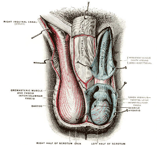
The septum of scrotum or scrotal septum is an incomplete vertical wall (septum) that divides the scrotum into two compartments –each containing a single testis. It consists of flexible connective tissue and nonstriated muscle (dartos fascia The dartos fascia, dartos tunic or simply dartos is a layer of connective tissue found in the penile shaft, foreskin and scrotum. The penile portion is referred to as the superficial fascia of penis or the subcutaneous tissue of penis, while the ...). The site of the median septum is apparent on the surface of the scrotum along a median longitudinal ridge called the scrotal raphe. The perineal raphe further extends forward to the undersurface of the penis and backward to the anal opening. The purpose of the median septum is to compartmentalize each testis in order to prevent friction or trauma. The scrotal skin is vascularized via the two external pudental arteries and the internal pudental artery. The central territory of the scrotum ... [...More Info...] [...Related Items...] OR: [Wikipedia] [Google] [Baidu] |
Septum
In biology, a septum (Latin language, Latin for ''something that encloses''; septa) is a wall, dividing a Body cavity, cavity or structure into smaller ones. A cavity or structure divided in this way may be referred to as septate. Examples Human anatomy * Interatrial septum, the wall of tissue that is a sectional part of the left and right atria of the heart * Interventricular septum, the wall separating the left and right ventricles of the heart * Lingual septum, a vertical layer of fibrous tissue that separates the halves of the tongue *Nasal septum: the cartilage wall separating the nostrils of the nose * Alveolar septum: the thin wall which separates the Pulmonary alveolus, alveoli from each other in the lungs * Orbital septum, a palpebral ligament in the upper and lower eyelids * Septum pellucidum or septum lucidum, a thin structure separating two fluid pockets in the brain * Uterine septum, a malformation of the uterus * Septum of the penis, Penile septum, a fibrous w ... [...More Info...] [...Related Items...] OR: [Wikipedia] [Google] [Baidu] |
Pudendal Arteries
The pudendal arteries are a group of arteries which supply many of the muscles and organs of the pelvic cavity. The arteries include the internal pudendal artery, the superficial external pudendal artery, and the deep external pudendal artery. The internal pudendal artery branches off the internal iliac artery, the main artery of the pelvis, and supplies blood to the sex organs. The internal pudendal artery gives rise to the perineal artery and the inferior rectal artery. The superficial external pudendal artery arises from the medial side of the femoral artery. It supplies the male scrotum and the female labia majora In primates, and specifically in humans, the labia majora (: labium majus), also known as the outer lips or outer labia, are two prominent Anatomical terms of location, longitudinal skin folds that extend downward and backward from the mons pubis .... References Arteries of the lower limb Arteries of the abdomen {{Portal bar, Anatomy ... [...More Info...] [...Related Items...] OR: [Wikipedia] [Google] [Baidu] |
Sebileau's Muscle
Sebileau's muscle is the deep muscle fibres of the dartos tunic which pass into the scrotal septum The septum of scrotum or scrotal septum is an incomplete vertical wall (septum) that divides the scrotum into two compartments –each containing a single testis. It consists of flexible connective tissue and nonstriated muscle (dartos fascia .... It is named after French anatomist Pierre Sebileau (1860–1953). References Muscles of the torso Scrotum Connective tissue {{Muscle-stub ... [...More Info...] [...Related Items...] OR: [Wikipedia] [Google] [Baidu] |
Pierre Sebileau
Pierre Sebileau (18 October 1860 – 4 October 1953) was a French surgeon born in Saint-Fort-sur-Gironde, a commune in Charente-Maritime. He was father-in-law to plastic surgeon Léon Dufourmentel (1884–1957). He served as an interne in hospitals of Bordeaux (from 1879) and Paris (from 1884), where he later worked as an anatomical prosector (1888). Subsequently, he became a surgeon at Lariboisière Hospital, Hôpital Lariboisière, specializing in the field of otorhinolaryngology. In 1893, he became an associate to the Faculty of Medicine in Paris. In addition to his work with ear, nose and throat concerns, Sebileau made contributions in his investigations involving diseases of the genitourinary system and the kidneys. His name is associated with "Sebileau's muscle", described as deep fibres of the Dartos fascia, dartos tunic which pass into the scrotal septum. Selected writings * ''Démonstrations d'anatomie; région temporale, région parotidienne, région sus-hyoïdienne, ... [...More Info...] [...Related Items...] OR: [Wikipedia] [Google] [Baidu] |
Perineal Raphe
The perineal raphe is a visible line or ridge of tissue on the body that extends from the anus through the perineum to the scrotum (male) or the vulva (female). It is found in both males and females, arises from the fusion of the urogenital folds, and is visible running medial through anteroposterior, to the anus where it resolves in a small knot of skin of varying size. In males, this structure continues through the midline of the scrotum (scrotal raphe) and upwards through the posterior midline aspect of the penis ( penile raphe). It also exists deeper through the scrotum where it is called the scrotal septum. It is the result of a fetal developmental phenomenon whereby the scrotum and penis close toward the midline and fuse. See also * Embryonic and prenatal development of the male reproductive system in humans * Frenulum of penis * Linea nigra * Raphe Raphe ( ; from ;Liddell, H.G. & Scott, R. (1940). ''A Greek-English Lexicon. revised and augmented throughout by Sir He ... [...More Info...] [...Related Items...] OR: [Wikipedia] [Google] [Baidu] |
Linea Nigra
Linea nigra (Latin for "black line"), colloquially known as the pregnancy line, manifests as a linear area of heightened pigmentation frequently observed on the abdominal region during pregnancy. Typically spanning approximately one centimeter (0.4 in) in width, this brownish streak extends vertically along the midline of the abdomen, spanning from the pubis to the umbilicus. Variably, it may traverse from the pubis to the upper abdominal region. For pregnant women, the emergence of linea nigra is attributed to an increased production of melanocyte-stimulating hormone by the placenta. This physiological phenomenon is concomitant with the occurrence of melasma and darkened nipples. Individuals with lighter skin pigmentation tend to exhibit this phenomenon less frequently in comparison to those possessing darker pigmentation. It is typical for the linea nigra to fade and dissipate within several months following childbirth. Although predominantly associated with pregnancy, it ca ... [...More Info...] [...Related Items...] OR: [Wikipedia] [Google] [Baidu] |
Pudendal Nerve
The pudendal nerve is the main nerve of the perineum. It is a Mixed nerve, mixed (motor and sensory) nerve and also conveys Sympathetic nervous system, sympathetic Autonomic nervous system, autonomic fibers. It carries sensation from the external genitalia of both sexes and the skin around the Human anus, anus and perineum, as well as the Motor neuron, motor supply to various pelvic muscles, including the external sphincter muscle of male urethra, male or external sphincter muscle of female urethra, female external urethral sphincter and the external anal sphincter. If damaged, most commonly by childbirth, loss of sensation or fecal incontinence may result. The nerve may be temporarily anesthetized, called pudendal anesthesia or pudendal block. The pudendal canal that carries the pudendal nerve is also known by the eponymous term "Alcock's canal", after Benjamin Alcock, an Irish anatomist who documented the canal in 1836. Structure Origin The pudendal nerve is paired, me ... [...More Info...] [...Related Items...] OR: [Wikipedia] [Google] [Baidu] |
Perineal Nerve
The perineal nerve is a nerve of the pelvis. It arises from the pudendal nerve in the pudendal canal. It gives superficial branches to the skin, and a deep branch to muscles. It supplies the skin and muscles of the perineum. Its latency is tested with electrodes. Structure The perineal nerve is a branch of the pudendal nerve. It lies below the internal pudendal artery. It accompanies the perineal artery. It passes through the pudendal canal for around 2 or 3 cm. Whilst still in the canal, it divides into superficial branches and a deep branch. The superficial branches of the perineal nerve become the posterior scrotal nerves in men,Essential Clinical Anatomy. K.L. Moore & A.M. Agur. Lippincott, 2 ed. 2002. Page 263 and the posterior labial nerves in women. The deep branch of the perineal nerve (also known as the "muscular" branch) travels to the muscles of the perineum. Both of these are superficial to the dorsal nerve of the penis or the dorsal nerve of the clitoris. ... [...More Info...] [...Related Items...] OR: [Wikipedia] [Google] [Baidu] |
Perineal Artery
The perineal artery (superficial perineal artery) arises from the internal pudendal artery, and turns upward, crossing either over or under the superficial transverse perineal muscle, and runs forward, parallel to the pubic arch, in the interspace between the bulbospongiosus and ischiocavernosus muscles, both of which it supplies, and finally divides into several posterior scrotal branches which are distributed to the skin and dartos tunic of the scrotum. As it crosses the superficial transverse perineal muscle it gives off the ''transverse perineal artery'' which runs transversely on the cutaneous surface of the muscle, and anastomoses with the corresponding vessel of the opposite side and with the perineal and inferior hemorrhoidal arteries. It supplies the transverse perineal muscles and the structures between the anus In mammals, invertebrates and most fish, the anus (: anuses or ani; from Latin, 'ring' or 'circle') is the external body orifice at the ''exit'' end ... [...More Info...] [...Related Items...] OR: [Wikipedia] [Google] [Baidu] |
Scrotal Arteries (other)
Scrotal arteries may refer to: * Anterior scrotal arteries, branches of the deep external pudendal artery * Posterior scrotal arteries, branches of the internal pudendal artery {{disambig ... [...More Info...] [...Related Items...] OR: [Wikipedia] [Google] [Baidu] |
Scrotum
In most terrestrial mammals, the scrotum (: scrotums or scrota; possibly from Latin ''scortum'', meaning "hide" or "skin") or scrotal sac is a part of the external male genitalia located at the base of the penis. It consists of a sac of skin containing the external spermatic fascia, testicles, epididymides, and vasa deferentia. The scrotum will usually tighten when exposed to cold temperatures. The scrotum is homologous to the labia majora in females. Structure In regards to humans, the scrotum is a suspended two-chambered sac of skin and muscular tissue containing the testicles and the lower part of the spermatic cords. It is located behind the penis and above the perineum. The perineal raphe is a small, vertical ridge of skin that expands from the anus and runs through the middle of the scrotum front to back. The scrotum is also a distention of the perineum and carries some abdominal tissues into its cavity including the testicular artery, testicular vein, and ... [...More Info...] [...Related Items...] OR: [Wikipedia] [Google] [Baidu] |
Internal Pudendal Artery
The internal pudendal artery is one of the three pudendal arteries. It branches off the internal iliac artery, and provides blood to the external genitalia. Structure The internal pudendal artery is the terminal branch of the anterior trunk of the internal iliac artery. It is smaller in the female than in the male. Path It arises from the anterior division of internal iliac artery. It runs on the lateral pelvic wall. It exits the pelvic cavity through the greater sciatic foramen, inferior to the piriformis muscle, to enter the gluteal region. It then curves around the sacrospinous ligament to enter the perineum through the lesser sciatic foramen. It travels through the pudendal canal with the internal pudendal veins and the pudendal nerve. Branches The internal pudendal artery gives off the following branches: The deep artery of clitoris is a branch of the internal pudendal artery and supplies the clitoral crura. Another branch of the internal pudendal artery is th ... [...More Info...] [...Related Items...] OR: [Wikipedia] [Google] [Baidu] |
