|
Saethre–Chotzen Syndrome
Saethre–Chotzen syndrome (SCS), also known as acrocephalosyndactyly type III, is a rare congenital disorder associated with craniosynostosis (premature closure of one or more of the sutures between the bones of the skull). This affects the shape of the head and face, resulting in a cone-shaped head and an asymmetrical face. Individuals with SCS also have droopy eyelids (ptosis), widely spaced eyes (hypertelorism), and minor abnormalities of the hands and feet (syndactyly). Individuals with more severe cases of SCS may have mild to moderate intellectual or learning disabilities. Depending on the level of severity, some individuals with SCS may require some form of medical or surgical intervention. Most individuals with SCS live fairly normal lives, regardless of whether medical treatment is needed or not. Signs and symptoms SCS presents in a variable fashion. The majority of individuals with SCS are moderately affected, with uneven facial features and a relatively flat face due to ... [...More Info...] [...Related Items...] OR: [Wikipedia] [Google] [Baidu] |
Acrocephalosyndactylia
Acrocephalosyndactyly is a group of autosomal dominant congenital disorders characterized by craniofacial (craniosynostosis) and hand and foot (syndactyly) abnormalities. When polydactyly is present, the classification is acrocephalopolysyndactyly. Acrocephalosyndactyly is mainly diagnosed postnatally, although prenatal diagnosis is possible if the mutation is known to be within the family genome. Treatment often involves surgery in early childhood to correct for craniosynostosis and syndactyly. Characteristics Acrocephalosyndactyly presents in numerous different subtypes, however, considerable overlap in symptoms occurs. Generally, all forms of acrocephalosyndactyly are characterized by craniofacial, hand, and foot abnormalities, such as premature closure of the fibrous joints in between certain bones of the skull, fusion of certain fingers or toes, and/or more than the normal number of digits. Some subtypes also involve structural heart deformations that are present at birth. ... [...More Info...] [...Related Items...] OR: [Wikipedia] [Google] [Baidu] |
Enamel Hypoplasia
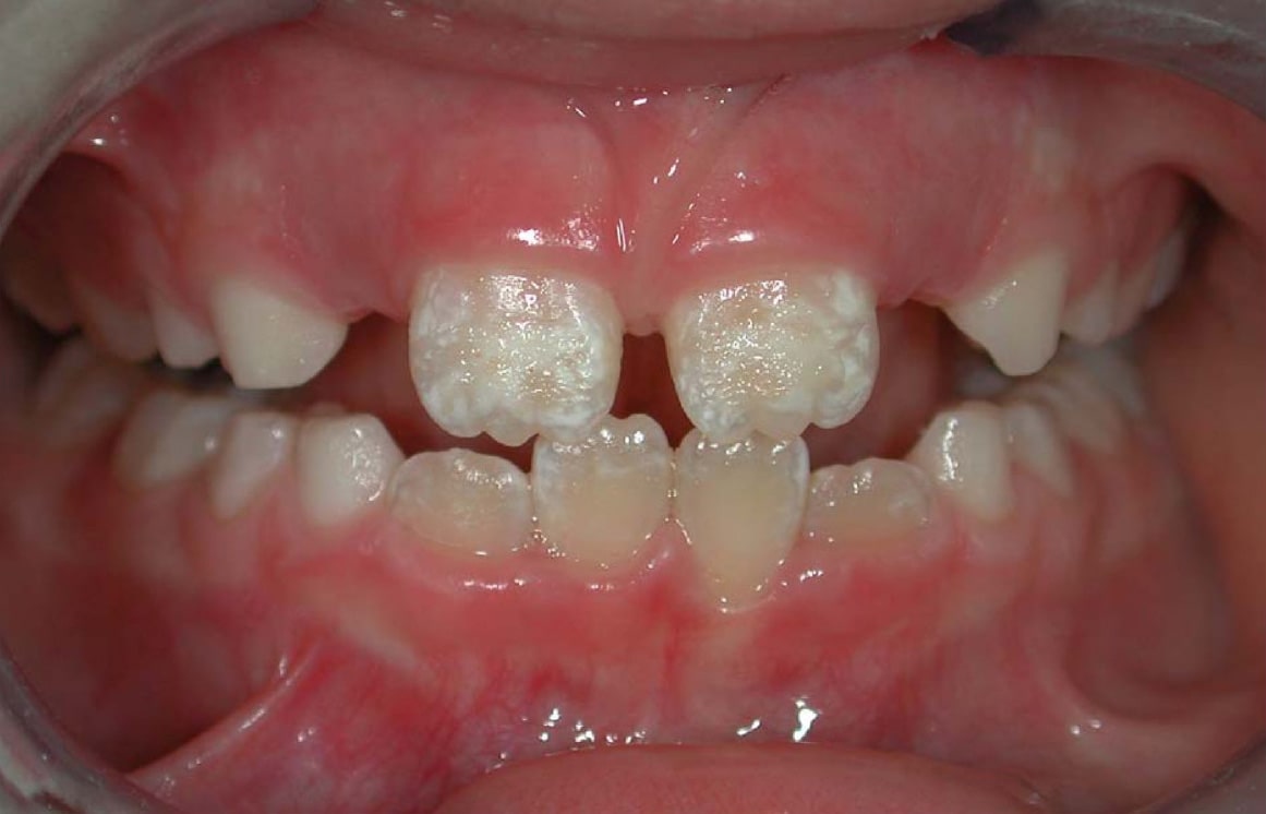
Enamel hypoplasia is a defect of the teeth in which the enamel is deficient in quantity, caused by defective enamel matrix formation during enamel development, as a result of inherited and acquired systemic condition(s). It can be identified as missing tooth structure and may manifest as pits or grooves in the crown of the affected teeth, and in extreme cases, some portions of the crown of the tooth may have no enamel, exposing the dentin. It may be generalized across the dentition or localized to a few teeth. Defects are categorized by shape or location. Common categories are pit-form, plane-form, linear-form, and localised enamel hypoplasia. Hypoplastic lesions are found in areas of the teeth where the enamel was being actively formed during a systemic or local disturbance. Since the formation of enamel extends over a long period of time, defects may be confined to one well-defined area of the affected teeth. Knowledge of chronological development of deciduous and permanent ... [...More Info...] [...Related Items...] OR: [Wikipedia] [Google] [Baidu] |
Malocclusion
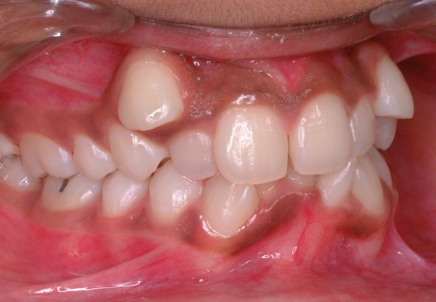
In orthodontics, a malocclusion is a misalignment or incorrect relation between the teeth of the upper and lower dental arches when they approach each other as the jaws close. The English-language term dates from 1864; Edward Angle (1855-1930), the "father of modern orthodontics", popularised it. The word "malocclusion" derives from ''occlusion'', and refers to the manner in which opposing teeth meet ('' mal-'' + ''occlusion'' = "incorrect closure"). The malocclusion classification is based on the relationship of the mesiobuccal cusp of the maxillary first molar and the buccal groove of the mandibular first molar. If this molar relationship exists, then the teeth can align into normal occlusion. According to Angle, malocclusion is any deviation of the occlusion from the ideal. However, assessment for malocclusion should also take into account aesthetics and the impact on functionality. If these aspects are acceptable to the patient despite meeting the formal definitio ... [...More Info...] [...Related Items...] OR: [Wikipedia] [Google] [Baidu] |
Nasal Septum Deviation
Nasal septum deviation is a physical disorder of the nose, involving a displacement of the nasal septum. Some displacement is common, affecting 80% of people, mostly without their knowledge. Signs and symptoms The nasal septum is the bone and cartilage in the nose that separates the nasal cavity into the two nostrils. The cartilage is called the quadrangular cartilage and the bones comprising the septum include the maxillary crest, vomer and the perpendicular plate of the ethmoid. Normally, the septum lies centrally, and thus the nasal passages are symmetrical. A deviated septum is an abnormal condition in which the top of the cartilaginous ridge leans to the left or the right, causing obstruction of the affected nasal passage. It is common for nasal septa to depart from the exact centerline; the septum is only considered deviated if the shift is substantial or causes problems. By itself, a deviated septum can go undetected for years and thus be without any need for corre ... [...More Info...] [...Related Items...] OR: [Wikipedia] [Google] [Baidu] |
Pinna (anatomy)
The auricle or auricula is the visible part of the ear that is outside the head. It is also called the pinna (Latin for " wing" or " fin", plural pinnae), a term that is used more in zoology. Structure The diagram shows the shape and location of most of these components: * '' antihelix'' forms a 'Y' shape where the upper parts are: ** ''Superior crus'' (to the left of the ''fossa triangularis'' in the diagram) ** ''Inferior crus'' (to the right of the ''fossa triangularis'' in the diagram) * '' Antitragus'' is below the ''tragus'' * ''Aperture'' is the entrance to the ear canal * ''Auricular sulcus'' is the depression behind the ear next to the head * ''Concha'' is the hollow next to the ear canal * Conchal angle is the angle that the back of the ''concha'' makes with the side of the head * ''Crus'' of the helix is just above the ''tragus'' * ''Cymba conchae'' is the narrowest end of the ''concha'' * External auditory meatus is the ear canal * ''Fossa triangularis'' is the ... [...More Info...] [...Related Items...] OR: [Wikipedia] [Google] [Baidu] |


