|
SORBS2
ArgBP2 protein, also referred to as Sorbin and SH3 domain-containing protein 2 is a protein that in humans is encoded by the ''SORBS2'' gene. ArgBP2 belongs to the a small family of adaptor proteins having sorbin homology (SOHO) domains. ArgBP2 is highly abundant in cardiac muscle cells at sarcomeric Z-disc structures, and is expressed in other cells at actin stress fibers and the nucleus. Structure ArgBP2 may exist in as many as 9 unique isoforms ranging from 52 kDa to 117 kDa (492 to 1100 amino acids). ArgBP2 belongs to the a small family of adaptor proteins having sorbin homology (SOHO) domains and three SH3 domains, which regulate cell adhesion, cytoskeletal organization and growth factor signaling; other members include CAP/ponsin and vinexin. The three SH3 domains are C-terminal, a serine-threonine rich domain resides in the middle, and the sorbin homology (SoHo) domain is N-terminal. The SH3 domains interact with Arg/Abl, vinculin. The SOHO domain interacts with flotill ... [...More Info...] [...Related Items...] OR: [Wikipedia] [Google] [Baidu] |
SORBS1
CAP/Ponsin protein, also known as Sorbin and SH3 domain-containing protein 1 is a protein that in humans is encoded by the ''SORBS1'' gene. It is part of a small family of adaptor proteins that regulate cell adhesion, growth factor signaling and cytoskeletal formation. It is mainly expressed in heart, skeletal muscle, liver, adipose tissue, and macrophages; in striated muscle tissue, it is localized to costamere structures. Structure CAP/Ponsin may exist as thirteen alternatively-spliced isoforms, ranging from 81 kDa to 142 kDa. It is part of an adaptor protein family, of which ArgBP2 and vinexin are also a part. These proteins contain a conserved sorbin homology (SOHO) domain and three SH3 domains, and CAP/Ponsin is expressed in heart, skeletal muscle, liver, adipose tissue, and macrophages. Function In muscle, CAP/Ponsin plays a role in the formation of mature costameres from focal adhesion-like contacts during assembly of the contractile apparatus, as overexpression of CA ... [...More Info...] [...Related Items...] OR: [Wikipedia] [Google] [Baidu] |
Pyk2
Protein tyrosine kinase 2 beta is an enzyme that in humans is encoded by the ''PTK2B'' gene. Function This gene encodes a cytoplasmic protein tyrosine kinase that is involved in calcium-induced regulation of ion channels and activation of the MAP kinase signaling pathway. The encoded protein may represent an important signaling intermediate between neuropeptide-activated receptors or neurotransmitters that increase calcium flux and the downstream signals that regulate neuronal activity. The encoded protein undergoes rapid tyrosine phosphorylation and activation in response to increases in the intracellular calcium concentration , nicotinic acetylcholine receptor activation, membrane depolarization, or protein kinase C activation. In addition, SOCE-induced Pyk2 activation mediates disassembly of endothelial adherens junctions, via tyrosine (Y1981-residue) phosphorylation of VE-PTP. This protein has been shown to bind a CRK-associated substrate, a nephrocystin, a GTPase re ... [...More Info...] [...Related Items...] OR: [Wikipedia] [Google] [Baidu] |
ABL1
Tyrosine-protein kinase ABL1 also known as ABL1 is a protein that, in humans, is encoded by the ''ABL1'' gene (previous symbol ''ABL'') located on chromosome 9. c-Abl is sometimes used to refer to the version of the gene found within the mammalian genome, while v-Abl refers to the viral gene, which was initially isolated from the Abelson murine leukemia virus. Function The ''ABL1'' proto-oncogene encodes a cytoplasmic and nuclear protein tyrosine kinase that has been implicated in processes of cell differentiation, cell division, cell adhesion, and stress response such as DNA repair. Activity of ABL1 protein is negatively regulated by its SH3 domain, and deletion of the SH3 domain turns ABL1 into an oncogene. The t(9;22) translocation results in the head-to-tail fusion of the '' BCR'' and ''ABL1'' genes, leading to a fusion gene present in many cases of chronic myelogenous leukemia. The DNA-binding activity of the ubiquitously expressed ABL1 tyrosine kinase is regulated by ... [...More Info...] [...Related Items...] OR: [Wikipedia] [Google] [Baidu] |
Cbl Gene
E3 ubiquitin-protein ligase CBL is an enzyme that is humans is encoded by the ''CBL'' (Casitas B-lineage Lymphoma) gene. ''CBL'' gene is the founding member the Cbl family. The protein CBL which is an Ubiquitin ligase, E3 ubiquitin-protein ligase involved in cell signalling and protein ubiquitination. Mutations to this gene have been implicated in a number of human cancers, particularly acute myeloid leukaemia. Discovery In 1989 a virally encoded portion of the chromosomal mouse ''Cbl'' gene was the first member of the Cbl family to be discovered and was named ''v-Cbl'' to distinguish it from normal mouse ''c-Cbl''. The virus used in the experiment was a mouse-tropic strain of Murine leukemia virus isolated from the brain of a mouse captured at Lake Casitas, California known as ''Cas-Br-M'', and was found to have excised approximately a third of the original ''c-Cbl'' gene from a mouse into which it was injected. Sequencing revealed that the portion carried by the retrovirus e ... [...More Info...] [...Related Items...] OR: [Wikipedia] [Google] [Baidu] |
Myofibril
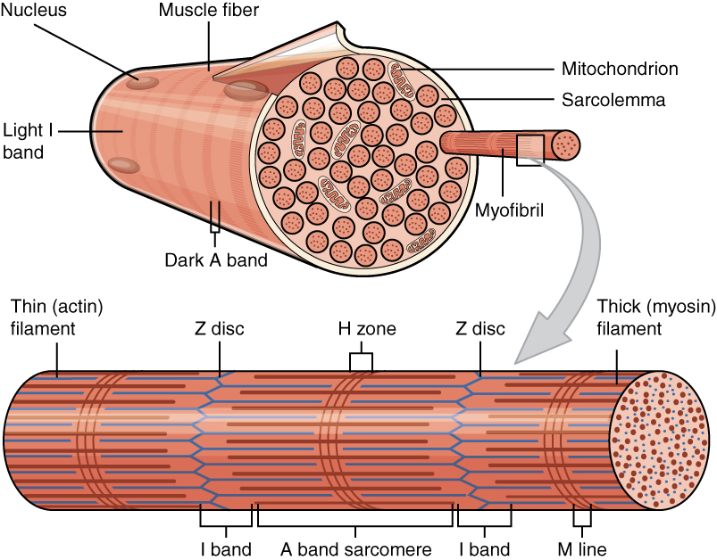
A myofibril (also known as a muscle fibril or sarcostyle) is a basic rod-like organelle of a muscle cell. Skeletal muscles are composed of long, tubular cells known as Skeletal muscle#Skeletal muscle cells, muscle fibers, and these cells contain many chains of myofibrils. Each myofibril has a diameter of 1–2 micrometres. They are created during embryogenesis, embryonic development in a process known as myogenesis. Myofibrils are composed of long proteins including actin, myosin, and titin, and other proteins that hold them together. These proteins are organized into Thick filaments, thick, thin filaments, thin, and elastic filament, elastic myofilaments, which repeat along the length of the myofibril in sections or units of contraction called sarcomeres. Muscle contraction, Muscles contract by sliding filament theory, sliding the thick myosin, and thin actin myofilaments along each other. Structure Each myofibril has a diameter of between 1 and 2 micrometres (μm). The filamen ... [...More Info...] [...Related Items...] OR: [Wikipedia] [Google] [Baidu] |
Vinculin
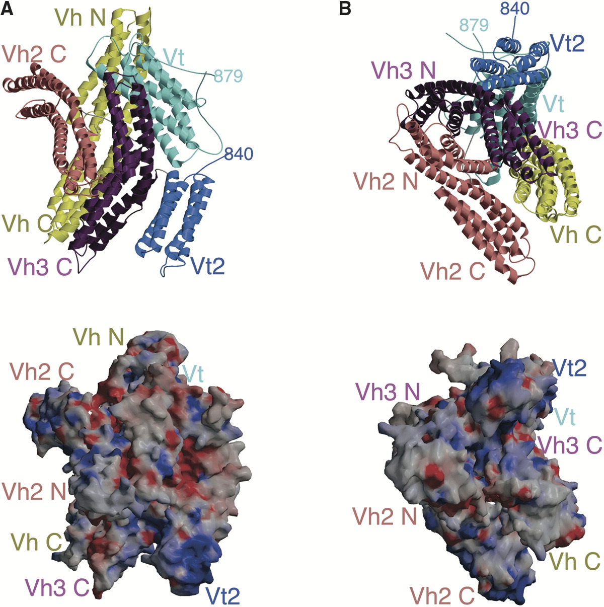
In mammalian cells, vinculin is a membrane-cytoskeletal protein in focal adhesion plaques that is involved in linkage of integrin adhesion molecules to the actin cytoskeleton. Vinculin is a cytoskeletal protein associated with cell-cell and cell-matrix junctions, where it is thought to function as one of several interacting proteins involved in anchoring F-actin to the membrane. Discovered independently by Benny Geiger and Keith Burridge, its sequence is 20%–30% similar to α-catenin, which serves a similar function. Binding alternately to talin or α-actinin, vinculin's shape and, as a consequence, its binding properties are changed. The vinculin gene occurs as a single copy and what appears to be no close relative to take over functions in its absence. Its splice variant metavinculin (see below) also needs vinculin to heterodimerize and work in a dependent fashion. Structure Vinculin is a 117-kDa cytoskeletal protein with 1066 amino acids. The protein contains an acidi ... [...More Info...] [...Related Items...] OR: [Wikipedia] [Google] [Baidu] |
SPEC
The Standard Performance Evaluation Corporation (SPEC) is a non-profit consortium that establishes and maintains standardized benchmarks and performance evaluation tools for new generations of computing systems. SPEC was founded in 1988 and its membership comprises over 120 computer hardware and software vendors, educational institutions, research organizations, and government agencies internationally. SPEC benchmarks and tools are widely used to evaluate the performance of computer systems; the test results are published on the SPEC website. External links * Official List of SPEC Benchmarks Computer performance Evaluation of computers Companies established in 1988 Companies based in Virginia Standards organizations in the United States {{Compu-stub ... [...More Info...] [...Related Items...] OR: [Wikipedia] [Google] [Baidu] |
Paxillin
Paxillin is a protein that in humans is encoded by the ''PXN'' gene. Paxillin is expressed at focal adhesions of non-striated cells and at costameres of striated muscle cells, and it functions to adhere cells to the extracellular matrix. Mutations in ''PXN'' as well as abnormal expression of paxillin protein has been implicated in the progression of various cancers. Structure Human paxillin is 64.5 kDa in molecular weight and 591 amino acids in length. The C-terminal region of paxillin is composed of four tandem double zinc finger LIM domains that are cysteine/histidine-rich with conserved repeats; these serve as binding sites for the protein tyrosine phosphatase-PEST, tubulin and serves as the targeting motif for focal adhesions. The N-terminal region of paxillin has five highly conserved leucine-rich sequences termed LD motifs, which mediate several interactions, including that with pp125FAK and vinculin. The LD motifs are predicted to form amphipathic alpha helices, w ... [...More Info...] [...Related Items...] OR: [Wikipedia] [Google] [Baidu] |
Protein
Proteins are large biomolecules and macromolecules that comprise one or more long chains of amino acid residue (biochemistry), residues. Proteins perform a vast array of functions within organisms, including Enzyme catalysis, catalysing metabolic reactions, DNA replication, Cell signaling, responding to stimuli, providing Cytoskeleton, structure to cells and Fibrous protein, organisms, and Intracellular transport, transporting molecules from one location to another. Proteins differ from one another primarily in their sequence of amino acids, which is dictated by the Nucleic acid sequence, nucleotide sequence of their genes, and which usually results in protein folding into a specific Protein structure, 3D structure that determines its activity. A linear chain of amino acid residues is called a polypeptide. A protein contains at least one long polypeptide. Short polypeptides, containing less than 20–30 residues, are rarely considered to be proteins and are commonly called pep ... [...More Info...] [...Related Items...] OR: [Wikipedia] [Google] [Baidu] |
Cardiac
The heart is a muscular organ found in humans and other animals. This organ pumps blood through the blood vessels. The heart and blood vessels together make the circulatory system. The pumped blood carries oxygen and nutrients to the tissue, while carrying metabolic waste such as carbon dioxide to the lungs. In humans, the heart is approximately the size of a closed fist and is located between the lungs, in the middle compartment of the chest, called the mediastinum. In humans, the heart is divided into four chambers: upper left and right atria and lower left and right ventricles. Commonly, the right atrium and ventricle are referred together as the right heart and their left counterparts as the left heart. In a healthy heart, blood flows one way through the heart due to heart valves, which prevent backflow. The heart is enclosed in a protective sac, the pericardium, which also contains a small amount of fluid. The wall of the heart is made up of three layers: epicardi ... [...More Info...] [...Related Items...] OR: [Wikipedia] [Google] [Baidu] |
Sarcomere
A sarcomere (Greek σάρξ ''sarx'' "flesh", μέρος ''meros'' "part") is the smallest functional unit of striated muscle tissue. It is the repeating unit between two Z-lines. Skeletal striated muscle, Skeletal muscles are composed of tubular muscle cells (called muscle fibers or myofibers) which are formed during embryonic development, embryonic myogenesis. Muscle fibers contain numerous tubular myofibrils. Myofibrils are composed of repeating sections of sarcomeres, which appear under the microscope as alternating dark and light bands. Sarcomeres are composed of long, fibrous proteins as filaments that slide past each other when a muscle contracts or relaxes. The costamere is a different component that connects the sarcomere to the sarcolemma. Two of the important proteins are myosin, which forms the thick filament, and actin, which forms the thin filament. Myosin has a long fibrous tail and a globular head that binds to actin. The myosin head also binds to Adenosine triphos ... [...More Info...] [...Related Items...] OR: [Wikipedia] [Google] [Baidu] |
Cardiac Hypertrophy
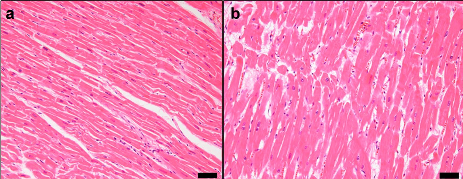
Ventricular hypertrophy (VH) is thickening of the walls of a ventricle (lower chamber) of the heart. Although left ventricular hypertrophy (LVH) is more common, right ventricular hypertrophy (RVH), as well as concurrent hypertrophy of both ventricles can also occur. Ventricular hypertrophy can result from a variety of conditions, both adaptive and maladaptive. For example, it occurs in what is regarded as a physiologic, adaptive process in pregnancy in response to increased blood volume; but can also occur as a consequence of ventricular remodeling following a heart attack. Importantly, pathologic and physiologic remodeling engage different cellular pathways in the heart and result in different gross cardiac phenotypes. Presentation In individuals with eccentric hypertrophy there may be little or no indication that hypertrophy has occurred as it is generally a healthy response to increased demands on the heart. Conversely, concentric hypertrophy can make itself known in a varie ... [...More Info...] [...Related Items...] OR: [Wikipedia] [Google] [Baidu] |


