|
Russell Van Gelder
Russell Van Gelder is an American clinician-scientist and board-certified ophthalmologist; he has served as the chair of the University of Washington Medicine Department of Ophthalmology since 2008 and Editor-in-Chief of the journal ''Ophthalmology'' since 2022. He is known for his research on the mechanisms of uveitis, non-visual photoreception in the eye, and vision-restoration methods for retinal degenerative disease, as well as his leadership and advisory positions in various American ophthalmological and medical societies. Education Van Gelder graduated from Northern Valley Regional High School at Old Tappan in New Jersey in 1981. He attended Stanford University for his Bachelors, and MD/Ph.D.: receiving his bachelors in Biological Sciences in 1985, and his MD/Ph.D. in Neurosciences in 1994 as part of the MSTP, during which time he studied the molecular basis for circadian rhythms. He then completed an internal medicine internship at Stanford before moving to Washington ... [...More Info...] [...Related Items...] OR: [Wikipedia] [Google] [Baidu] |
Ophthalmology
Ophthalmology (, ) is the branch of medicine that deals with the diagnosis, treatment, and surgery of eye diseases and disorders. An ophthalmologist is a physician who undergoes subspecialty training in medical and surgical eye care. Following a medical degree, a doctor specialising in ophthalmology must pursue additional postgraduate residency training specific to that field. In the United States, following graduation from medical school, one must complete a four-year residency in ophthalmology to become an ophthalmologist. Following residency, additional specialty training (or fellowship) may be sought in a particular aspect of eye pathology. Ophthalmologists prescribe medications to treat ailments, such as eye diseases, implement laser therapy, and perform surgery when needed. Ophthalmologists provide both primary and specialty eye care—medical and surgical. Most ophthalmologists participate in academic research on eye diseases at some point in their training and many inc ... [...More Info...] [...Related Items...] OR: [Wikipedia] [Google] [Baidu] |
Seasonal Affective Disorder
Seasonal affective disorder (SAD) is a mood disorder subset in which people who typically have normal mental health throughout most of the year exhibit depressive symptoms at the same time each year. It is commonly, but not always, associated with the reductions or increases in total daily sunlight hours that occur during the winter or summer. Common symptoms include sleeping too much, having little to no energy, and overeating. The condition in the summer can include heightened anxiety.Seasonal affective disorder (SAD): Symptoms MayoClinic.com (September 22, 2011). Retrieved on March 24, 2013. However, there are significant differences in the duration, severity, and symptoms of each individual's experience of SAD. For instance, in a fifth of patients, the disor ... [...More Info...] [...Related Items...] OR: [Wikipedia] [Google] [Baidu] |
OPN1SW
Blue-sensitive opsin is a protein that in humans is encoded by the ''OPN1SW'' gene. The OPN1SW gene provides instructions for making a protein that is essential for normal color vision. This protein is found in the retina, which is the light-sensitive tissue at the back of the eye. The OPN1SW gene provides instructions for making an opsin pigment that is more sensitive to light in the blue/violet part of the visible spectrum (short-wavelength light). Cones with this pigment are called short-wavelength-sensitive or S cones. In response to light, the photopigment triggers a series of chemical reactions within an S cone. These reactions ultimately alter the cell's electrical charge, generating a signal that is transmitted to the brain. The brain combines input from all three types of cones to produce normal color vision. See also * Opsin Animal opsins are G-protein-coupled receptors and a group of proteins made light-sensitive via a chromophore, typically retinal. When bound to r ... [...More Info...] [...Related Items...] OR: [Wikipedia] [Google] [Baidu] |
Melanopsin
Melanopsin is a type of photopigment belonging to a larger family of light-sensitive retinylidene protein, retinal proteins called opsins and encoded by the gene ''Opn4''. In the mammalian retina, there are two additional categories of opsins, both involved in the formation of visual images: rhodopsin and photopsin (types I, II, and III) in the Rod cell, rod and Cone cell, cone photoreceptor cells, respectively. In humans, melanopsin is found in intrinsically photosensitive retinal ganglion cells (ipRGCs). It is also found in the iris of mice and primates. Melanopsin is also found in rats, amphioxus, and other chordates. ipRGCs are photoreceptor cells which are particularly sensitive to the absorption of short-wavelength (blue) visible light and communicate information directly to the area of the brain called the suprachiasmatic nucleus (SCN), also known as the central "body clock", in mammals. Melanopsin plays an important non-image-forming role in the Entrainment (chronobiology ... [...More Info...] [...Related Items...] OR: [Wikipedia] [Google] [Baidu] |
Suprachiasmatic Nucleus
The suprachiasmatic nucleus or nuclei (SCN) is a small region of the brain in the hypothalamus, situated directly above the optic chiasm. It is responsible for regulating sleep cycles in animals. Reception of light inputs from photosensitive retinal ganglion cells allow it to coordinate the subordinate cellular clocks of the body and entrain to the environment. The neuronal and hormonal activities it generates regulate many different body functions in an approximately 24-hour cycle. The SCN also interacts with many other regions of the brain. It contains several cell types, neurotransmitters and peptides, including vasopressin and vasoactive intestinal peptide. Disruptions or damage to the SCN has been associated with different mood disorders and sleep disorders, suggesting the significance of the SCN in regulating circadian timing. Neuroanatomy The SCN is situated in the anterior part of the hypothalamus immediately dorsal, or ''superior'' (hence supra) to the optic c ... [...More Info...] [...Related Items...] OR: [Wikipedia] [Google] [Baidu] |
OPN5
Opsin-5, also known as G-protein coupled receptor 136 or neuropsin is a protein that in humans is encoded by the ''OPN5'' gene. Opsin-5 is a member of the opsin subfamily of the G protein-coupled receptors. It is a photoreceptor protein sensitive to ultraviolet (UV) light. The OPN5 gene was discovered in mouse and human genomes and its mRNA expression was also found in neural tissues. Neuropsin is bistable at 0 °C and activates a UV-sensitive, heterotrimeric G protein Gi-mediated pathway in mammalian and avian tissues. Function Human neuropsin is expressed in the eye, brain, testes, and spinal cord. Neuropsin belongs to the seven-exon subfamily of mammalian opsin genes that includes peropsin (RRH) and retinal G protein coupled receptor (RGR). Neuropsin has different isoforms created by alternative splicing. Photochemistry When reconstituted with 11-cis-retinal, mouse and human neuropsins absorb maximally at 380 nm. When illuminated these neuropsins are converted i ... [...More Info...] [...Related Items...] OR: [Wikipedia] [Google] [Baidu] |
Murinae
The Old World rats and mice, part of the subfamily Murinae in the family Muridae, comprise at least 519 species. Members of this subfamily are called murines. In terms of species richness, this subfamily is larger than all mammal families except the Cricetidae and Muridae, and is larger than all mammal orders except the bats and the remainder of the rodents. Description The Murinae are native to Africa, Europe, Asia, and Australia. They are terrestrial placental mammals. They have also been introduced to all continents except Antarctica, and are serious pest animals. This is particularly true in island communities where they have contributed to the endangerment and extinction of many native animals. Two prominent murine species have become vital laboratory animals: the brown rat and house mouse are both used as medical subjects. The murines have a distinctive molar pattern that involves three rows of cusps instead of two, the primitive pattern seen most frequently ... [...More Info...] [...Related Items...] OR: [Wikipedia] [Google] [Baidu] |
Entrainment (chronobiology)
In the study of chronobiology, entrainment refers to the synchronization of a biological clock to an environmental cycle. An example is the interaction between circadian rhythms and environmental cues, such as light and temperature. Entrainment helps organisms adapt their bodily processes according to the timing of a changing environment. For example, entrainment is manifested during travel between time zones, hence why humans experience jet lag. Biological rhythms are endogenous; they persist even in the absence of environmental cues as they are driven by an internal mechanism, most notably the circadian clock. Of the several possible cues (known as '' zeitgebers,'' German for 'time-givers') that can contribute to entrainment of the circadian clock, light has the greatest impact. Units of circadian time (CT) are used to describe entrainment to refer to the relationship between the rhythm and the light signal/pulse. Modes of entrainment There are two general modes of entrainm ... [...More Info...] [...Related Items...] OR: [Wikipedia] [Google] [Baidu] |
Intrinsically Photosensitive Retinal Ganglion Cell
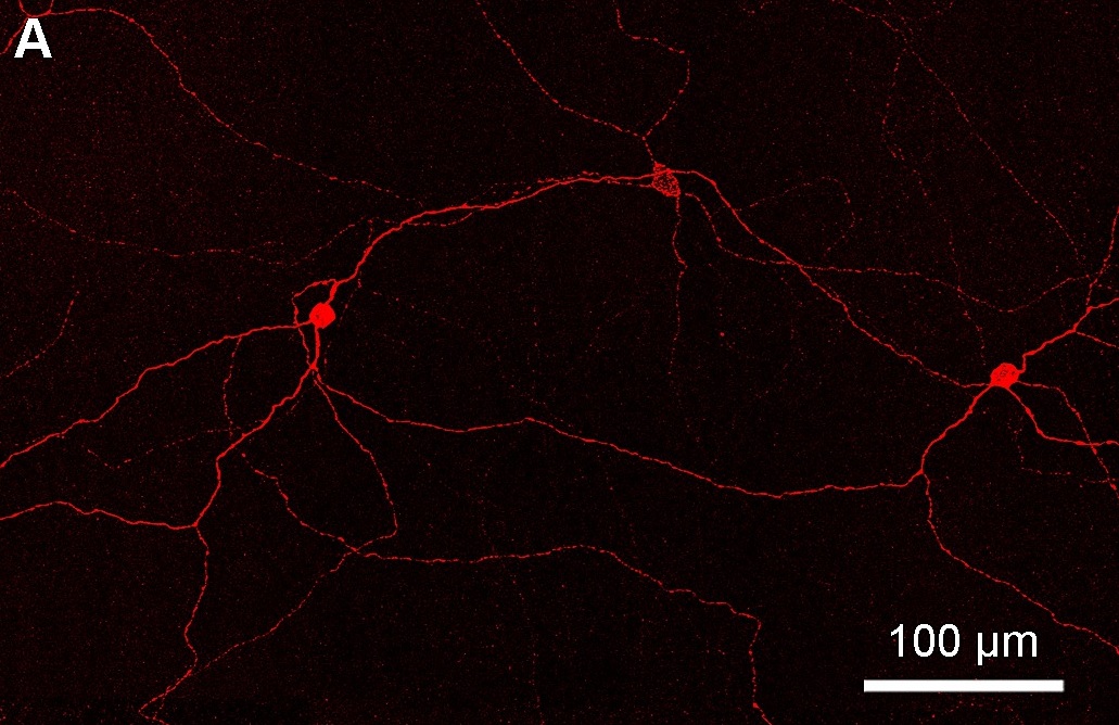
Intrinsically photosensitive retinal ganglion cells (ipRGCs), also called photosensitive retinal ganglion cells (pRGC), or melanopsin-containing retinal ganglion cells (mRGCs), are a type of neuron in the retina of the mammalian eye. The presence of an additional photoreceptor was first suspected in 1927 when mice lacking rod and cone cells still responded to changing light levels through pupil constriction; this suggested that rods and cones are not the only light-sensitive tissue. However, it was unclear whether this light sensitivity arose from an additional retinal photoreceptor or elsewhere in the body. Recent research has shown that these retinal ganglion cells, unlike other retinal ganglion cells, are intrinsically photosensitive due to the presence of melanopsin, a light-sensitive protein. Therefore, they constitute a third class of photoreceptors, in addition to rod and cone cells. Overview Compared to the rods and cones, the ipRGCs respond more sluggishly and signal ... [...More Info...] [...Related Items...] OR: [Wikipedia] [Google] [Baidu] |

