|
Rhadinosuchus
''Rhadinosuchus'' is an extinct genus of proterochampsian archosauriform reptile from the Late Triassic. It is known only from the type species ''Rhadinosuchus gracilis'', reposited in Munich, Germany. The fossil includes an incomplete skull and fragments of post-cranial material. Hosffstetter (1955), Kuhn (1966), Reig (1970) and Bonaparte (1971) hypothesized it to be synonymous with '' Cerritosaurus'', but other characteristics suggest it is closer to '' Chanaresuchus'' and '' Gualosuchus'', while it is certainly different from '' Proterochampsa'' and '' Barberenachampsa''. The small size indicates it is a young animal, making it hard to classify. The fossil was collected at the Sanga 6 site (part of the Santa Maria Formation), in Santa Maria, Paleorrota, Brazil. It was collected by Friedrich von Huene in 1938. The remains are dated to the Triassic Period. Description Skull The skull of ''Rhadinosuchus'' had an estimated length of 11.0 centimeters (4.3 inches), with miss ... [...More Info...] [...Related Items...] OR: [Wikipedia] [Google] [Baidu] |
Santa Maria Formation
The Santa Maria Formation is a sedimentary rock formation found in Rio Grande do Sul, Brazil. It is primarily Carnian in age ( Late Triassic), and is notable for its fossils of cynodonts, " rauisuchian" pseudosuchians, and early dinosaurs and other dinosauromorphs, including the herrerasaurid ''Staurikosaurus'', the basal sauropodomorphs '' Buriolestes'' and ''Saturnalia,'' and the lagerpetid '' Ixalerpeton''. The formation is named after the city of Santa Maria in the central region of Rio Grande do Sul, where outcrops were first studied. The Santa Maria Formation makes up the majority of the Santa Maria Supersequence, which extends through the entire Late Triassic. The Santa Maria Supersequence is divided into four geological sequences, separated from each other by short unconformities. The first two of these sequences (Pinheiros-Chiniquá and Santa Cruz sequences) lie entirely within the Santa Maria Formation, while the third (the Candelária sequence) is shared with ... [...More Info...] [...Related Items...] OR: [Wikipedia] [Google] [Baidu] |
Proterochampsia
Proterochampsia is a clade of early archosauriform reptiles from the Triassic period. It includes the Proterochampsidae (e.g. ''Proterochampsa'', ''Chanaresuchus'' and ''Tropidosuchus'') and probably also the Doswelliidae. Nesbitt (2011) defines Proterochampsia as a stem-based taxon that includes ''Proterochampsa'' and all forms more closely related to it than ''Euparkeria'', ''Erythrosuchus'', ''Passer domesticus'' (the House Sparrow), or ''Crocodylus niloticus'' (the Nile crocodile). Therefore, the inclusion of Doswelliidae in it is dependent upon whether ''Doswellia'' and ''Proterochampsa'' form a monophyletic group to the exclusion of Archosauria and other related groups. Description Nesbitt (2011) found that Proterochampsians share several distinguishing characteristics, or synapomorphies. A prominent ridge runs along the length of the jugal, a bone below the eye. Another ridge is present on the quadratojugal, a bone positioned toward the back of the skull behind the juga ... [...More Info...] [...Related Items...] OR: [Wikipedia] [Google] [Baidu] |
Doswelliidae
Doswelliidae is an extinct family of carnivorous archosauriform reptiles that lived in North America and Europe during the Middle to Late Triassic period. Long represented solely by the heavily-armored reptile ''Doswellia'', the family's composition has expanded since 2011, although two supposed South American doswelliids (''Archeopelta'' and ''Tarjadia'') were later redescribed as erpetosuchids. Doswelliids were not true archosaurs, but they were close relatives and some studies have considered them among the most derived non-archosaurian archosauriforms. They may have also been related to the Proterochampsidae, a South American family of crocodile-like archosauriforms. Description Doswelliids are believed to be semiaquatic carnivores similar to crocodilians in appearance, as evidenced by their short legs and eyes and nostrils which are set high on the head, though the putative member '' Scleromochlus'' has been interpreted as a frog-like hopper by one study. They had long ... [...More Info...] [...Related Items...] OR: [Wikipedia] [Google] [Baidu] |
Friedrich Von Huene
Friedrich von Huene, born Friedrich Richard von Hoinigen, (March 22, 1875 – April 4, 1969) was a German paleontologist who renamed more dinosaurs in the early 20th century than anyone else in Europe. He also made key contributions about various Permo-Carboniferous limbed vertebrates. Biography Huene was born in Tübingen, Kingdom of Württemberg. His discoveries include the skeletons of more than 35 individuals of ''Plateosaurus'' in the famous Trossingen quarry, the early proto-dinosaur ''Saltopus'' in 1910, ''Proceratosaurus'' in 1926, the giant ''Antarctosaurus'' in 1929, and numerous other dinosaurs and fossilized animals like pterosaurs. He also was the first to naming several higher taxa, including Prosauropoda and Sauropodomorpha. In 1941 he found a stone that had petrified wood in it, sadly, He thought that it was a dinosaur. However a couple Polish paleontologists. The “dinosaur” was called the Succinodon He visited the Geopark of Paleorrota in 1928, and the ... [...More Info...] [...Related Items...] OR: [Wikipedia] [Google] [Baidu] |
Antorbital Fenestra
An antorbital fenestra (plural: fenestrae) is an opening in the skull that is in front of the eye sockets. This skull character is largely associated with archosauriforms, first appearing during the Triassic Period. Among extant archosaurs, birds still possess antorbital fenestrae, whereas crocodylians have lost them. The loss in crocodylians is believed to be related to the structural needs of their skulls for the bite force and feeding behaviours that they employ.Preushscoft, H., Witzel, U. 2002. Biomechanical Investigations on the Skulls of Reptiles and Mammals. Senckenbergiana Lethaea 82:207–222.Rayfield, E.J., Milner, A.C., Xuan, V.B., Young, P.G. 2007. Functional Morphology of Spinosaur "Crocodile Mimic" Dinosaurs. JVP. 27(4):892–901. In some archosaur species, the opening has closed but its location is still marked by a depression, or fossa, on the surface of the skull called the antorbital fossa. The antorbital fenestra houses a paranasal sinus that is confluent with ... [...More Info...] [...Related Items...] OR: [Wikipedia] [Google] [Baidu] |
Gastralium
Gastralia (singular gastralium) are dermal bones found in the ventral body wall of modern crocodilians and tuatara, and many prehistoric tetrapods. They are found between the sternum and pelvis, and do not articulate with the vertebrae. In these reptiles, gastralia provide support for the abdomen and attachment sites for abdominal muscles. The possession of gastralia may be ancestral for Tetrapoda and were possibly derived from the ventral scales found in animals like rhipidistians, labyrinthodonts, and '' Acanthostega'', and may be related to ventral elements of turtle plastrons. Similar, but not homologous cartilagenous elements, are found in the ventral body walls of lizards and anurans. These structures have been referred to as inscriptional ribs, based on their alleged association with the inscriptiones tendinae (the tendons that form the six pack in humans). However, the terminology for these gastral-like structures remains confused. Both types, along with ster ... [...More Info...] [...Related Items...] OR: [Wikipedia] [Google] [Baidu] |
Cervical Rib
A cervical rib in humans is an extra rib which arises from the seventh cervical vertebra. Their presence is a congenital abnormality located above the normal first rib. A cervical rib is estimated to occur in 0.2% to 0.5% (1 in 200 to 500) of the population. People may have a cervical rib on the right, left or both sides. Most cases of cervical ribs are not clinically relevant and do not have symptoms; cervical ribs are generally discovered incidentally, most often during x-rays and CT scans. However, they vary widely in size and shape, and in rare cases, they may cause problems such as contributing to thoracic outlet syndrome, because of pressure on the nerves that may be caused by the presence of the rib. A cervical rib represents a persistent ossification of the C7 lateral costal element. During early development, this ossified costal element typically becomes re-absorbed. Failure of this process results in a variably elongated transverse process or complete rib that can be ... [...More Info...] [...Related Items...] OR: [Wikipedia] [Google] [Baidu] |
Doswellia
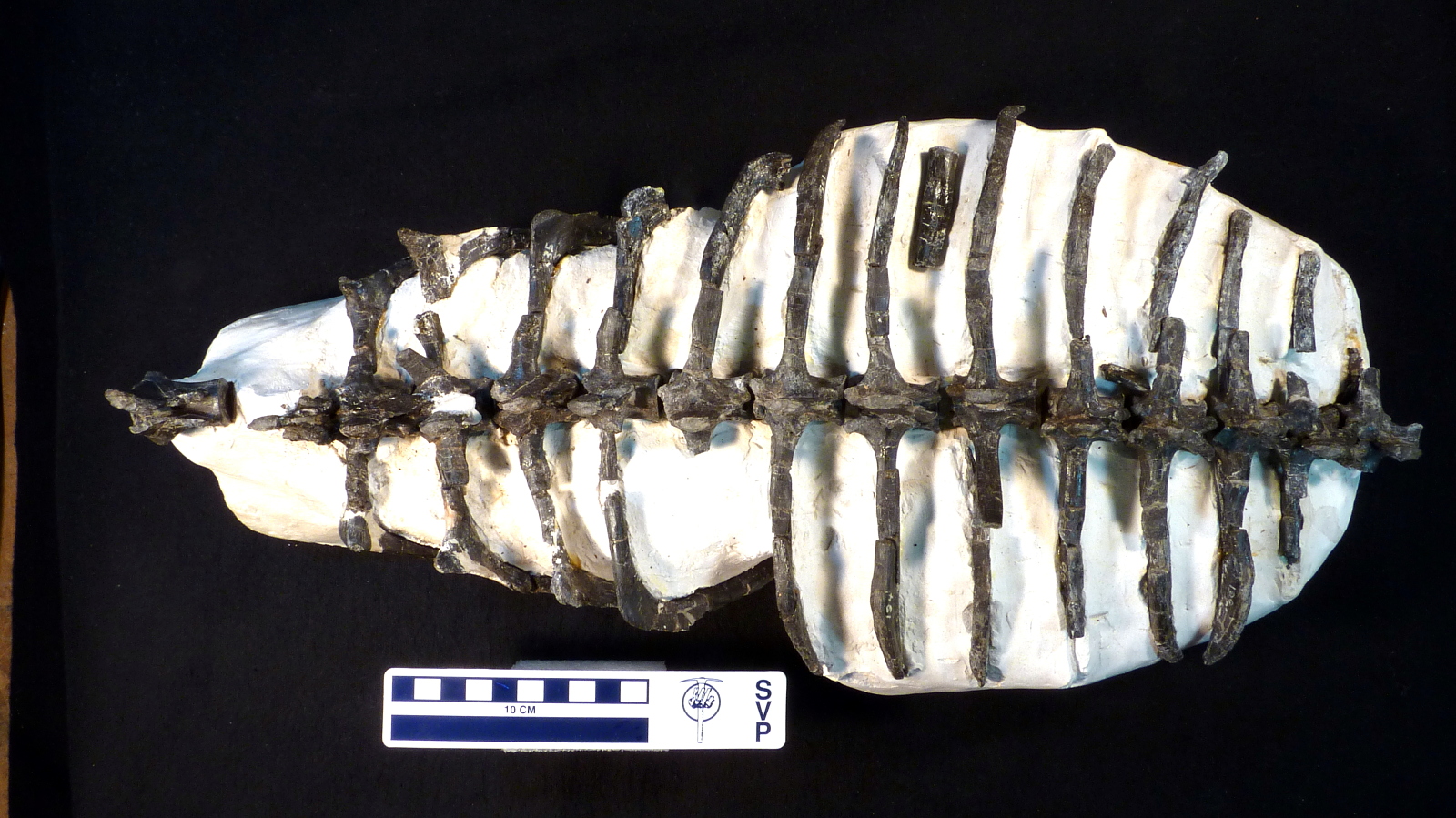
''Doswellia'' is an extinct genus of archosauriform from the Late Triassic of North America. It is the most notable member of the family Doswelliidae, related to the proterochampsids. ''Doswellia'' was a low and heavily built carnivore which lived during the Carnian stage of the Late Triassic. It possesses many unusual features including a wide, flattened head with narrow jaws and a box-like rib cage surrounded by many rows of bony plates. The type species ''Doswellia kaltenbachi'' was named in 1980 from fossils found within the Vinita member of the Doswell Formation (formerly known as the Falling Creek Formation) in Virginia. The formation, which is found in the Taylorsville Basin, is part of the larger Newark Supergroup. ''Doswellia'' is named after Doswell, the town from which much of the taxon's remains have been found. A second species, ''D. sixmilensis,'' was described in 2012 from the Bluewater Creek Formation of the Chinle Group in New Mexico; however, this species w ... [...More Info...] [...Related Items...] OR: [Wikipedia] [Google] [Baidu] |
Autapomorphy
In phylogenetics, an autapomorphy is a distinctive feature, known as a derived trait, that is unique to a given taxon. That is, it is found only in one taxon, but not found in any others or outgroup taxa, not even those most closely related to the focal taxon (which may be a species, family or in general any clade). It can therefore be considered an apomorphy in relation to a single taxon. The word ''autapomorphy'', first introduced in 1950 by German entomologist Willi Hennig, is derived from the Greek words αὐτός, ''autos'' "self"; ἀπό, ''apo'' "away from"; and μορφή, ''morphḗ'' = "shape". Discussion Because autapomorphies are only present in a single taxon, they do not convey information about relationship. Therefore, autapomorphies are not useful to infer phylogenetic relationships. However, autapomorphy, like synapomorphy and plesiomorphy is a relative concept depending on the taxon in question. An autapomorphy at a given level may well be a synapomorphy a ... [...More Info...] [...Related Items...] OR: [Wikipedia] [Google] [Baidu] |
Hypoglossal Nerve
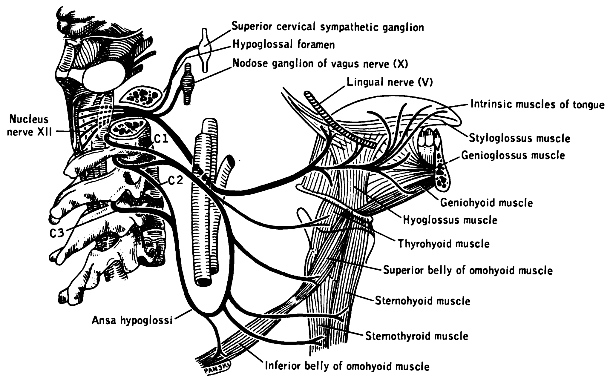
The hypoglossal nerve, also known as the twelfth cranial nerve, cranial nerve XII, or simply CN XII, is a cranial nerve that innervates all the extrinsic and intrinsic muscles of the tongue except for the palatoglossus, which is innervated by the vagus nerve. CN XII is a nerve with a solely motor function. The nerve arises from the hypoglossal nucleus in the medulla as a number of small rootlets, passes through the hypoglossal canal and down through the neck, and eventually passes up again over the tongue muscles it supplies into the tongue. The nerve is involved in controlling tongue movements required for speech and swallowing, including sticking out the tongue and moving it from side to side. Damage to the nerve or the neural pathways which control it can affect the ability of the tongue to move and its appearance, with the most common sources of damage being injury from trauma or surgery, and motor neuron disease. The first recorded description of the nerve is by Her ... [...More Info...] [...Related Items...] OR: [Wikipedia] [Google] [Baidu] |
Quadrate Bone
The quadrate bone is a skull bone in most tetrapods, including amphibians, sauropsids (reptiles, birds), and early synapsids. In most tetrapods, the quadrate bone connects to the quadratojugal and squamosal bones in the skull, and forms upper part of the jaw joint. The lower jaw articulates at the articular bone, located at the rear end of the lower jaw. The quadrate bone forms the lower jaw articulation in all classes except mammals. Evolutionarily, it is derived from the hindmost part of the primitive cartilaginous upper jaw. Function in reptiles In certain extinct reptiles, the variation and stability of the morphology of the quadrate bone has helped paleontologists in the species-level taxonomy and identification of mosasaur squamates and spinosaurine dinosaurs. In some lizards and dinosaurs, the quadrate is articulated at both ends and movable. In snakes, the quadrate bone has become elongated and very mobile, and contributes greatly to their ability to swallow ve ... [...More Info...] [...Related Items...] OR: [Wikipedia] [Google] [Baidu] |
Quadratojugal Bone
The quadratojugal is a skull bone present in many vertebrates, including some living reptiles and amphibians. Anatomy and function In animals with a quadratojugal bone, it is typically found connected to the jugal (cheek) bone from the front and the squamosal bone from above. It is usually positioned at the rear lower corner of the cranium. Many modern tetrapods lack a quadratojugal bone as it has been lost or fused to other bones. Modern examples of tetrapods without a quadratojugal include salamanders, mammals, birds, and squamates (lizards and snakes). In tetrapods with a quadratojugal bone, it often forms a portion of the jaw joint. Developmentally, the quadratojugal bone is a dermal bone in the temporal series, forming the original braincase. The squamosal and quadratojugal bones together form the cheek region and may provide muscular attachments for facial muscles. In reptiles and amphibians In most modern reptiles and amphibians, the quadratojugal is a prominent, str ... [...More Info...] [...Related Items...] OR: [Wikipedia] [Google] [Baidu] |


