|
Retinaculum
A retinaculum (: retinacula) is a band of thickened deep fascia around tendons that holds them in place. It is not part of any muscle and primarily functions to stabilize tendons. The term retinaculum is Neo-Latin, derived from the Latin verb ''retinere'' (to retain). Specific retinacula include: * In the wrist: ** Flexor retinaculum of the hand ** Extensor retinaculum of the hand * In the ankle: ** Flexor retinaculum of foot ** Superior extensor retinaculum of foot ** Inferior extensor retinaculum of foot The inferior extensor retinaculum of the foot (cruciate crural ligament, lower part of anterior annular ligament) is a Y-shaped band placed in front of the ankle-joint, the stem of the Y being attached laterally to the upper surface of the calca ... ** Superior fibular retinaculum ** Inferior fibular retinaculum * In the knee: ** Lateral retinaculum ** Medial patellar retinaculum References {{reflist Tendons ... [...More Info...] [...Related Items...] OR: [Wikipedia] [Google] [Baidu] |
Flexor Retinaculum Of The Hand
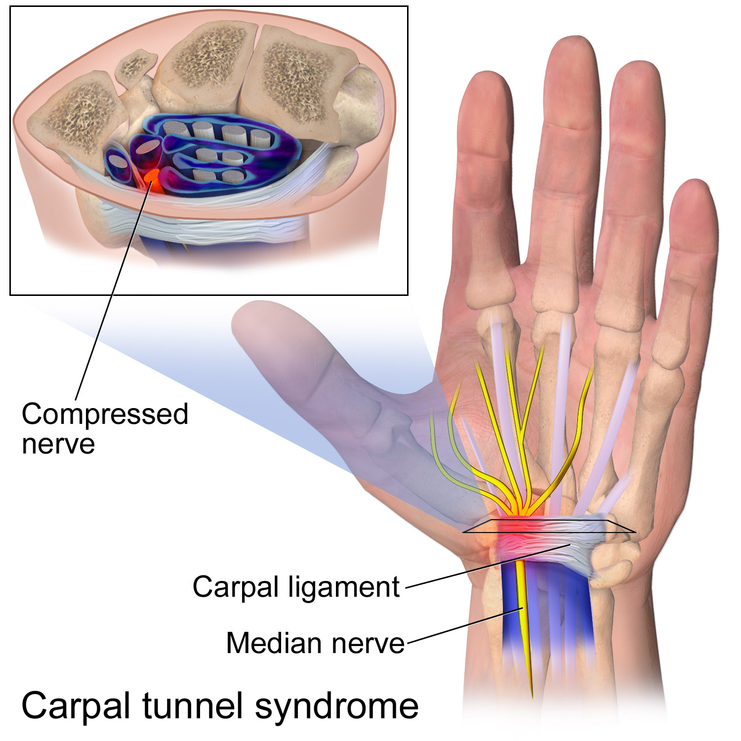
The flexor retinaculum (transverse carpal ligament or anterior annular ligament) is a fibrous band on the palmar side of the hand near the wrist. It arches over the carpal bones of the hands, covering them and forming the carpal tunnel. Structure The flexor retinaculum is a strong, fibrous band that covers the carpal bones on the palmar side of the hand near the wrist. It attaches to the bones near the radius and ulna. On the ulnar side, the flexor retinaculum attaches to the pisiform bone and the hook of the hamate bone. On the radial side, it attaches to the tubercle of the scaphoid bone, and to the medial part of the palmar surface and the ridge of the trapezium bone. The flexor retinaculum is continuous with the palmar carpal ligament, and deeper with the palmar aponeurosis. The ulnar artery and ulnar nerve, and the cutaneous branches of the median and ulnar nerves, pass on top of the flexor retinaculum. On the radial side of the retinaculum is the tendon of the flexor car ... [...More Info...] [...Related Items...] OR: [Wikipedia] [Google] [Baidu] |
Extensor Retinaculum Of The Hand
The extensor retinaculum (dorsal carpal ligament, or posterior annular ligament) is a thickened portion of the antebrachial fascia that holds the tendons of the extensor muscles in place. It is located on the back of the forearm, just proximal to the hand. It is continuous with the palmar carpal ligament (which is located on the anterior side of the forearm). Structure The extensor retinaculum is a strong, fibrous band, extending obliquely downward and medialward across the back of the wrist. It consists of part of the deep fascia of the back of the forearm, strengthened by the addition of some transverse fibers. Relations There are six separate synovial sheaths run beneath the extensor retinaculum: (1st) abductor pollicis longus and extensor pollicis brevis tendons, (2nd) extensor carpi radialis longus and brevis tendons, (3rd) extensor pollicis longus tendon, (4th) extensor digitorium communis and extensor indicis proprius tendons, (5th) extensor digiti minimi tendon and (6 ... [...More Info...] [...Related Items...] OR: [Wikipedia] [Google] [Baidu] |
Superior Extensor Retinaculum Of Foot
The superior extensor retinaculum of the foot (transverse crural ligament) is the upper part of the extensor retinaculum of foot which extends from the ankle to the heelbone. The superior extensor retinaculum binds down the tendons of extensor digitorum longus, extensor hallucis longus, peroneus tertius, and tibialis anterior as they descend on the front of the tibia and fibula; under it are found also the anterior tibial vessels and deep peroneal nerve. It is found on the lateral side of the lower leg, attached laterally to the lower end of the fibula, and medially to the tibia; above it is continuous with the fascia A fascia (; : fasciae or fascias; adjective fascial; ) is a generic term for macroscopic membranous bodily structures. Fasciae are classified as superficial, visceral or deep, and further designated according to their anatomical location. ... of the leg. Additional images File:Gray437.png, Muscles of the front of the leg. See also * Peroneal ... [...More Info...] [...Related Items...] OR: [Wikipedia] [Google] [Baidu] |
Flexor Retinaculum Of Foot
The flexor retinaculum of the foot (laciniate ligament, internal annular ligament) is a strong fibrous band in the foot. Structure The flexor retinaculum of the foot extends from the medial malleolus above, to the calcaneus below. This converts a series of bony grooves into canals for the passage of the tendons of the flexor muscles and the posterior tibial vessels and tibial nerve into the sole of the foot, known as the tarsal tunnel. It is continuous by its upper border with the deep fascia of the leg, and by its lower border with the plantar aponeurosis and the fibers of origin of the abductor hallucis muscle. Enumerated from the medial side, the four canals which it forms transmit the tendons of the tibialis posterior and flexor digitorum longus muscles; the posterior tibial artery and tibial nerve, which run through a broad space beneath the ligament; and lastly, in a canal formed partly by the talus, the tendon of the flexor hallucis longus. Clinical significance ... [...More Info...] [...Related Items...] OR: [Wikipedia] [Google] [Baidu] |
Inferior Extensor Retinaculum Of Foot
The inferior extensor retinaculum of the foot (cruciate crural ligament, lower part of anterior annular ligament) is a Y-shaped band placed in front of the ankle-joint, the stem of the Y being attached laterally to the upper surface of the calcaneus, in front of the depression for the interosseous talocalcaneal ligament The interosseous talocalcaneal ligament forms the chief bond of union between the talus and calcaneus. It is a portion of the united capsules of the talocalcaneonavicular and the talocalcaneal joints, and consists of two partially united layer ...; it is directed medialward as a double layer, one lamina passing in front of, and the other behind, the tendons of the peroneus tertius and extensor digitorum longus. At the medial border of the latter tendon, these two layers join, forming a compartment in which the tendons are enclosed. From the medial extremity of this sheath, the two limbs of the Y diverge: one is directed upward and medialward, to be attach ... [...More Info...] [...Related Items...] OR: [Wikipedia] [Google] [Baidu] |
Lateral Retinaculum
The lateral retinaculum is the fibrous tissue on the lateral (outer) side of the kneecap ( patella). The kneecap has both a medial (on the inner aspect) and a lateral (on the outer side) retinaculum, and these help to support the kneecap in its position in relation to the femur The femur (; : femurs or femora ), or thigh bone is the only long bone, bone in the thigh — the region of the lower limb between the hip and the knee. In many quadrupeds, four-legged animals the femur is the upper bone of the hindleg. The Femo ... bone underneath it. The lateral retinaculum is an extension of the fibrous 'aponeurosis' of the vastus lateralis muscle (itself a part of the quadriceps muscles making up the 'lap'). External links Patello-femoral Pain Syndrome {{Authority control Tissues (biology) ... [...More Info...] [...Related Items...] OR: [Wikipedia] [Google] [Baidu] |
Knee
In humans and other primates, the knee joins the thigh with the leg and consists of two joints: one between the femur and tibia (tibiofemoral joint), and one between the femur and patella (patellofemoral joint). It is the largest joint in the human body. The knee is a modified hinge joint, which permits flexion and extension (kinesiology), extension as well as slight internal and external rotation. The knee is vulnerable to injury and to the development of osteoarthritis. It is often termed a ''compound joint'' having tibiofemoral and patellofemoral components. (The fibular collateral ligament is often considered with tibiofemoral components.) Structure The knee is a modified hinge joint, a type of synovial joint, which is composed of three functional compartments: the patellofemoral articulation, consisting of the patella, or "kneecap", and the patellar groove on the front of the femur through which it slides; and the medial and lateral tibiofemoral articulations linking the ... [...More Info...] [...Related Items...] OR: [Wikipedia] [Google] [Baidu] |
Deep Fascia
Deep fascia (or investing fascia) is a fascia, a layer of dense connective tissue that can surround individual muscles and groups of muscles to separate into fascial compartments. This fibrous connective tissue interpenetrates and surrounds the muscles, bones, nerves, and blood vessels of the body. It provides connection and communication in the form of aponeuroses, ligaments, tendons, retinaculum, retinacula, joint capsules, and septum, septa. The deep fasciae envelop all bone (periosteum and endosteum); cartilage (perichondrium), and blood vessels (tunica externa) and become specialized in muscles (epimysium, perimysium, and endomysium) and nerves (epineurium, perineurium, and endoneurium). The high density of collagen fibers gives the deep fascia its strength and integrity. The amount of elastin fiber determines how much extensibility and resilience it will have. Examples Examples include: * Fascia lata * Deep fascia of leg * Brachial fascia * Buck's fascia Fascial dynamics D ... [...More Info...] [...Related Items...] OR: [Wikipedia] [Google] [Baidu] |
Tendon
A tendon or sinew is a tough band of fibrous connective tissue, dense fibrous connective tissue that connects skeletal muscle, muscle to bone. It sends the mechanical forces of muscle contraction to the skeletal system, while withstanding tension (physics), tension. Tendons, like ligaments, are made of collagen. The difference is that ligaments connect bone to bone, while tendons connect muscle to bone. There are about 4,000 tendons in the adult human body. Structure A tendon is made of dense regular connective tissue, whose main cellular components are special fibroblasts called tendon cells (tenocytes). Tendon cells synthesize the tendon's extracellular matrix, which abounds with densely-packed collagen fibers. The collagen fibers run parallel to each other and are grouped into fascicles. Each fascicle is bound by an endotendineum, which is a delicate loose connective tissue containing thin collagen fibrils and elastic fibers. A set of fascicles is bound by an epitenon, whi ... [...More Info...] [...Related Items...] OR: [Wikipedia] [Google] [Baidu] |
Skeletal Muscle
Skeletal muscle (commonly referred to as muscle) is one of the three types of vertebrate muscle tissue, the others being cardiac muscle and smooth muscle. They are part of the somatic nervous system, voluntary muscular system and typically are attached by tendons to bones of a skeleton. The skeletal muscle cells are much longer than in the other types of muscle tissue, and are also known as ''muscle fibers''. The tissue of a skeletal muscle is striated muscle tissue, striated – having a striped appearance due to the arrangement of the sarcomeres. A skeletal muscle contains multiple muscle fascicle, fascicles – bundles of muscle fibers. Each individual fiber and each muscle is surrounded by a type of connective tissue layer of fascia. Muscle fibers are formed from the cell fusion, fusion of developmental myoblasts in a process known as myogenesis resulting in long multinucleated cells. In these cells, the cell nucleus, nuclei, termed ''myonuclei'', are located along the inside ... [...More Info...] [...Related Items...] OR: [Wikipedia] [Google] [Baidu] |
Neo-Latin
Neo-LatinSidwell, Keith ''Classical Latin-Medieval Latin-Neo Latin'' in ; others, throughout. (also known as New Latin and Modern Latin) is the style of written Latin used in original literary, scholarly, and scientific works, first in Italy during the Italian Renaissance of the fourteenth and fifteenth centuries, and then across northern Europe after about 1500, as a key feature of the humanist movement. Through comparison with Classical Latin, Latin of the Classical period, scholars from Petrarch onwards promoted a standard of Latin closer to that of the ancient Romans, especially in grammar, style, and spelling. The term ''Neo-Latin'' was however coined much later, probably in Germany in the late eighteenth century, as ''Neulatein'', spreading to French and other languages in the nineteenth century. Medieval Latin had diverged quite substantially from the classical standard and saw notable regional variation and influence from vernacular languages. Neo-Latin attempts to retur ... [...More Info...] [...Related Items...] OR: [Wikipedia] [Google] [Baidu] |
Latin Language
Latin ( or ) is a classical language belonging to the Italic languages, Italic branch of the Indo-European languages. Latin was originally spoken by the Latins (Italic tribe), Latins in Latium (now known as Lazio), the lower Tiber area around Rome, Italy. Through the expansion of the Roman Republic, it became the dominant language in the Italian Peninsula and subsequently throughout the Roman Empire. It has greatly influenced many languages, Latin influence in English, including English, having contributed List of Latin words with English derivatives, many words to the English lexicon, particularly after the Christianity in Anglo-Saxon England, Christianization of the Anglo-Saxons and the Norman Conquest. Latin Root (linguistics), roots appear frequently in the technical vocabulary used by fields such as theology, List of Latin and Greek words commonly used in systematic names, the sciences, List of medical roots, suffixes and prefixes, medicine, and List of Latin legal terms ... [...More Info...] [...Related Items...] OR: [Wikipedia] [Google] [Baidu] |


