|
QRS Concordance
Precordial concordance, also known as QRS concordance, is a pattern in which all precordial leads on an electrocardiogram are either positive (positive concordance: all the major spikes point upwards from the baseline) or negative (negative concordance: point downwards). When there is a negative concordance, it almost always represents a life-threatening condition called ventricular tachycardia because there is no other condition that suggests any abnormal conduction from the apex of the heart to the upper parts. However, in positive concordance another rare conditions such as left side accessory pathway In cardiology, an accessory pathway is an additional electrical connection between two parts of the heart. These pathways can lead to abnormal heart rhythms ( arrhythmias) associated with symptoms of palpitations. Some pathways may activate a regi ...s or blocks are also possible. References Electrophysiology Physiology {{med-imaging-stub ... [...More Info...] [...Related Items...] OR: [Wikipedia] [Google] [Baidu] |
Precordial
In anatomy, the precordium or praecordium is the portion of the body over the heart and lower chest. Cited 2 Dec 2009 (UTC) anatomically, it is the area of the anterior chest wall over the . It is therefore usually on the left side, except in conditions like , where the individual's heart is on the right side. In such a case, the precordium is on the right side as well. The ... [...More Info...] [...Related Items...] OR: [Wikipedia] [Google] [Baidu] |
Electrocardiography
Electrocardiography is the process of producing an electrocardiogram (ECG or EKG), a recording of the heart's electrical activity through repeated cardiac cycles. It is an electrogram of the heart which is a graph of voltage versus time of the electrical activity of the heart using electrodes placed on the skin. These electrodes detect the small electrical changes that are a consequence of cardiac muscle depolarization followed by repolarization during each cardiac cycle (heartbeat). Changes in the normal ECG pattern occur in numerous cardiac abnormalities, including: * Cardiac rhythmicity, Cardiac rhythm disturbances, such as atrial fibrillation and ventricular tachycardia; * Inadequate coronary artery blood flow, such as myocardial ischemia and myocardial infarction; * and electrolyte disturbances, such as hypokalemia. Traditionally, "ECG" usually means a 12-lead ECG taken while lying down as discussed below. However, other devices can record the electrical activity of ... [...More Info...] [...Related Items...] OR: [Wikipedia] [Google] [Baidu] |
QRS Complex
The QRS complex is the combination of three of the graphical deflections seen on a typical electrocardiogram (ECG or EKG). It is usually the central and most visually obvious part of the tracing. It corresponds to the depolarization of the right and left ventricles of the heart and contraction of the large ventricular muscles. In adults, the QRS complex normally lasts ; in children it may be shorter. The Q, R, and S waves occur in rapid succession, do not all appear in all leads, and reflect a single event and thus are usually considered together. A Q wave is any downward deflection immediately following the P wave. An R wave follows as an upward deflection, and the S wave is any downward deflection after the R wave. The T wave follows the S wave, and in some cases, an additional U wave follows the T wave. To measure the QRS interval start at the end of the PR interval (or beginning of the Q wave) to the end of the S wave. Normally this interval is 0.08 to 0.10 seconds. W ... [...More Info...] [...Related Items...] OR: [Wikipedia] [Google] [Baidu] |
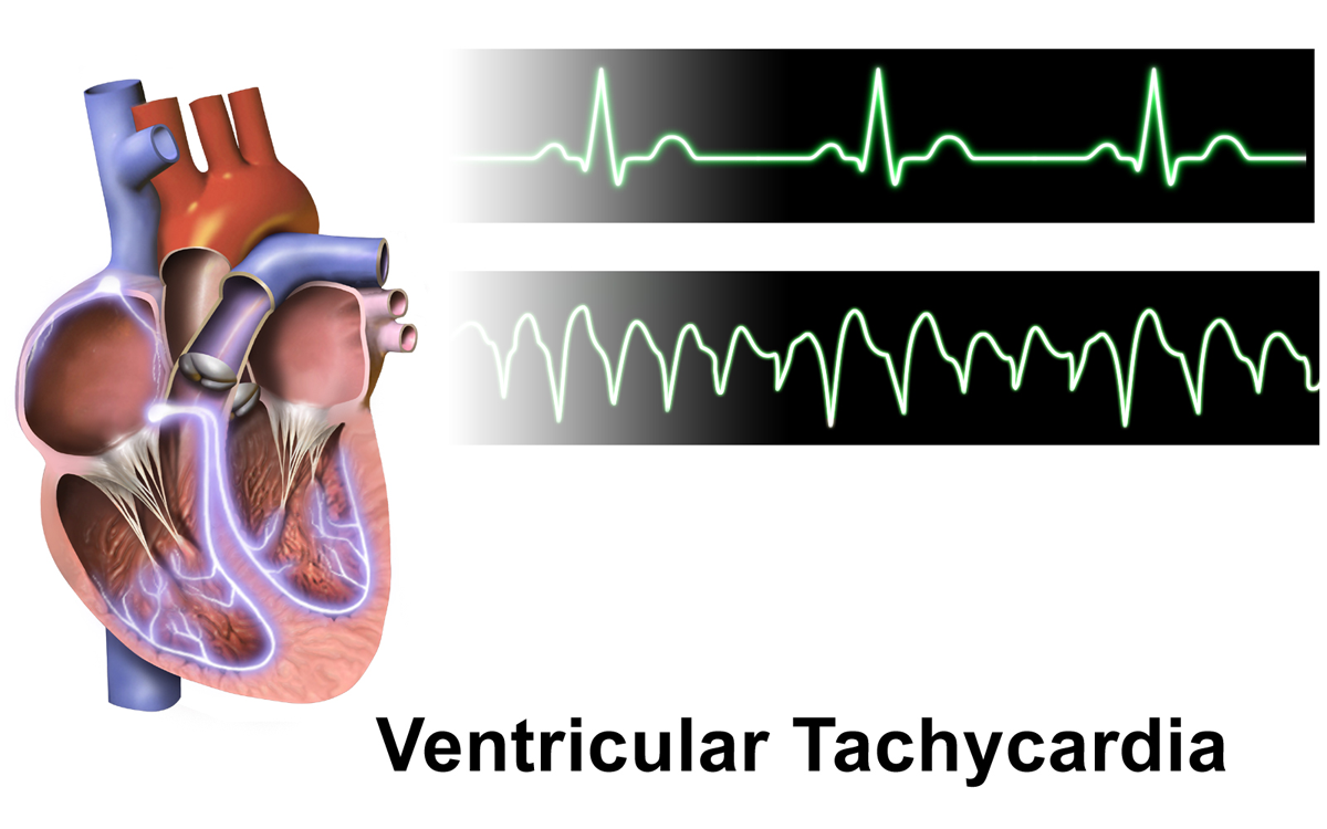
Ventricular Tachycardia
Ventricular tachycardia (V-tach or VT) is a cardiovascular disorder in which fast heart rate occurs in the ventricles of the heart. Although a few seconds of VT may not result in permanent problems, longer periods are dangerous; and multiple episodes over a short period of time are referred to as an electrical storm. Short periods may occur without symptoms, or present with lightheadedness, palpitations, shortness of breath, chest pain, and decreased level of consciousness. Ventricular tachycardia may lead to coma and persistent vegetative state due to lack of blood and oxygen to the brain. Ventricular tachycardia may result in ventricular fibrillation (VF) and turn into cardiac arrest. This conversion of the VT into VF is called the degeneration of the VT. It is found initially in about 7% of people in cardiac arrest. Ventricular tachycardia can occur due to coronary heart disease, aortic stenosis, cardiomyopathy, electrolyte imbalance, or a heart attack. Diagnosis is ... [...More Info...] [...Related Items...] OR: [Wikipedia] [Google] [Baidu] |
Electrical Conduction System Of The Heart
The cardiac conduction system (CCS, also called the electrical conduction system of the heart) transmits the Cardiac action potential, signals generated by the sinoatrial node – the heart's Cardiac pacemaker, pacemaker, to cause the heart muscle to Muscle contraction, contract, and pump blood through the body's circulatory system. The Cardiac pacemaker, pacemaking signal travels through the right atrium to the atrioventricular node, along the bundle of His, and through the bundle branches to Purkinje fibers in the Ventricle (heart), walls of the ventricles. The Purkinje fibers transmit the signals more rapidly to stimulate contraction of the ventricles. The conduction system consists of specialized Cardiomyocyte, heart muscle cells, situated within the myocardium. There is a cardiac skeleton, skeleton of fibrous tissue that surrounds the conduction system which can be seen on an ECG. Dysfunction of the conduction system can cause Heart arrhythmia, irregular heart rhythms includ ... [...More Info...] [...Related Items...] OR: [Wikipedia] [Google] [Baidu] |
Accessory Pathway
In cardiology, an accessory pathway is an additional electrical connection between two parts of the heart. These pathways can lead to abnormal heart rhythms ( arrhythmias) associated with symptoms of palpitations. Some pathways may activate a region of ventricular muscle earlier than would normally occur, referred to as pre-excitation, and this may be seen on an electrocardiogram. The combination of an accessory pathway that causes pre-excitation with arrhythmias is known as Wolff–Parkinson–White syndrome. Accessory pathways are often diagnosed using an electrocardiogram, but characterisation and location of the pathway may require an electrophysiological study. Accessory pathways may not require any treatment, but those causing symptoms may be treated with medication including calcium channel antagonists, beta blockers or flecainide. Alternatively, the electrical conduction through an accessory pathways can be abolished using catheter ablation, potentially offering ... [...More Info...] [...Related Items...] OR: [Wikipedia] [Google] [Baidu] |
Heart Block
Heart block (HB) is a disorder in the heart's rhythm due to a fault in the natural pacemaker. This is caused by an obstruction – a block – in the electrical conduction system of the heart. Sometimes a disorder can be inherited. Despite the severe-sounding name, heart block may cause no symptoms at all in some cases, or occasional missed heartbeats in other cases (which can cause light-headedness, syncope (fainting), and palpitations), or may require the implantation of an artificial pacemaker, depending upon exactly where in the heart conduction is being impaired and how significantly it is affected. Heart block should not be confused with other conditions, which may or may not be co-occurring, relating to the heart and/or other nearby organs that are or can be serious, including angina (heart-related chest pain), heart attack (myocardial infarction), any type of heart failure, cardiogenic shock or other types of shock, different types of abnormal heart rhythms (arrhythm ... [...More Info...] [...Related Items...] OR: [Wikipedia] [Google] [Baidu] |
Electrophysiology
Electrophysiology (from [see the Electron#Etymology, etymology of "electron"]; ; and ) is the branch of physiology that studies the electrical properties of biological cell (biology), cells and tissues. It involves measurements of voltage changes or electric current or manipulations on a wide variety of scales from single ion channel proteins to whole organs like the heart. In neuroscience, it includes measurements of the electrical activity of neurons, and, in particular, action potential activity. Recordings of large-scale electric signals from the nervous system, such as electroencephalography, may also be referred to as electrophysiological recordings. They are useful for electrodiagnostic medicine, electrodiagnosis and monitoring (medicine), monitoring. Definition and scope Classical electrophysiological techniques Principle and mechanisms Electrophysiology is the branch of physiology that pertains broadly to the flow of ions (ion current) in biological tissues and, in p ... [...More Info...] [...Related Items...] OR: [Wikipedia] [Google] [Baidu] |

