|
Pyramidalis Muscle
The pyramidalis muscle is a small triangular muscle, anterior to the rectus abdominis muscle, and contained in the rectus sheath. Structure The pyramidalis muscle is part of the anterior abdominal wall. Inferiorly, the pyramidalis muscle attaches to the pelvis in two places: the pubic symphysis and pubic crest, arising by tendinous fibers from the anterior part of the pubis and the anterior pubic ligament. Superiorly, the fleshy portion of the pyramidalis muscle passes upward, diminishing in size as it ascends, and ends by a pointed extremity which is inserted into the linea alba, midway between the umbilicus and pubis. Nerve supply The pyramidalis muscle is innervated by the ventral portion of T12. Blood supply The inferior and superior epigastric arteries supply blood to the pyramidalis muscle. Variation The pyramidalis muscle is present in 80% of human population. It may be absent on one or both sides; the lower end of the rectus then becomes proportionately increas ... [...More Info...] [...Related Items...] OR: [Wikipedia] [Google] [Baidu] |
Pubic Symphysis
The pubic symphysis (: symphyses) is a secondary cartilaginous joint between the left and right superior rami of the pubis of the hip bones. It is in front of and below the urinary bladder. In males, the suspensory ligament of the penis attaches to the pubic symphysis. In females, the pubic symphysis is attached to the suspensory ligament of the clitoris. In most adults, it can be moved roughly 2 mm and with 1 degree rotation. This increases for women at the time of childbirth. The name comes from the Greek word ''symphysis'', meaning 'growing together'. Structure The pubic symphysis is a nonsynovial amphiarthrodial joint. The width of the pubic symphysis at the front is 3–5 mm greater than its width at the back. This joint is connected by fibrocartilage and may contain a fluid-filled cavity; the center is avascular, possibly due to the nature of the compressive forces passing through this joint, which may lead to harmful vascular disease. The ends of both pubi ... [...More Info...] [...Related Items...] OR: [Wikipedia] [Google] [Baidu] |
Pubic Crest
Medial to the pubic tubercle is the pubic crest, which extends from this process to the medial end of the pubis (bone), pubic bone. It gives attachment to the conjoint tendon, the rectus abdominis, the abdominal external oblique muscle, and the pyramidalis muscle. The point of junction of the crest with the medial border of the bone is called the ''angle'' to it, as well as to the symphysis, the superior crus of the subcutaneous inguinal ring is attached. References External links * Bones of the pelvis Pubis (bone) {{musculoskeletal-stub ... [...More Info...] [...Related Items...] OR: [Wikipedia] [Google] [Baidu] |
Linea Alba (abdomen)
The linea alba (Latin for: white line) is a strong fibrous midline structure of the anterior abdominal wall situated between the two recti abdominis muscles (one on either side). The umbilicus (navel) is present on the linea alba through which foetal umbilical vessels pass before birth. The linea alba is formed by the union of aponeuroses (of the muscles of the anterior abdominal wall) that collectively make up the rectus sheath. The linea alba attaches to the xiphoid process superiorly, and to the pubic symphysis inferiorly. It is narrow inferiorly where the two recti abdominis muscles are in contact with each other posterior to it, and broadens superior-ward from just inferior to the umbilicus. The name means ''white line'' as it is composed mostly of collagen connective tissue, which has a white appearance. In sufficiently muscular individuals, its presence can be seen on the skin, forming the depression between the left and right halves of a " six pack". Function The l ... [...More Info...] [...Related Items...] OR: [Wikipedia] [Google] [Baidu] |
Subcostal Nerve
The subcostal nerve (anterior division of the twelfth thoracic nerve) is a mixed motor and sensory nerve contributing to the lumbar plexus. It runs along the lower border of the twelfth rib, often gives a communicating branch to the first lumbar nerve, and passes under the lateral lumbocostal arch. It then runs in front of the quadratus lumborum, innervates the transversus, and passes forward between it and the abdominal internal oblique to be distributed in the same manner as the lower intercostal nerves. It communicates with the iliohypogastric nerve and the ilioinguinal nerve of the lumbar plexus, and gives a branch to the pyramidalis muscle The pyramidalis muscle is a small triangular muscle, anterior to the rectus abdominis muscle, and contained in the rectus sheath. Structure The pyramidalis muscle is part of the anterior abdominal wall. Inferiorly, the pyramidalis muscle attache ... and the quadratus lumborum muscle. It also gives off a lateral cutaneous branch ... [...More Info...] [...Related Items...] OR: [Wikipedia] [Google] [Baidu] |
Muscle
Muscle is a soft tissue, one of the four basic types of animal tissue. There are three types of muscle tissue in vertebrates: skeletal muscle, cardiac muscle, and smooth muscle. Muscle tissue gives skeletal muscles the ability to muscle contraction, contract. Muscle tissue contains special Muscle contraction, contractile proteins called actin and myosin which interact to cause movement. Among many other muscle proteins, present are two regulatory proteins, troponin and tropomyosin. Muscle is formed during embryonic development, in a process known as myogenesis. Skeletal muscle tissue is striated consisting of elongated, multinucleate muscle cells called muscle fibers, and is responsible for movements of the body. Other tissues in skeletal muscle include tendons and perimysium. Smooth and cardiac muscle contract involuntarily, without conscious intervention. These muscle types may be activated both through the interaction of the central nervous system as well as by innervation ... [...More Info...] [...Related Items...] OR: [Wikipedia] [Google] [Baidu] |
Rectus Abdominis Muscle
The rectus abdominis muscle, () also known as the "abdominal muscle" or simply better known as the "abs", is a pair of segmented skeletal muscle on the ventral aspect of a person's abdomen. The paired muscle is separated at the midline by a band of dense connective tissue called the linea alba, and the connective tissue defining each lateral margin of the rectus abdominus is the linea semilunaris. The muscle extends from the pubic symphysis, pubic crest and pubic tubercle inferiorly, to the xiphoid process and costal cartilages of the 5th–7th ribs superiorly. The rectus abdominis muscle is contained in the rectus sheath, which consists of the aponeuroses of the lateral abdominal muscles. Each rectus abdominus is traversed by bands of connective tissue called the tendinous intersections, which interrupt it into distinct muscle bellies. Structure The rectus abdominis is a very long flat muscle, which extends along the whole length of the front of the abdomen, and is s ... [...More Info...] [...Related Items...] OR: [Wikipedia] [Google] [Baidu] |
Rectus Sheath
The rectus sheath (also called the rectus fascia.) is a tough fibrous compartment formed by the aponeuroses of the transverse abdominal, transverse abdominal muscle, and the internal oblique, internal and external oblique muscles. It contains the Rectus abdominis muscle, rectus abdominis and Pyramidalis muscle, pyramidalis muscles, as well as vessels and nerves. Structure The rectus sheath extends between the inferior costal margin and costal cartilages of ribs 5-7 superiorly, and the pubic crest inferiorly. Studies indicate that all three aponeuroses constituting the rectus sheath are in fact bilaminar. Below the costal margin Superficial/anterior to the anterior layer of the rectus sheath are the following two layers: # Camper's fascia (anterior part of superficial fascia) # Scarpa's fascia (posterior part of the superficial fascia) Deep/posterior posterior layer of the rectus sheath (where present) are the following three layers: # transversalis fascia # extraperitoneal ... [...More Info...] [...Related Items...] OR: [Wikipedia] [Google] [Baidu] |
Pelvis
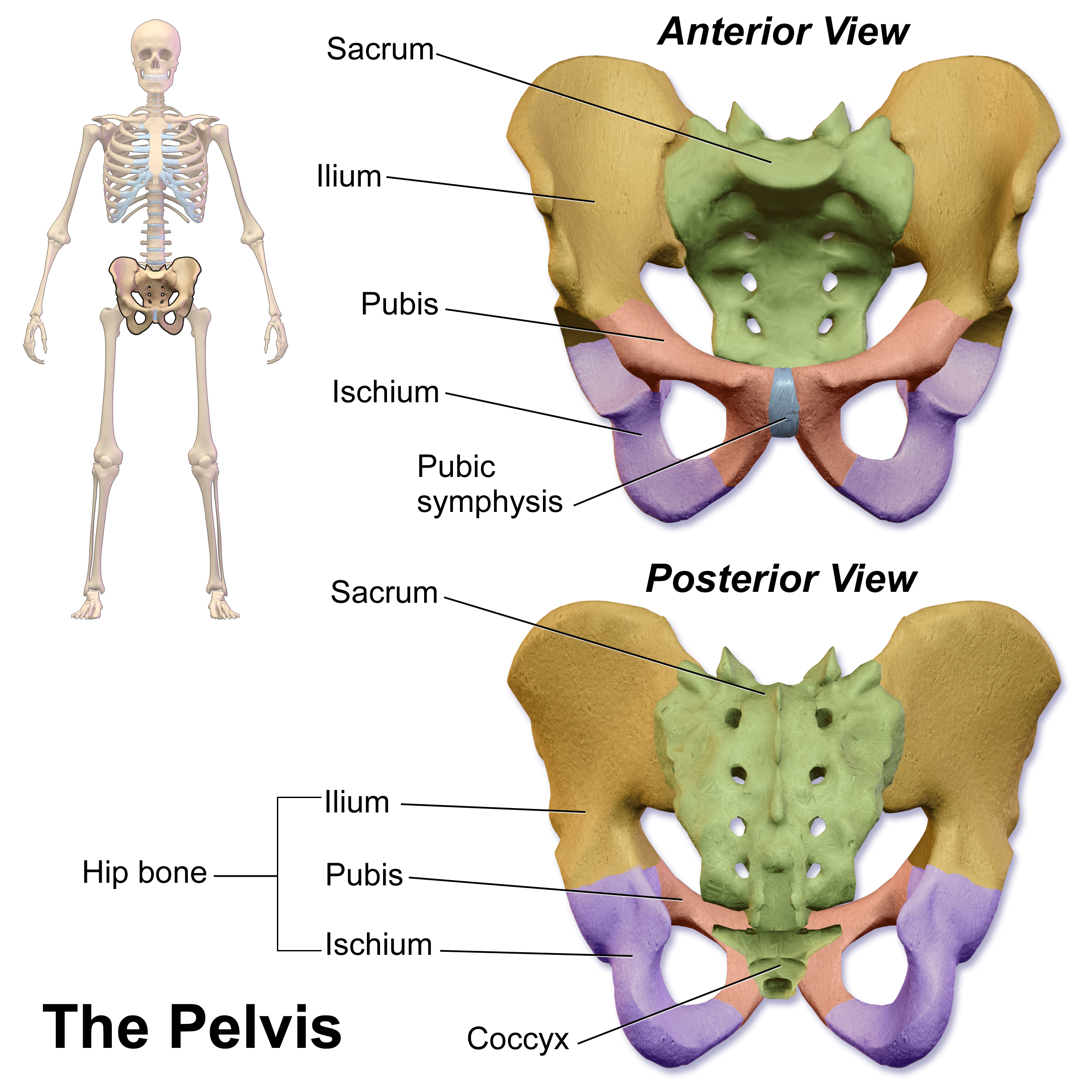
The pelvis (: pelves or pelvises) is the lower part of an Anatomy, anatomical Trunk (anatomy), trunk, between the human abdomen, abdomen and the thighs (sometimes also called pelvic region), together with its embedded skeleton (sometimes also called bony pelvis or pelvic skeleton). The pelvic region of the trunk includes the bony pelvis, the pelvic cavity (the space enclosed by the bony pelvis), the pelvic floor, below the pelvic cavity, and the perineum, below the pelvic floor. The pelvic skeleton is formed in the area of the back, by the sacrum and the coccyx and anteriorly and to the left and right sides, by a pair of hip bones. The two hip bones connect the spine with the lower limbs. They are attached to the sacrum posteriorly, connected to each other anteriorly, and joined with the two femurs at the hip joints. The gap enclosed by the bony pelvis, called the pelvic cavity, is the section of the body underneath the abdomen and mainly consists of the reproductive organs and ... [...More Info...] [...Related Items...] OR: [Wikipedia] [Google] [Baidu] |
Pubic Symphysis
The pubic symphysis (: symphyses) is a secondary cartilaginous joint between the left and right superior rami of the pubis of the hip bones. It is in front of and below the urinary bladder. In males, the suspensory ligament of the penis attaches to the pubic symphysis. In females, the pubic symphysis is attached to the suspensory ligament of the clitoris. In most adults, it can be moved roughly 2 mm and with 1 degree rotation. This increases for women at the time of childbirth. The name comes from the Greek word ''symphysis'', meaning 'growing together'. Structure The pubic symphysis is a nonsynovial amphiarthrodial joint. The width of the pubic symphysis at the front is 3–5 mm greater than its width at the back. This joint is connected by fibrocartilage and may contain a fluid-filled cavity; the center is avascular, possibly due to the nature of the compressive forces passing through this joint, which may lead to harmful vascular disease. The ends of both pubi ... [...More Info...] [...Related Items...] OR: [Wikipedia] [Google] [Baidu] |
Pubis (bone)
In vertebrates, the pubis or pubic bone () forms the lower and anterior part of each side of the hip bone. The pubis is the most forward-facing ( ventral and anterior) of the three bones that make up the hip bone. The left and right pubic bones are each made up of three sections; a superior ramus, an inferior ramus, and a body. Structure The pubic bone is made up of a ''body'', ''superior ramus'', and ''inferior ramus'' (). The left and right coxal bones join at the pubic symphysis. It is covered by a layer of fat – the mons pubis. The pubis is the lower limit of the suprapubic region. In the female, the pubis is anterior to the urethral sponge. Body The body of pubis has: * a superior border or the pubic crest * a pubic tubercle at the lateral end of the pubic crest * three surfaces (anterior, posterior and medial). The body forms the wide, strong, middle and flat part of the pubic bone. The bodies of the left and right pubic bones join at the pubic symphysis. The r ... [...More Info...] [...Related Items...] OR: [Wikipedia] [Google] [Baidu] |
Navel
The navel (clinically known as the umbilicus; : umbilici or umbilicuses; also known as the belly button or tummy button) is a protruding, flat, or hollowed area on the abdomen at the attachment site of the umbilical cord. Structure The umbilicus is used to visually separate the abdomen into quadrants. The umbilicus is a prominent Scar#Umbilical, scar on the abdomen, with its position being relatively consistent among humans. The skin around the waist at the level of the umbilicus is supplied by the tenth thoracic spinal nerve (T10 dermatome (anatomy), dermatome). The umbilicus itself typically lies at a vertical level corresponding to the junction between the L3 and L4 vertebrae, with a normal variation among people between the L3 and L5 vertebrae. Parts of the adult navel include the "umbilical cord remnant" or "umbilical tip", which is the often protruding scar left by the detachment of the umbilical cord. This is located in the center of the navel, sometimes described ... [...More Info...] [...Related Items...] OR: [Wikipedia] [Google] [Baidu] |
Thoracic Spinal Nerve 12
The thoracic spinal nerve 12 (T12) is a spinal nerve of the thoracic segment. It originates from the spinal column from below the thoracic vertebra 12 (T12). It may also be known as the subcostal nerve The subcostal nerve (anterior division of the twelfth thoracic nerve) is a mixed motor and sensory nerve contributing to the lumbar plexus. It runs along the lower border of the twelfth rib, often gives a communicating branch to the first lumbar .... References Spinal nerves {{neuroanatomy-stub ... [...More Info...] [...Related Items...] OR: [Wikipedia] [Google] [Baidu] |


