|
Probainognathid
Probainognathidae is an extinct family of insectivorous cynodonts which lived in what is now South America during the Middle to Late Triassic. The family was established by Alfred Romer in 1973 and includes two genera, ''Probainognathus'' from the Chañares Formation of Argentina and ''Bonacynodon'' from the ''Dinodontosaurus'' Assemblage Zone of Brazil. Probainognathids were closely related to the clade Prozostrodontia, which includes mammals and their close relatives. Description Members of Probainognathidae were relatively small-bodied animals, with skull lengths of around . The temporal region (area behind the eye sockets) was rather wide, and longer than the snout. The secondary palate was well-developed compared to earlier cynodonts, and the portion made up by the maxilla was larger than the part made from the palatine bone. The dentary, the tooth-bearing bone of the lower jaw, was quite tall when seen from the side. The mandibular symphysis (the joint between the two h ... [...More Info...] [...Related Items...] OR: [Wikipedia] [Google] [Baidu] |
Bonacynodon
''Bonacynodon'' is an extinct genus of cynodonts that lived in what is now southern Brazil during the Triassic period (Ladinian–Carnian ages). The genus is monotypic, containing only the type species ''Bonacynodon schultzi''. ''B. schultzi'' is known from two specimens, consisting of two partial skulls and some badly preserved parts of the postcranium. Both specimens were recovered from the Pinheiros-Chiniquá Sequence, part of the Santa Maria Supersequence of the Paraná Basin. This sequence preserves a faunal association known as the Dinodontosaurus Assemblage Zone, ''Dinodontosaurus'' Assemblage Zone, which contains numerous other species of cynodonts, dicynodonts and reptiles. ''Bonacynodon'' was a small, likely insectivorous cynodont, whose length has been estimated at around . It can be distinguished from other cynodonts by its large, serrated (saw-like) canine teeth. Together with the genus ''Probainognathus'' of Argentina, it made up the family Probainognathidae, one of ... [...More Info...] [...Related Items...] OR: [Wikipedia] [Google] [Baidu] |
Probainognathus
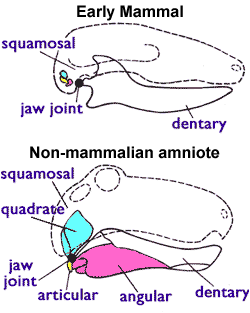
''Probainognathus'' meaning “progressive jaw” is an extinct genus of cynodonts that lived around 235 to 221.5 million years ago, during the Late Triassic in what is now Argentina. Together with the genus ''Bonacynodon'' from Brazil, ''Probainognathus'' forms the family Probainognathidae. ''Probainognathus'' was a relatively small, carnivorous or insectivorous cynodont. Like all cynodonts, it was a relative of mammals, and it possessed several mammal-like features. Like some other cynodonts, ''Probainognathus'' had a double jaw joint, which not only included the quadrate bone, quadrate and articular bones like in more basal synapsids, but also the squamosal and surangular bones. A joint between the Dentary bone, dentary and squamosal bones, as seen in modern mammals, was however absent in ''Probainognathus''. Discovery and naming The first specimens of ''Probainognathus jenseni'' were discovered in the Chañares Formation in La Rioja Province, Argentina, La Rioja Province, ... [...More Info...] [...Related Items...] OR: [Wikipedia] [Google] [Baidu] |
Bonacynodon Witout Visible Ears
''Bonacynodon'' is an extinct genus of cynodonts that lived in what is now southern Brazil during the Triassic period (Ladinian–Carnian ages). The genus is monotypic, containing only the type species ''Bonacynodon schultzi''. ''B. schultzi'' is known from two specimens, consisting of two partial skulls and some badly preserved parts of the postcranium. Both specimens were recovered from the Pinheiros-Chiniquá Sequence, part of the Santa Maria Supersequence of the Paraná Basin. This sequence preserves a faunal association known as the ''Dinodontosaurus'' Assemblage Zone, which contains numerous other species of cynodonts, dicynodonts and reptiles. ''Bonacynodon'' was a small, likely insectivorous cynodont, whose length has been estimated at around . It can be distinguished from other cynodonts by its large, serrated (saw-like) canine teeth. Together with the genus ''Probainognathus'' of Argentina, it made up the family Probainognathidae, one of the earliest-diverging lineages ... [...More Info...] [...Related Items...] OR: [Wikipedia] [Google] [Baidu] |
Chañares Formation
The Chañares Formation is a Carnian-age geologic formation of the Ischigualasto-Villa Unión Basin, located in La Rioja Province, Argentina. It is characterized by drab-colored fine-grained volcaniclastic claystones, siltstones, and sandstones which were deposited in a fluvial to lacustrine environment. The formation is most prominently exposed within Talampaya National Park, a UNESCO World Heritage Site within La Rioja Province. The Chañares formation is the lowermost stratigraphic unit of the Agua de la Peña Group, overlying the Tarjados Formation of the Paganzo Group, and underlying the Los Rastros Formation. Though previously considered Ladinian in age, U-Pb dating has determined that most or all of the Chañares Formation dates to the early Carnian stage of the Late Triassic.Kent et al, 2014, p.7959 The Chañares Formation has provided a diverse and well-preserved faunal assemblage which has been studied intensively since the 1960s. The most common reptiles were ... [...More Info...] [...Related Items...] OR: [Wikipedia] [Google] [Baidu] |
Dinodontosaurus Assemblage Zone
The Santa Maria Formation is a sedimentary rock formation found in Rio Grande do Sul, Brazil. It is primarily Carnian in age (Late Triassic), and is notable for its fossils of cynodonts, "rauisuchian" pseudosuchians, and early dinosaurs and other dinosauromorphs, including the herrerasaurid ''Staurikosaurus'', the basal sauropodomorphs ''Buriolestes'' and ''Saturnalia,'' and the lagerpetid ''Ixalerpeton''. The formation is named after the city of Santa Maria in the central region of Rio Grande do Sul, where outcrops were first studied. The Santa Maria Formation makes up the majority of the Santa Maria Supersequence, which extends through the entire Late Triassic. The Santa Maria Supersequence is divided into four geological sequences, separated from each other by short unconformities. The first two of these sequences (Pinheiros-Chiniquá and Santa Cruz sequences) lie entirely within the Santa Maria Formation, while the third (the Candelária sequence) is shared with the overlyin ... [...More Info...] [...Related Items...] OR: [Wikipedia] [Google] [Baidu] |
Articular Bone
The articular bone is part of the lower jaw of most vertebrates, including most jawed fish, amphibians, birds and various kinds of reptiles, as well as ancestral mammals. Anatomy In most vertebrates, the articular bone is connected to two other lower jaw bones, the suprangular and the angular. Developmentally, it originates from the embryonic mandibular cartilage. The most caudal portion of the mandibular cartilage ossifies to form the articular bone, while the remainder of the mandibular cartilage either remains cartilaginous or disappears. In snakes In snakes, the articular, surangular, and prearticular bones have fused to form the compound bone. The mandible is suspended from the quadrate bone and articulates at this compound bone. Function In amphibians and reptiles In most tetrapods, the articular bone forms the lower portion of the jaw joint. The upper jaw articulates at the quadrate bone. In mammals In mammals, the articular bone evolves to form the malleu ... [...More Info...] [...Related Items...] OR: [Wikipedia] [Google] [Baidu] |
Squamosal
The squamosal is a skull bone found in most reptiles, amphibians, and birds. In fishes, it is also called the pterotic bone. In most tetrapods, the squamosal and quadratojugal bones form the cheek series of the skull. The bone forms an ancestral component of the dermal roof and is typically thin compared to other skull bones. The squamosal bone lies ventral to the temporal series and otic notch, and is bordered anteriorly by the postorbital. Posteriorly, the squamosal articulates with the quadrate and pterygoid bones. The squamosal is bordered anteroventrally by the jugal and ventrally by the quadratojugal. Function in reptiles In reptiles, the quadrate and articular bones of the skull articulate to form the jaw joint. The squamosal bone lies anterior to the quadrate bone. Anatomy in synapsids Non-mammalian synapsids In non-mammalian synapsids, the jaw is composed of four bony elements and referred to as a quadro-articular jaw because the joint is between the ... [...More Info...] [...Related Items...] OR: [Wikipedia] [Google] [Baidu] |
Surangular
The surangular or suprangular is a jaw bone found in most land vertebrates, except mammals. Usually in the back of the jaw, on the upper edge, it is connected to all other jaw bones: dentary, angular bone, angular, splenial and articular. It is often a muscle attachment site. It has been noted in dinosaurs. References Skull bones {{Vertebrate anatomy-stub ... [...More Info...] [...Related Items...] OR: [Wikipedia] [Google] [Baidu] |
Canine Teeth
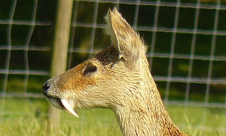
In mammalian oral anatomy, the canine teeth, also called cuspids, dogteeth, eye teeth, vampire teeth, or fangs, are the relatively long, pointed teeth. In the context of the upper jaw, they are also known as '' fangs''. They can appear more flattened, however, causing them to resemble incisors and leading them to be called ''incisiform''. They developed and are used primarily for firmly holding food in order to tear it apart, and occasionally as weapons. They are often the largest teeth in a mammal's mouth. Individuals of most species that develop them normally have four, two in the upper jaw and two in the lower, separated within each jaw by incisors; humans and dogs are examples. In most species, canines are the anterior-most teeth in the maxillary bone. The four canines in humans are the two upper maxillary canines and the two lower mandibular canines. They are specially prominent in dogs (Canidae), hence the name. Details There are generally four canine teeth: two in t ... [...More Info...] [...Related Items...] OR: [Wikipedia] [Google] [Baidu] |
Serrated
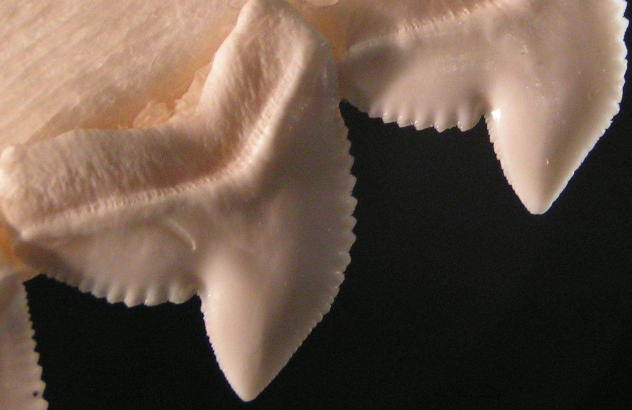
Serration is a saw-like appearance or a row of sharp or tooth-like projections. A serrated cutting edge has many small points of contact with the material being cut. By having less contact area than a smooth blade or other edge, the applied pressure at each point of contact is greater, and the points of contact are at a sharper angle to the material being cut. This causes a cutting action that involves many small splits in the surface of the material being cut, which cumulatively serve to cut the material along the line of the blade. Serration in nature In nature, serration is commonly seen in the cutting edge on the teeth of some species, usually sharks. However, it also appears on non-cutting surfaces, for example, in botany where a toothed leaf margin or other plant part, such as the edge of a carnation petal, is described as being serrated. A serrated leaf edge may reduce the force of wind and other natural elements. Probably the largest serrations on Earth occur on the s ... [...More Info...] [...Related Items...] OR: [Wikipedia] [Google] [Baidu] |
Mandibular Symphysis
In human anatomy, the facial skeleton of the skull the external surface of the mandible is marked in the median line by a faint ridge, indicating the mandibular symphysis (Latin: ''symphysis menti'') or line of junction where the two lateral halves of the mandible typically fuse in the first year of life (6–9 months after birth). It is not a true symphysis as there is no cartilage between the two sides of the mandible. This ridge divides below and encloses a triangular eminence, the mental protuberance, the base of which is depressed in the center but raised on either side to form the mental tubercle. The lowest (most inferior) end of the mandibular symphysis — the point of the chin — is called the "menton". It serves as the origin for the geniohyoid and the genioglossus muscles. Other animals Solitary mammalian carnivores that rely on a powerful canine bite to subdue their prey have a strong mandibular symphysis, while pack hunters delivering shallow bites have a ... [...More Info...] [...Related Items...] OR: [Wikipedia] [Google] [Baidu] |
Postcanine
Cheek teeth or postcanines comprise the molar and premolar teeth in mammals. Cheek teeth are multicuspidate (having many folds or tubercles). Mammals have multicuspidate molars (three in placentals, four in marsupials, in each jaw quadrant) and premolars situated between canines and molars whose shape and number varies considerably among particular groups. For example, many modern Carnivora possess carnassials, or secodont teeth. This scissor-like pairing of the last upper premolar and first lower molar is adapted for shearing meat. In contrast, the cheek teeth of deer and cattle are selenodont. Viewed from the side, these teeth have a series of triangular cusps or ridges, enabling the ruminants' sideways jaw motions to break down tough vegetable matter. Cheek teeth are sometimes separated from the incisors by a gap called a diastema. Cheek teeth in reptiles are much simpler as compared to mammals. Roles and significance Apart from helping grind the food to properly reduce the s ... [...More Info...] [...Related Items...] OR: [Wikipedia] [Google] [Baidu] |


