|
Pringle Maneuver
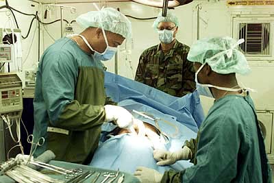
The Pringle manoeuvre is a surgical technique used in some abdominal operations and in liver trauma. The hepatoduodenal ligament is clamped either with a surgical tool called a haemostat, an umbilical tape or by hand. This limits blood inflow through the hepatic artery and the portal vein, controlling bleeding from the liver. It was first published by and named after James Hogarth Pringle in 1908. Uses The Pringle manoeuvre is used during liver surgery and in some cases of severe liver trauma to minimize blood loss. For short durations of use, it is very effective at reducing intraoperative blood loss. The Pringle manoeuvre is applied during closure of a vena cava injury when an atriocaval shunt is placed. Limits The Pringle manoeuvre is more effective in preventing blood loss during liver surgery if central venous pressure is maintained at 5 mmHg or lower. This is due to the fact that Pringle manoeuver technique aims at controlling the blood inflow into the liver, hav ... [...More Info...] [...Related Items...] OR: [Wikipedia] [Google] [Baidu] |
General Surgery
General surgery is a Surgical specialties, surgical specialty that focuses on alimentary canal and Abdomen, abdominal contents including the esophagus, stomach, small intestine, large intestine, liver, pancreas, gallbladder, Appendix (anatomy), appendix and bile ducts, and often the thyroid gland. General surgeons also deal with diseases involving the human skin, skin, breast, soft tissue, trauma (medicine), trauma, peripheral artery disease and hernias and perform endoscopic as such as gastroscopy, colonoscopy and laparoscopic procedures. Scope General surgeons may sub-specialise into one or more of the following disciplines: Trauma surgery In many parts of the world including North America, Australia and the UK, United Kingdom, the overall responsibility for major trauma, trauma care falls under the auspices of general surgery. Some general surgeons obtain advanced training in this field (most commonly surgical critical care) and specialty certification surgical critical ... [...More Info...] [...Related Items...] OR: [Wikipedia] [Google] [Baidu] |
Bleeding
Bleeding, hemorrhage, haemorrhage or blood loss, is blood escaping from the circulatory system from damaged blood vessels. Bleeding can occur internally, or externally either through a natural opening such as the mouth, nose, ear, urethra, vagina, or anus, or through a puncture in the skin. Hypovolemia is a massive decrease in blood volume, and death by excessive loss of blood is referred to as exsanguination. Typically, a healthy person can endure a loss of 10–15% of the total blood volume without serious medical difficulties (by comparison, blood donation typically takes 8–10% of the donor's blood volume). The stopping or controlling of bleeding is called hemostasis and is an important part of both first aid and surgery. Types * Upper head ** Intracranial hemorrhage — bleeding in the skull. ** Cerebral hemorrhage — a type of intracranial hemorrhage, bleeding within the brain tissue itself. ** Intracerebral hemorrhage — bleeding in the brain caused by ... [...More Info...] [...Related Items...] OR: [Wikipedia] [Google] [Baidu] |
Hemostat
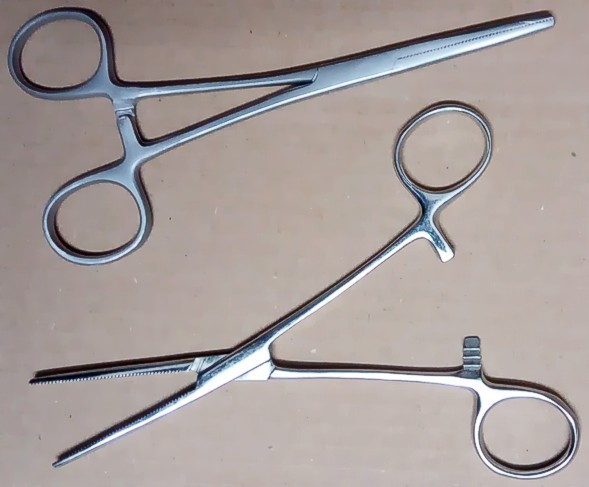
A hemostat (also called a hemostatic clamp; arterial forceps; and pean, after Jules-Émile Péan) is a tool used to control bleeding during surgery. Similar in design to both pliers and scissors, it is used to clamp exposed blood vessels shut. Hemostats belong to a group of instruments that pivot (similar to scissors, and including needle holders, tissue holders, and some other clamps) where the structure of the tip determines the tool's function. A hemostat has handles that can be held in place by their locking mechanism, which usually is a series of interlocking teeth, a few on each handle, that allow the user to adjust the clamping force of the pliers. When the tips are locked together, the force between them is about 40 N (9 lbf). Often in the first phases of surgery, the incision is lined with hemostats on blood vessels that are awaiting ligation. History The earliest known drawing of a pivoting surgical instrument dates from 1500 B.C. and is on a tomb at Thebes, Eg ... [...More Info...] [...Related Items...] OR: [Wikipedia] [Google] [Baidu] |
Blood
Blood is a body fluid in the circulatory system of humans and other vertebrates that delivers necessary substances such as nutrients and oxygen to the cells, and transports metabolic waste products away from those same cells. Blood is composed of blood cells suspended in blood plasma. Plasma, which constitutes 55% of blood fluid, is mostly water (92% by volume), and contains proteins, glucose, mineral ions, and hormones. The blood cells are mainly red blood cells (erythrocytes), white blood cells (leukocytes), and (in mammals) platelets (thrombocytes). The most abundant cells are red blood cells. These contain hemoglobin, which facilitates oxygen transport by reversibly binding to it, increasing its solubility. Jawed vertebrates have an adaptive immune system, based largely on white blood cells. White blood cells help to resist infections and parasites. Platelets are important in the clotting of blood. Blood is circulated around the body through blood vessels by the ... [...More Info...] [...Related Items...] OR: [Wikipedia] [Google] [Baidu] |
Lesser Omentum
The lesser omentum (small omentum or gastrohepatic omentum) is the double layer of peritoneum that extends from the liver to the lesser curvature of the stomach, and to the first part of the duodenum. The lesser omentum is usually divided into these two connecting parts: the hepatogastric ligament, and the hepatoduodenal ligament. Structure The lesser omentum is extremely thin, and is continuous with the two layers of peritoneum which cover respectively the antero-superior and postero-inferior surfaces of the stomach and first part of the duodenum. When these two layers reach the lesser curvature of the stomach and the upper border of the duodenum, they join and ascend as a double fold to the porta hepatis. To the left of the porta, the fold is attached to the bottom of the fossa for the ductus venosus, along which it is carried to the diaphragm, where the two layers separate to embrace the end of the esophagus. At the right border of the lesser omentum, the two layer ... [...More Info...] [...Related Items...] OR: [Wikipedia] [Google] [Baidu] |
Reperfusion Injury
Reperfusion injury, sometimes called ischemia-reperfusion injury (IRI) or reoxygenation injury, is the tissue damage caused when blood supply returns to tissue ('' re-'' + ''perfusion'') after a period of ischemia or lack of oxygen (anoxia or hypoxia). The absence of oxygen and nutrients from blood during the ischemic period creates a condition in which the restoration of circulation results in inflammation and oxidative damage through the induction of oxidative stress rather than (or along with) restoration of normal function. Reperfusion injury is distinct from cerebral hyperperfusion syndrome (sometimes called "Reperfusion syndrome"), a state of abnormal cerebral vasodilation. Mechanisms Reperfusion of ischemic tissues is often associated with microvascular injury, particularly due to increased permeability of capillaries and arterioles that lead to an increase of diffusion and fluid filtration across the tissues. Activated endothelial cells produce more reactive oxygen sp ... [...More Info...] [...Related Items...] OR: [Wikipedia] [Google] [Baidu] |
Hepatic Vein
In human anatomy, the hepatic veins are the veins that drain venous blood from the liver into the inferior vena cava (as opposed to the hepatic portal vein which conveys blood from the gastrointestinal organs to the liver). There are usually three large upper hepatic veins draining from the left, middle, and right parts of the liver, as well as a number (6-20) of lower hepatic veins. All hepatic veins are valveless. Structure All the hepatic veins drain into the inferior vena cava. The hepatic veins are divided into an upper and a lower group. Upper group The upper group consists of three hepatic veins - the right, middle, and left hepatic veins - draining the central veins from the right, middle, and left regions of the liver and are larger than the lower group of veins. The veins of the upper group drain into the suprahepatic part of the inferior vena cava (i.e. part superior to the liver). Right hepatic vein The right hepatic vein is the longest and largest of all the he ... [...More Info...] [...Related Items...] OR: [Wikipedia] [Google] [Baidu] |
Inferior Vena Cava
The inferior vena cava is a large vein that carries the deoxygenated blood from the lower and middle body into the right atrium of the heart. It is formed by the joining of the right and the left common iliac veins, usually at the level of the fifth Lumbar vertebrae, lumbar vertebra. The inferior vena cava is the lower ("anatomical terms of location#Superior and inferior, inferior") of the two venae cavae, the two large veins that carry deoxygenated blood from the body to the right atrium of the heart: the inferior vena cava carries blood from the lower half of the body whilst the superior vena cava carries blood from the upper half of the body. Together, the venae cavae (in addition to the coronary sinus, which carries blood from the muscle of the heart itself) form the venous counterparts of the aorta. It is a large retroperitoneal vein that lies Posterior (anatomy), posterior to the abdominal cavity and runs along the right side of the vertebral column. It enters the right a ... [...More Info...] [...Related Items...] OR: [Wikipedia] [Google] [Baidu] |
Millimetre Of Mercury
A millimetre of mercury is a manometric unit of pressure, formerly defined as the extra pressure generated by a column of mercury one millimetre high. Currently, it is defined as exactly , or approximately 1 torr = atmosphere = pascals.Council Directive 80/181/EEC of 20 December 1979 on the approximation of the laws of the Member States relating to units of measurement and on the repeal of Directive 71/354/EEC of the It is denoted mmHg or mm Hg ... [...More Info...] [...Related Items...] OR: [Wikipedia] [Google] [Baidu] |
Atriocaval Shunt
An atriocaval shunt (ACS) is an intraoperative surgical shunt between the atrium of the heart and the inferior vena cava. It is used during the repair of larger juxtahepatic (next to the liver) vascular injuries such as an injury to the local vena cava. Injuries to the inferior vena cava are challenging, those behind the liver being the most difficult to repair. __TOC__ Procedure and results Injury to the vena cava adjacent to the liver and/or connected hepatic veins leads to often fatal bleeding. Patients may be admitted already in hemorrhagic shock with death occurring even before the bleeding area is localized. Surgically, the area is difficult to access as it is largely covered by the liver. In 1968 Schrock et al. reported on the first use of the ACS. They devised this approach after observing that above the renal veins only the right adrenal vein, the hepatic veins, and the inferior phrenic veins enter the inferior vena cava. A 1988 review by Burch et al. analyzed their exper ... [...More Info...] [...Related Items...] OR: [Wikipedia] [Google] [Baidu] |
Abdominal Surgery
The term abdominal surgery broadly covers surgical procedures that involve opening the abdomen (laparotomy). Surgery of each abdominal organ is dealt with separately in connection with the description of that organ (see stomach, kidney, liver, etc.) Diseases affecting the abdominal cavity are dealt with generally under their own names. Types The most common abdominal surgeries are described below. *Appendectomy: surgical opening of the abdominal cavity and removal of the appendix. Typically performed as definitive treatment for appendicitis, although sometimes the appendix is prophylactically removed incidental to another abdominal procedure. *Caesarean section (also known as C-section): a surgical procedure in which one or more incisions are made through a mother's abdomen (laparotomy) and uterus ( hysterotomy) to deliver one or more babies, or, rarely, to remove a dead fetus. * Inguinal hernia surgery: the repair of an inguinal hernia. *Exploratory laparotomy: the openin ... [...More Info...] [...Related Items...] OR: [Wikipedia] [Google] [Baidu] |
Chronic Venous Insufficiency
Chronic venous insufficiency (CVI) is a medical condition characterized by blood pooling in the veins, leading to increased pressure and strain on the vein walls. The most common cause of CVI is superficial venous reflux, which often results in the formation of varicose veins, a treatable condition. Since functional venous valves are necessary to facilitate efficient Venous return, blood return from the lower extremities, CVI primarily affects the legs. When impaired vein function leads to significant symptoms such as oedema (swelling) or venous ulcer formation, the condition is referred to as chronic venous disease. It is also known as ''chronic peripheral venous insufficiency'' and should not be confused with post-thrombotic syndrome, a separate condition caused by damage to the deep veins following deep vein thrombosis (DVT). Most cases of CVI can be managed or improved through treatments targeting the superficial venous system or stenting the deep venous system. For instanc ... [...More Info...] [...Related Items...] OR: [Wikipedia] [Google] [Baidu] |


