|
Potter Sequence
Potter sequence is the atypical physical appearance of a baby due to oligohydramnios experienced when in the uterus. It includes clubbed feet, pulmonary hypoplasia and cranial anomalies related to the oligohydramnios. Oligohydramnios is the decrease in amniotic fluid volume sufficient to cause deformations in morphogenesis of the baby. Oligohydramnios is the cause of Potter sequence, but there are many things that can lead to oligohydramnios. It can be caused by renal diseases such as bilateral renal agenesis (BRA), atresia of the ureter or urethra causing obstruction of the urinary tract, polycystic or multicystic kidney diseases, renal hypoplasia, amniotic rupture, toxemia, or uteroplacental insufficiency from maternal hypertension. The term ''Potter sequence'' was initially intended to only refer to cases caused by BRA; however, it is now commonly used by many clinicians and researchers to refer to any case that presents with oligohydramnios or anhydramnios regardless of ... [...More Info...] [...Related Items...] OR: [Wikipedia] [Google] [Baidu] |
Oligohydramnios
Oligohydramnios is a medical condition in pregnancy characterized by a deficiency of amniotic fluid, the fluid that surrounds the fetus in the abdomen, in the amniotic sac. The limiting case is anhydramnios, where there is a complete absence of amniotic fluid. It is typically diagnosed by ultrasound when the amniotic fluid index (AFI) measures less than 5 cm or when the single deepest pocket (SDP) of amniotic fluid measures less than 2 cm. Amniotic fluid is necessary to allow for normal fetal movement, lung development, and cushioning from uterine compression. Low amniotic fluid can be attributed to a maternal, fetal, placental or idiopathic cause and can result in poor fetal outcomes including death. The prognosis of the fetus is dependent on the etiology, gestational age at diagnosis, and the severity of the oligohydramnios. The opposite of oligohydramnios is polyhydramnios, or an excess of amniotic fluid. Background Amniotic fluid is a clear, watery substance that surrounds ... [...More Info...] [...Related Items...] OR: [Wikipedia] [Google] [Baidu] |
Autosomal Recessive Polycystic Kidney Disease
Autosomal recessive polycystic kidney disease (ARPKD) is the recessive form of polycystic kidney disease. It is associated with a group of congenital fibrocystic syndromes. Mutations in the '' PKHD1'' (chromosomal locus 6p12.2) cause ARPKD. Signs and symptoms Symptoms and signs include abdominal discomfort, polyuria, polydipsia, incidental discovery of hypertension, and abdominal mass. The classic presentation for ARPKD is systemic hypertension with progression to end-stage kidney disease (ESKD) by the age of 15. In a typical presentation, a small number of individuals with ARPKD live to adulthood with some kidney function; but with significant deterioration in liver function. This outcome is postulated to result from expression of the polycystic kidney and hepatic disease gene PKHD1, which is located on chromosome 6p. In severe cases, a fetus will present with oligohydramnios and as a result, may present with Potter sequence. Genetics The cause of ARPKD is linked to mutations ... [...More Info...] [...Related Items...] OR: [Wikipedia] [Google] [Baidu] |
Urinary Bladder
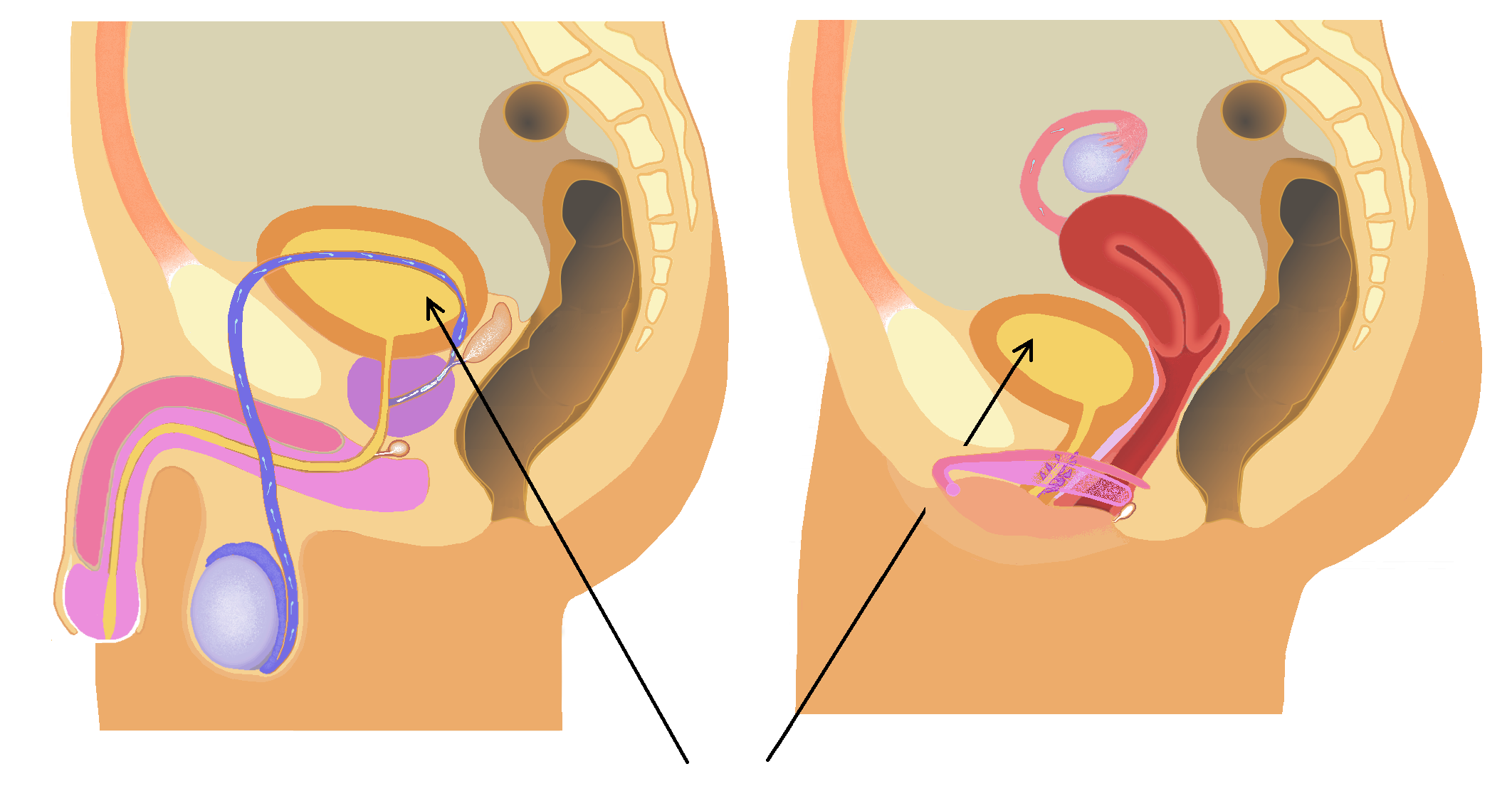
The bladder () is a hollow organ in humans and other vertebrates that stores urine from the Kidney (vertebrates), kidneys. In placental mammals, urine enters the bladder via the ureters and exits via the urethra during urination. In humans, the bladder is a distensible organ that sits on the pelvic floor. The typical adult human bladder will hold between 300 and (10 and ) before the urge to empty occurs, but can hold considerably more. The Latin phrase for "urinary bladder" is ''vesica urinaria'', and the term ''vesical'' or prefix ''vesico-'' appear in connection with associated structures such as vesical veins. The modern Latin word for "bladder" – ''cystis'' – appears in associated terms such as cystitis (inflammation of the bladder). Structure In humans, the bladder is a hollow muscular organ situated at the base of the pelvis. In gross anatomy, the bladder can be divided into a broad (base), a body, an apex, and a neck. The apex (also called the vertex) is directed ... [...More Info...] [...Related Items...] OR: [Wikipedia] [Google] [Baidu] |
Apoptosis
Apoptosis (from ) is a form of programmed cell death that occurs in multicellular organisms and in some eukaryotic, single-celled microorganisms such as yeast. Biochemistry, Biochemical events lead to characteristic cell changes (Morphology (biology), morphology) and death. These changes include Bleb (cell biology), blebbing, Plasmolysis, cell shrinkage, Karyorrhexis, nuclear fragmentation, Pyknosis, chromatin condensation, Apoptotic DNA fragmentation, DNA fragmentation, and mRNA decay. The average adult human loses 50 to 70 1,000,000,000, billion cells each day due to apoptosis. For the average human child between 8 and 14 years old, each day the approximate loss is 20 to 30 billion cells. In contrast to necrosis, which is a form of traumatic cell death that results from acute cellular injury, apoptosis is a highly regulated and controlled process that confers advantages during an organism's life cycle. For example, the separation of fingers and toes in a developing human embryo ... [...More Info...] [...Related Items...] OR: [Wikipedia] [Google] [Baidu] |
Mesenchyme
Mesenchyme () is a type of loosely organized animal embryonic connective tissue of undifferentiated cells that give rise to most tissues, such as skin, blood, or bone. The interactions between mesenchyme and epithelium help to form nearly every organ in the developing embryo. Vertebrates Structure Mesenchyme is characterized morphologically by a prominent ground substance matrix containing a loose aggregate of reticular fibers and unspecialized mesenchymal stem cells. Mesenchymal cells can migrate easily (in contrast to epithelial cells, which lack mobility, are organized into closely adherent sheets, and are polarized in an apical- basal orientation). Development The mesenchyme originates from the mesoderm. From the mesoderm, the mesenchyme appears as an embryologically primitive "soup". This "soup" exists as a combination of the mesenchymal cells plus serous fluid plus the many different tissue proteins. Serous fluid is typically stocked with the many serous elements, ... [...More Info...] [...Related Items...] OR: [Wikipedia] [Google] [Baidu] |
Physical Trauma
Injury is physiology, physiological damage to the living tissue of any organism, whether Injury in humans, in humans, Injury in animals, in other animals, or Injury in plants, in plants. Injuries can be caused in many ways, including mechanically with penetrating trauma, penetration by sharp objects such as Tooth, teeth or blunt trauma, with blunt objects, by heat or cold, or by venoms and biotoxins. Injury prompts an Inflammation, inflammatory response in many taxa of animals; this prompts wound healing. In both plants and animals, substances are often released to help to occlude the wound, limiting loss of fluids and the entry of pathogens such as bacteria. Many organisms secrete antimicrobial chemicals which limit wound infection; in addition, animals have a variety of immune responses for the same purpose. Both plants and animals have regrowth mechanisms which may result in complete or partial healing over the injury. Cells too can Cell damage, repair damage to a certain de ... [...More Info...] [...Related Items...] OR: [Wikipedia] [Google] [Baidu] |
Amniotic Sac
The amniotic sac, also called the bag of waters or the membranes, is the sac in which the embryo and later fetus develops in amniotes. It is a thin but tough transparent pair of biological membrane, membranes that hold a developing embryo (and later fetus) until shortly before birth. The inner of these membranes, the amnion, encloses the amniotic cavity, containing the amniotic fluid and the embryo. The outer membrane, the chorion, contains the amnion and is part of the placenta. On the outer side, the amniotic sac is connected to the yolk sac, the allantois, and via the umbilical cord, the placenta. The yolk sac, amnion, chorion, and allantois are the four extraembryonic membranes that lie outside of the embryo and are involved in providing nutrients and protection to the developing embryo. They form from the inner cell mass; the first to form is the yolk sac followed by the amnion which grows over the developing embryo. The amnion remains an important extraembryonic membrane th ... [...More Info...] [...Related Items...] OR: [Wikipedia] [Google] [Baidu] |
Hydronephrosis
Hydronephrosis is the hydrostatic dilation of the renal pelvis and Renal calyx, calyces as a result of obstruction to urine flow downstream. Alternatively, hydroureter describes the dilation of the ureter, and hydronephroureter describes the dilation of the entire upper urinary tract (both the renal pelvicalyceal system and the ureter). Signs and symptoms The signs and symptoms of hydronephrosis depend upon whether the obstruction is Acute (medicine), acute or Chronic (medicine), chronic, partial or complete, unilateral or bilateral. Hydronephrosis that occurs acutely with sudden onset (as caused by a kidney stone) can cause intense pain in the flank area (between the hips and ribs) known as a renal colic. Historically, this type of pain has been described as "Dietl's crisis". Conversely, hydronephrosis that develops gradually over time will generally cause either a dull discomfort or no pain. Nausea and vomiting may also occur. An obstruction that occurs at the urethra or bla ... [...More Info...] [...Related Items...] OR: [Wikipedia] [Google] [Baidu] |
Ureter
The ureters are tubes composed of smooth muscle that transport urine from the kidneys to the urinary bladder. In an adult human, the ureters typically measure 20 to 30 centimeters in length and about 3 to 4 millimeters in diameter. They are lined with urothelial cells, a form of transitional epithelium, and feature an extra layer of smooth muscle in the lower third to aid in peristalsis. The ureters can be affected by a number of diseases, including urinary tract infections and kidney stone. is when a ureter is narrowed, due to for example chronic inflammation. Congenital abnormalities that affect the ureters can include the development of two ureters on the same side or abnormally placed ureters. Additionally, reflux of urine from the bladder back up the ureters is a condition commonly seen in children. The ureters have been identified for at least two thousand years, with the word "ureter" stemming from the stem relating to urinating and seen in written records since at ... [...More Info...] [...Related Items...] OR: [Wikipedia] [Google] [Baidu] |
PKD2
Polycystin-2 (PC2) is a protein that in humans is encoded by the ''PKD2'' gene. The gene ''PKD2'' also known as TRPP2, encodes a member of the polycystin protein family, called TRPP, and contains multiple transmembrane domains, and cytoplasmic N- and C-termini. The protein may be an integral membrane protein involved in cell-cell/matrix interactions. TRPP2 may function in renal tubular development, morphology, and function, and may modulate intracellular calcium homeostasis and other signal transduction pathways. This protein interacts with polycystin 1 (TRPP1) to produce Ion channel, cation-permeable currents. It was discovered by Stefan Somlo at Yale University. Clinical significance Mutations in this gene have been associated with autosomal dominant polycystic kidney disease. Interactions Polycystin 2 has been shown to Protein-protein interaction, interact with the proteins TRPC1, PKD1 and TNNI3. See also * HAX1 * TRPP References Further reading * * * * * ... [...More Info...] [...Related Items...] OR: [Wikipedia] [Google] [Baidu] |
PKD1
Polycystin 1 (PC1) is a protein that in humans is encoded by the ''PKD1'' gene. Mutations of ''PKD1'' are associated with most cases of autosomal dominant polycystic kidney disease, a severe hereditary disorder of the kidneys characterised by the development of renal cysts and severe kidney dysfunction. Protein structure and function PC1 is a membrane-bound protein 4303 amino acids in length expressed largely upon the primary cilium, as well as apical membranes, adherens junctions, and desmosomes. It has 11 transmembrane domains, a large extracellular N-terminal domain, and a short (about 200 amino acid) cytoplasmic C-terminal domain. This intracellular domain contains a coiled-coil domain through which PC1 interacts with polycystin 2 (PC2), a membrane-bound Ca2+-permeable ion channel. PC1 has been proposed to act as a G protein–coupled receptor. The C-terminal domain may be cleaved in a number of different ways. In one instance, a ~35 kDa portion of the tail h ... [...More Info...] [...Related Items...] OR: [Wikipedia] [Google] [Baidu] |


