|
Portacaval Anastomosis
A portacaval anastomosis or portocaval anastomosis is a specific type of circulatory anastomosis that occurs between the veins of the portal circulation and the vena cava, thus forming one of the principal types of portasystemic anastomosis or portosystemic anastomosis, as it connects the portal circulation to the systemic circulation, providing an alternative pathway for the blood. When there is a blockage of the portal system, portocaval anastomosis enables the blood to still reach the systemic venous circulation. The inferior end of the esophagus and the superior part of the rectum are potential sites of a harmful portocaval anastomosis. In portal hypertension, as in the case of cirrhosis of the liver, the anastomoses become congested and form venous dilatations. Such dilatation can lead to esophageal varices and anorectal varices. Caput medusae can also result.'' Gray's Anatomy for Students'' Gray H, Drake R, Vogl W, Mitchell A, Tibbitts R, Richardson P. Philadelphia: Elsevi ... [...More Info...] [...Related Items...] OR: [Wikipedia] [Google] [Baidu] |
Circulatory Anastomosis
A circulatory anastomosis is a connection (an anastomosis) between two blood vessels, such as between arteries (arterio-arterial anastomosis), between veins (veno-venous anastomosis) or between an artery and a vein (arterio-venous anastomosis). Anastomoses between arteries and between veins result in a multitude of arteries and veins, respectively, serving the same volume of tissue. Such anastomoses occur normally in the body in the circulatory system, serving as back-up routes in a collateral circulation that allow blood to flow if one link is blocked or otherwise compromised, but may also occur pathologically. Physiologic Arterio-arterial anastomoses include actual (e.g., palmar and plantar arches) and potential varieties (e.g., coronary arteries and cortical branch of cerebral arteries). There are many examples of normal arterio-arterial anastomoses in the body. Clinically important examples include: * Circle of Willis (in the brain) * Coronary: anterior interventricul ... [...More Info...] [...Related Items...] OR: [Wikipedia] [Google] [Baidu] |
Esophageal Arteries
Esophageal (oesophageal in British English) arteries are a group of arteries from disparate sources supplying the esophagus. The blood supply to the esophagus can roughly be divided into thirds, with anastamoses between each area of supply. More specifically, it can refer to: * Esophageal branches of inferior thyroid artery (top third) * Esophageal branches of thoracic part of aorta The esophageal arteries four or five in number, arise from the front of the aorta, and pass obliquely downward to the esophagus, forming a chain of anastomoses along that tube, anastomosing with the esophageal branches of the inferior thyroid art ... (middle third) * Esophageal branches of left gastric artery (bottom third) Arteries {{circulatory-stub ... [...More Info...] [...Related Items...] OR: [Wikipedia] [Google] [Baidu] |
Splenic Vein
In human anatomy, the splenic vein (formerly the lienal vein) is a blood vessel that drains blood from the spleen, the stomach fundus and part of the pancreas. It is part of the hepatic portal system. Structure The splenic vein is formed from small venules that leave the spleen. It travels above the pancreas, alongside the splenic artery. It collects branches from the stomach and pancreas, and most notably from the large intestine (also drained by the superior mesenteric vein) via the inferior mesenteric vein, which drains in the splenic vein shortly before the origin of the hepatic portal vein. The splenic vein ends in the portal vein, formed when the splenic vein joins the superior mesenteric vein. Clinical significance The splenic vein can be affected by thrombosis, presenting some of the characteristics of portal vein thrombosis and portal hypertension but localized to part of the territory drained by the splenic vein. These include varices in the stomach wall due to ... [...More Info...] [...Related Items...] OR: [Wikipedia] [Google] [Baidu] |
Retroperitoneal
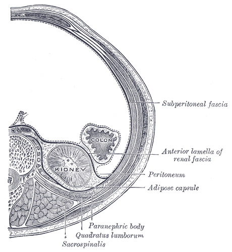
The retroperitoneal space (retroperitoneum) is the anatomical space (sometimes a potential space) behind (''retro'') the peritoneum. It has no specific delineating anatomical structures. Organs are retroperitoneal if they have peritoneum on their anterior side only. Structures that are not suspended by mesentery in the abdominal cavity and that lie between the parietal peritoneum and abdominal wall are classified as retroperitoneal. This is different from organs that are not retroperitoneal, which have peritoneum on their posterior side and are suspended by mesentery in the abdominal cavity. The retroperitoneum can be further subdivided into the following: *Perirenal (or perinephric) space *Anterior pararenal (or paranephric) space *Posterior pararenal (or paranephric) space Retroperitoneal structures Structures that lie behind the peritoneum are termed "retroperitoneal". Organs that were once suspended within the abdominal cavity by mesentery but migrated posterior to the pe ... [...More Info...] [...Related Items...] OR: [Wikipedia] [Google] [Baidu] |
Superficial Epigastric Vein
The superficial epigastric vein is a vein Veins () are blood vessels in the circulatory system of humans and most other animals that carry blood towards the heart. Most veins carry deoxygenated blood from the tissues back to the heart; exceptions are those of the pulmonary and feta ... which travels with the superficial epigastric artery. It joins the accessory saphenous vein near the fossa ovalis. Additional images File:Gray393.png, The subcutaneous inguinal ring File:Gray581.png, The great saphenous vein and its tributaries File:Gray584.png, The femoral vein and its tributaries File:Slide2por.JPG, Superficial veins of lower limb. Superficial dissection. Anterior view. External links * - "Anterior Abdominal Wall: Blood Vessels in the Superficial Fascia" * Veins of the lower limb {{circulatory-stub ... [...More Info...] [...Related Items...] OR: [Wikipedia] [Google] [Baidu] |
Paraumbilical Veins
In the course of the round ligament of the liver, small paraumbilical veins are found which establish an anastomosis between the veins of the anterior abdominal wall and the portal vein, hypogastric, and iliac veins. These veins include Burrow's veins, and the veins of Sappey – superior veins of Sappey and the inferior veins of Sappey. The best marked of these small veins is one which commences at the navel (umbilicus) and runs backward and upward in, or on the surface of, the round ligament (ligamentum teres) between the layers of the falciform ligament to end in the left portal vein. Pathophysiology In cases of portal hypertension, the paraumbilical veins may become enlarged in order to reduce hepatic portal vein pressure by shunting blood to the superficial epigastric vein. The superficial epigastric vein drains to the femoral vein which ultimately drains into the inferior vena cava directly through the external iliac and common iliac vein, thereby bypassing the liver. Dil ... [...More Info...] [...Related Items...] OR: [Wikipedia] [Google] [Baidu] |
Navel
The navel (clinically known as the umbilicus; : umbilici or umbilicuses; also known as the belly button or tummy button) is a protruding, flat, or hollowed area on the abdomen at the attachment site of the umbilical cord. Structure The umbilicus is used to visually separate the abdomen into quadrants. The umbilicus is a prominent Scar#Umbilical, scar on the abdomen, with its position being relatively consistent among humans. The skin around the waist at the level of the umbilicus is supplied by the tenth thoracic spinal nerve (T10 dermatome (anatomy), dermatome). The umbilicus itself typically lies at a vertical level corresponding to the junction between the L3 and L4 vertebrae, with a normal variation among people between the L3 and L5 vertebrae. Parts of the adult navel include the "umbilical cord remnant" or "umbilical tip", which is the often protruding scar left by the detachment of the umbilical cord. This is located in the center of the navel, sometimes described ... [...More Info...] [...Related Items...] OR: [Wikipedia] [Google] [Baidu] |
Inferior Rectal Veins
The lower part of the external hemorrhoidal plexus is drained by the inferior rectal veins (or inferior hemorrhoidal veins) into the internal pudendal vein. Veins superior to the middle rectal vein in the colon and rectum drain via the portal system to the liver. Veins inferior, and including, the middle rectal vein drain into systemic circulation and are returned to the heart, bypassing the liver. Pathologies involving the Inferior rectal veins may cause lower GI bleeding. Depending on the degree of inflammation, they are given a grade level ranging from 1 through 4. Additional images File:Gray405.png, The perineum. The integument and superficial layer of superficial fascia reflected. References Veins of the torso {{circulatory-stub ... [...More Info...] [...Related Items...] OR: [Wikipedia] [Google] [Baidu] |
Middle Rectal Veins
The middle rectal veins (or middle hemorrhoidal vein) take origin in the hemorrhoidal plexus and receive tributaries from the bladder, prostate, and seminal vesicle. They run lateralward on the pelvic surface of the levator ani The levator ani is a broad, thin muscle group, situated on either side of the pelvis. It is formed from three muscle components: the pubococcygeus, the iliococcygeus, and the puborectalis. It is attached to the inner surface of each side of the ... to end in the internal iliac vein. Veins superior to the middle rectal vein in the colon and rectum drain via the portal system to the liver. Veins inferior, and including, the middle rectal vein drain into systemic circulation and are returned to the heart, bypassing the liver. References Veins of the torso {{circulatory-stub ... [...More Info...] [...Related Items...] OR: [Wikipedia] [Google] [Baidu] |
Superior Rectal Vein
The inferior mesenteric vein begins in the rectum as the superior rectal vein (superior hemorrhoidal vein), which has its origin in the hemorrhoidal plexus, and through this plexus communicates with the middle and inferior hemorrhoidal veins. The superior rectal vein leaves the lesser pelvis and crosses the left common iliac vessels with the superior rectal artery, and is continued upward as the inferior mesenteric vein In human anatomy, the inferior mesenteric vein (IMV) is a blood vessel that drains blood from the large intestine. It usually terminates when reaching the splenic vein, which goes on to form the portal vein with the superior mesenteric vein (SMV) .... References Veins of the torso Rectum {{circulatory-stub ... [...More Info...] [...Related Items...] OR: [Wikipedia] [Google] [Baidu] |
Rectal Varices
Anorectal varices are collateral submucosal blood vessels dilated by backflow in the veins of the rectum. Typically this occurs due to portal hypertension which shunts venous blood from the portal system through the portosystemic anastomosis present at this site into the systemic venous system. This can also occur in the esophagus, causing esophageal varices, and at the level of the umbilicus, causing caput medusae. Between 44% and 78% of patients with portal hypertension get anorectal varices. Signs and symptoms Pathogenesis Blood from the superior portion of the rectum normally drains into the superior rectal vein and via the inferior mesenteric vein to the liver as part of the portal venous system. Blood from the middle and inferior portions of the rectum is drained via the middle and inferior rectal veins. In portal hypertension, venous resistance is increased within the portal venous system; when the pressure in the portal venous system increases above that of the sys ... [...More Info...] [...Related Items...] OR: [Wikipedia] [Google] [Baidu] |
Rectal
The rectum (: rectums or recta) is the final straight portion of the large intestine in humans and some other mammals, and the Gastrointestinal tract, gut in others. Before expulsion through the anus or cloaca, the rectum stores the feces temporarily. The adult human rectum is about long, and begins at the rectosigmoid junction (the end of the sigmoid colon) at the level of the third sacral vertebra or the sacral promontory depending upon what definition is used. Its diameter is similar to that of the sigmoid colon at its commencement, but it is dilated near its termination, forming the rectal ampulla. It terminates at the level of the anorectal ring (the level of the puborectalis sling) or the dentate line, again depending upon which definition is used. In humans, the rectum is followed by the anal canal, which is about long, before the gastrointestinal tract terminates at the anal verge. The word rectum comes from the Latin ''Wikt:rectum, rēctum Wikt:intestinum, intestīn ... [...More Info...] [...Related Items...] OR: [Wikipedia] [Google] [Baidu] |


