|
Pontine Nuclei
The pontine nuclei (or griseum pontis) are all the neurons of the ventral pons. Corticopontine fibres project from the primary motor cortex to the ipsilateral pontine nucleus; pontocerebellar fibers then relay the information to the contralateral cerebellum via the middle cerebellar peduncle. They are involved in motor function: the pontine nuclei are involved in adjusting movements according to their outcome (movement error correction), and are therefore important in learning motor skills. Anatomy The pontine nuclei encompass all of the about 20 million neurons scattered throughout the basilar part of pons. The pontine nuclei nuclei extend caudally into the medulla oblongata as the arcuate nucleus which is functionally homologous with the pontine nuclei. Afferents Corticopontine fibres arise primarily from the neocortex layer V of the premotor, somatosensory, non- striate visual, posterior parietal, and cingulate cerebral cortex; there are also a few fibers originating ... [...More Info...] [...Related Items...] OR: [Wikipedia] [Google] [Baidu] |
Pons
The pons (from Latin , "bridge") is part of the brainstem that in humans and other mammals, lies inferior to the midbrain, superior to the medulla oblongata and anterior to the cerebellum. The pons is also called the pons Varolii ("bridge of Varolius"), after the Italian anatomist and surgeon Costanzo Varolio (1543–75). This region of the brainstem includes neural pathways and tracts that conduct signals from the brain down to the cerebellum and medulla, and tracts that carry the sensory signals up into the thalamus. Structure The pons in humans measures about in length. It is the part of the brainstem situated between the midbrain and the medulla oblongata. The horizontal ''medullopontine sulcus'' demarcates the boundary between the pons and medulla oblongata on the ventral aspect of the brainstem, and the roots of cranial nerves VI/VII/VIII emerge from the brainstem along this groove. The junction of pons, medulla oblongata, and cerebellum forms the cerebellopontine ... [...More Info...] [...Related Items...] OR: [Wikipedia] [Google] [Baidu] |
Visual Cortex
The visual cortex of the brain is the area of the cerebral cortex that processes visual information. It is located in the occipital lobe. Sensory input originating from the eyes travels through the lateral geniculate nucleus in the thalamus and then reaches the visual cortex. The area of the visual cortex that receives the sensory input from the lateral geniculate nucleus is the primary visual cortex, also known as visual area 1 ( V1), Brodmann area 17, or the striate cortex. The extrastriate areas consist of visual areas 2, 3, 4, and 5 (also known as V2, V3, V4, and V5, or Brodmann area 18 and all Brodmann area 19). Both hemispheres of the brain include a visual cortex; the visual cortex in the left hemisphere receives signals from the right visual field, and the visual cortex in the right hemisphere receives signals from the left visual field. Introduction The primary visual cortex (V1) is located in and around the calcarine fissure in the occipital lobe. Each h ... [...More Info...] [...Related Items...] OR: [Wikipedia] [Google] [Baidu] |
Trigeminal Nuclei
The sensory trigeminal nerve nuclei are the largest of the cranial nerve nuclei, and extend through the whole of the midbrain, pons and medulla, and into the upper cervical spinal cord. The nucleus is divided into three parts, from rostral to caudal (top to bottom in humans): * The mesencephalic nucleus * The principal sensory nucleus * The spinal trigeminal nucleus ::The spinal trigeminal nucleus is further subdivided into three parts, from rostral to caudal: :* Pars oralis (from the Pons to the Hypoglossal nucleus) :* Pars interpolaris (from the Hypoglossal nucleus to the obex) :* Pars caudalis (from the obex to C2) There is also a distinct trigeminal motor nucleus that is medial to the principal sensory nucleus. See also * Photic sneeze reflex * Trigeminal nerve In neuroanatomy, the trigeminal nerve (literal translation, lit. ''triplet'' nerve), also known as the fifth cranial nerve, cranial nerve V, or simply CN V, is a cranial nerve responsible for Sense, sensat ... [...More Info...] [...Related Items...] OR: [Wikipedia] [Google] [Baidu] |
Cerebellum
The cerebellum (: cerebella or cerebellums; Latin for 'little brain') is a major feature of the hindbrain of all vertebrates. Although usually smaller than the cerebrum, in some animals such as the mormyrid fishes it may be as large as it or even larger. In humans, the cerebellum plays an important role in motor control and cognition, cognitive functions such as attention and language as well as emotion, emotional control such as regulating fear and pleasure responses, but its movement-related functions are the most solidly established. The human cerebellum does not initiate movement, but contributes to motor coordination, coordination, precision, and accurate timing: it receives input from sensory systems of the spinal cord and from other parts of the brain, and integrates these inputs to fine-tune motor activity. Cerebellar damage produces disorders in fine motor skill, fine movement, sense of balance, equilibrium, list of human positions, posture, and motor learning in humans. ... [...More Info...] [...Related Items...] OR: [Wikipedia] [Google] [Baidu] |
Mammillary Body
The mammillary bodies also mamillary bodies, are a pair of small round brainstem nuclei. They are located on the undersurface of the brain that, as part of the diencephalon, form part of the limbic system. They are located at the ends of the anterior arches of the fornix. They consist of two groups of nuclei, the medial mammillary nuclei and the lateral mammillary nuclei. Neuroanatomists have often categorized the mammillary bodies as part of the posterior part of hypothalamus. Structure Connections They are connected to other parts of the brain (as shown in the schematic, below left), and act as a relay for impulses coming from the amygdalae and hippocampi, via the mamillothalamic tract to the thalamus. The lateral mammillary nucleus has bidirectional connections with the dorsal tegmental nucleus. The medial mammillary nucleus connects with the ventral tegmental nucleus. Function File:Slide5dd.JPG, Mammillary body Mammillary bodies, and their projections to th ... [...More Info...] [...Related Items...] OR: [Wikipedia] [Google] [Baidu] |
Hypothalamus
The hypothalamus (: hypothalami; ) is a small part of the vertebrate brain that contains a number of nucleus (neuroanatomy), nuclei with a variety of functions. One of the most important functions is to link the nervous system to the endocrine system via the pituitary gland. The hypothalamus is located below the thalamus and is part of the limbic system. It forms the Basal (anatomy), basal part of the diencephalon. All vertebrate brains contain a hypothalamus. In humans, it is about the size of an Almond#Nut, almond. The hypothalamus has the function of regulating certain metabolic biological process, processes and other activities of the autonomic nervous system. It biosynthesis, synthesizes and secretes certain neurohormones, called releasing hormones or hypothalamic hormones, and these in turn stimulate or inhibit the secretion of hormones from the pituitary gland. The hypothalamus controls thermoregulation, body temperature, hunger (physiology), hunger, important aspects o ... [...More Info...] [...Related Items...] OR: [Wikipedia] [Google] [Baidu] |
Pretectal Area
In neuroanatomy, the pretectal area, or pretectum, is a midbrain structure composed of seven nuclei and comprises part of the subcortical visual system. Through reciprocal bilateral projections from the retina, it is involved primarily in mediating behavioral responses to acute changes in ambient light such as the pupillary light reflex, the optokinetic reflex, and temporary changes to the circadian rhythm. In addition to the pretectum's role in the visual system, the anterior pretectal nucleus has been found to mediate somatosensory and nociceptive information. Location and structure The pretectum is a bilateral group of highly interconnected nuclei located near the junction of the midbrain and forebrain. The pretectum is generally classified as a midbrain structure, although because of its proximity to the forebrain it is sometimes classified as part of the caudal diencephalon (forebrain). Within vertebrates, the pretectum is located directly anterior to the superior col ... [...More Info...] [...Related Items...] OR: [Wikipedia] [Google] [Baidu] |
Superior Colliculus
In neuroanatomy, the superior colliculus () is a structure lying on the tectum, roof of the mammalian midbrain. In non-mammalian vertebrates, the Homology (biology), homologous structure is known as the optic tectum or optic lobe. The adjective form ''tectum, tectal'' is commonly used for both structures. In mammals, the superior colliculus forms a major component of the midbrain. It is a paired structure and together with the paired inferior colliculi forms the corpora quadrigemina. The superior colliculus is a layered structure, with a pattern that is similar in all mammals. The layers can be grouped into the superficial layers (retinal nerve fiber layer, stratum opticum and above) and the deeper remaining layers. Neurons in the superficial layers receive direct input from the retina and respond almost exclusively to visual stimuli. Many neurons in the deeper layers also respond to other modalities, and some respond to stimuli in multiple modalities. The deeper layers also conta ... [...More Info...] [...Related Items...] OR: [Wikipedia] [Google] [Baidu] |
Lateral Geniculate Nucleus
In neuroanatomy, the lateral geniculate nucleus (LGN; also called the lateral geniculate body or lateral geniculate complex) is a structure in the thalamus and a key component of the mammalian visual pathway. It is a small, ovoid, Anatomical terms of location#Dorsal_and_ventral, ventral projection of the thalamus where the thalamus connects with the optic nerve. There are two LGNs, one on the left and another on the right side of the thalamus. In humans, both LGNs have six layers of neurons (grey matter) alternating with optic fibers (white matter). The LGN receives information directly from the ascending retinal ganglion cells via the optic tract and from the reticular activating system. Neurons of the LGN send their axons through the optic radiation, a direct pathway to the primary visual cortex. In addition, the LGN receives many strong feedback connections from the primary visual cortex. In humans as well as other mammals, the two strongest pathways linking the eye to the bra ... [...More Info...] [...Related Items...] OR: [Wikipedia] [Google] [Baidu] |
Striate Cortex
The visual cortex of the brain is the area of the cerebral cortex that processes visual information. It is located in the occipital lobe. Sensory input originating from the eyes travels through the lateral geniculate nucleus in the thalamus and then reaches the visual cortex. The area of the visual cortex that receives the sensory input from the lateral geniculate nucleus is the primary visual cortex, also known as visual area 1 ( V1), Brodmann area 17, or the striate cortex. The extrastriate areas consist of visual areas 2, 3, 4, and 5 (also known as V2, V3, V4, and V5, or Brodmann area 18 and all Brodmann area 19). Both hemispheres of the brain include a visual cortex; the visual cortex in the left hemisphere receives signals from the right visual field, and the visual cortex in the right hemisphere receives signals from the left visual field. Introduction The primary visual cortex (V1) is located in and around the calcarine fissure in the occipital lobe. Each hemisphere' ... [...More Info...] [...Related Items...] OR: [Wikipedia] [Google] [Baidu] |
Temporal Cortex
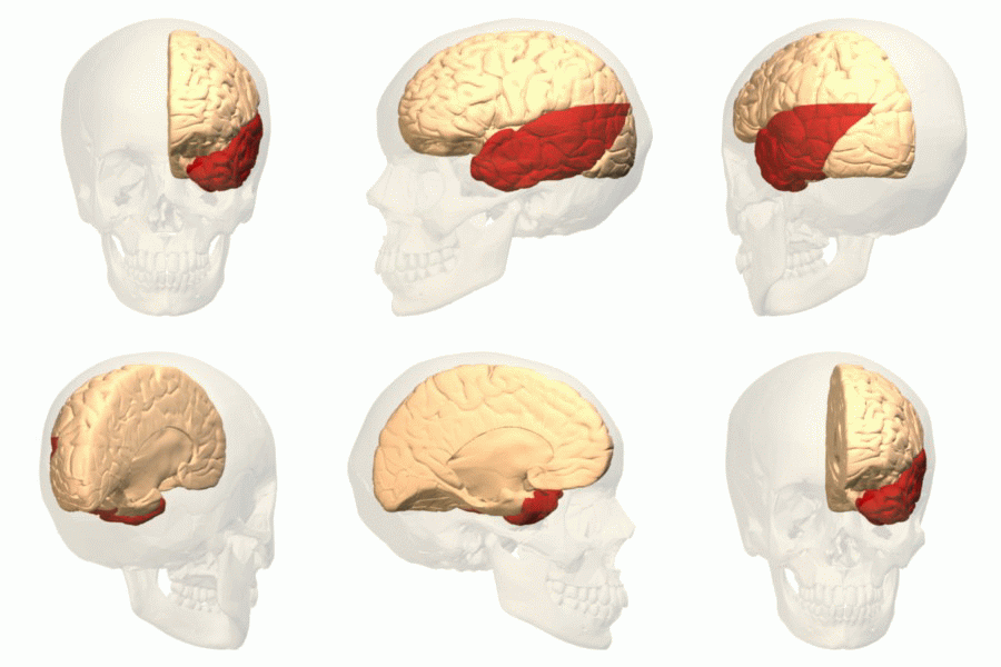
The temporal lobe is one of the four major lobes of the cerebral cortex in the brain of mammals. The temporal lobe is located beneath the lateral fissure on both cerebral hemispheres of the mammalian brain. The temporal lobe is involved in processing sensory input into derived meanings for the appropriate retention of visual memory, language comprehension, and emotion association. ''Temporal'' refers to the head's temples. Structure The temporal lobe consists of structures that are vital for declarative or long-term memory. Declarative (denotative) or explicit memory is conscious memory divided into semantic memory (facts) and episodic memory (events). The medial temporal lobe structures are critical for long-term memory, and include the hippocampal formation, perirhinal cortex, parahippocampal, and entorhinal neocortical regions. The hippocampus is critical for memory formation, and the surrounding medial temporal cortex is currently theorized to be critical for mem ... [...More Info...] [...Related Items...] OR: [Wikipedia] [Google] [Baidu] |
Prefrontal Cortex
In mammalian brain anatomy, the prefrontal cortex (PFC) covers the front part of the frontal lobe of the cerebral cortex. It is the association cortex in the frontal lobe. The PFC contains the Brodmann areas BA8, BA9, BA10, BA11, BA12, BA13, BA14, BA24, BA25, BA32, BA44, BA45, BA46, and BA47. This brain region is involved in a wide range of higher-order cognitive functions, including speech formation (Broca's area), gaze ( frontal eye fields), working memory ( dorsolateral prefrontal cortex), and risk processing (e.g. ventromedial prefrontal cortex). The basic activity of this brain region is considered to be orchestration of thoughts and actions in accordance with internal goals. Many authors have indicated an integral link between a person's will to live, personality, and the functions of the prefrontal cortex. This brain region has been implicated in executive functions, such as planning, decision making, working memory, personality expression, moderating ... [...More Info...] [...Related Items...] OR: [Wikipedia] [Google] [Baidu] |


