|
Podomonas Kaiyoae
''Podomonas'' is a genus of apusomonads, a group of small zooflagellates that glide on their posterior cilium. The genus was identified in 2010 as an independent lineage from '' Apusomonas'' and ''Amastigomonas'', and consequently were described as their own separate taxon, containing many species previously assigned to ''Amastigomonas''. Cell structure ''Podomonas'' are ovoid or fusiform apusomonads, not divided into cell body and mastigophore. They lack a dense rod associated with the 8-microtubule band, just like '' Thecamonas'' but unlike '' Apusomonas'' and '' Manchomonas''. The ciliary sleeve is inconspicuous, around 1.5 μm long, seemingly triangular and merging into the ciliary base, pointing anteriorly or slightly to the left. The anterior cilium is strongly acronematic, unlike ''Manchomonas'', but has a non-acronematic base extending several microns beyond the sleeve tip. The posterior cilium is acronematic or tapering, with a non-acronematic portion that extends visi ... [...More Info...] [...Related Items...] OR: [Wikipedia] [Google] [Baidu] |
Scanning Electron Microscope
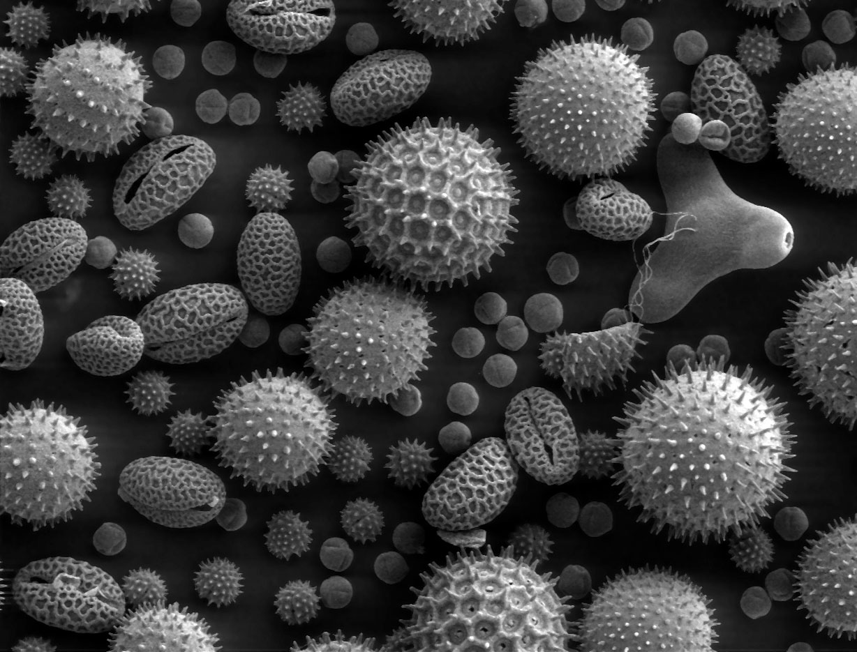
A scanning electron microscope (SEM) is a type of electron microscope that produces images of a sample by scanning the surface with a focused beam of electrons. The electrons interact with atoms in the sample, producing various signals that contain information about the surface topography and composition. The electron beam is scanned in a raster scan pattern, and the position of the beam is combined with the intensity of the detected signal to produce an image. In the most common SEM mode, secondary electrons emitted by atoms excited by the electron beam are detected using a secondary electron detector ( Everhart–Thornley detector). The number of secondary electrons that can be detected, and thus the signal intensity, depends, among other things, on specimen topography. Some SEMs can achieve resolutions better than 1 nanometer. Specimens are observed in high vacuum in a conventional SEM, or in low vacuum or wet conditions in a variable pressure or environmental SEM, an ... [...More Info...] [...Related Items...] OR: [Wikipedia] [Google] [Baidu] |
Thecamonas
Thecamonadinae is a subfamily of heterotrophic protists. It is a monophyletic group, or clade, of apusomonads, a group of protozoa with two flagella closely related to the eukaryotic supergroup Opisthokonta. The subfamily contains two genera ''Chelonemonas'' and '' Thecamonas'', which are found in marine habitats. Morphology Thecamonadinae are unicellular eukaryotes, exhibiting cells smaller than 10 μm, and an "''Amastigomonas''-type" cell body shape: plastic, oval to oblong, with a prominent proboscis that measures around ¼ of the cell body length. They have a rigid "tusk" of between 200 and 250 nm in diameter, that arises to the right of the anterior flagellum and extends around 0.5–1.0 μm. This tusk can be visible under optimal conditions of light microscopy. Aside from the flagella, they often present thin pseudopodia trailing behind the moving cell. Systematics History of taxonomy Thecamonadinae was initially a family-level taxon, Thecamonadidae, described in ... [...More Info...] [...Related Items...] OR: [Wikipedia] [Google] [Baidu] |
Cytoplasm
The cytoplasm describes all the material within a eukaryotic or prokaryotic cell, enclosed by the cell membrane, including the organelles and excluding the nucleus in eukaryotic cells. The material inside the nucleus of a eukaryotic cell and contained within the nuclear membrane is termed the nucleoplasm. The main components of the cytoplasm are the cytosol (a gel-like substance), the cell's internal sub-structures, and various cytoplasmic inclusions. In eukaryotes the cytoplasm also includes the nucleus, and other membrane-bound organelles.The cytoplasm is about 80% water and is usually colorless. The submicroscopic ground cell substance, or cytoplasmic matrix, that remains after the exclusion of the cell organelles and particles is groundplasm. It is the hyaloplasm of light microscopy, a highly complex, polyphasic system in which all resolvable cytoplasmic elements are suspended, including the larger organelles such as the ribosomes, mitochondria, plant plasti ... [...More Info...] [...Related Items...] OR: [Wikipedia] [Google] [Baidu] |
Pseudopod
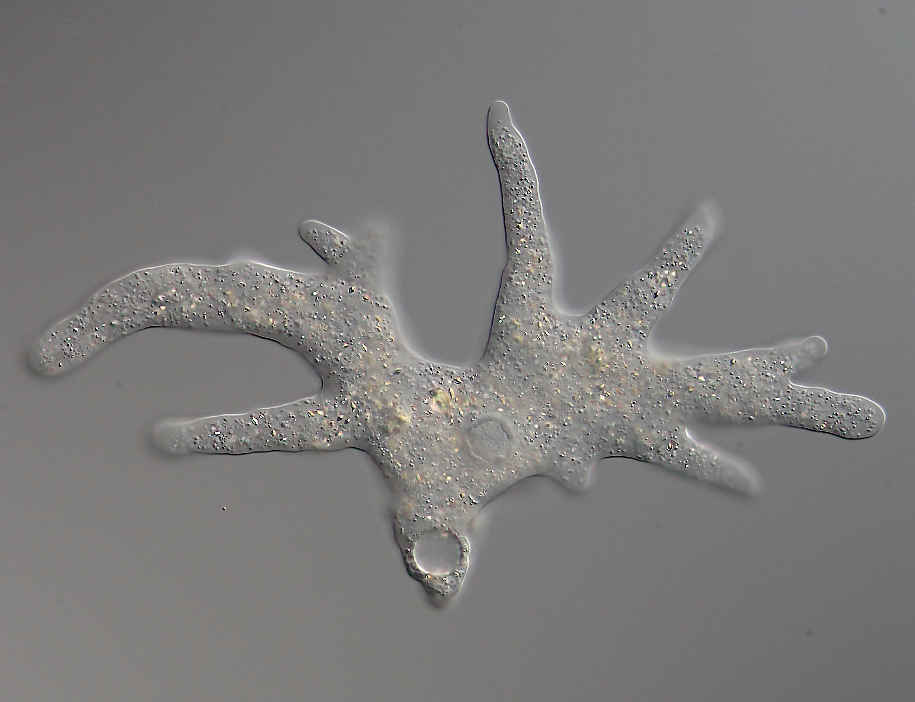
A pseudopod or pseudopodium (: pseudopods or pseudopodia) is a temporary arm-like projection of a eukaryotic cell membrane that is emerged in the direction of movement. Filled with cytoplasm, pseudopodia primarily consist of actin filaments and may also contain microtubules and intermediate filaments. Pseudopods are used for motility and ingestion. They are often found in amoebas. Different types of pseudopodia can be classified by their distinct appearances. Lamellipodia are broad and thin. Filopodia are slender, thread-like, and are supported largely by microfilaments. Lobopodia are bulbous and amoebic. Reticulopodia are complex structures bearing individual pseudopodia which form irregular nets. Axopodia are the phagocytosis type with long, thin pseudopods supported by complex microtubule arrays enveloped with cytoplasm; they respond rapidly to physical contact. Generally, several pseudopodia arise from the surface of the body, (''polypodial'', for example, '' Amoeba p ... [...More Info...] [...Related Items...] OR: [Wikipedia] [Google] [Baidu] |
Reticulopodia
{{Short pages monitor ... [...More Info...] [...Related Items...] OR: [Wikipedia] [Google] [Baidu] |
Centriole
In cell biology a centriole is a cylindrical organelle composed mainly of a protein called tubulin. Centrioles are found in most eukaryotic cells, but are not present in conifers ( Pinophyta), flowering plants ( angiosperms) and most fungi, and are only present in the male gametes of charophytes, bryophytes, seedless vascular plants, cycads, and ''Ginkgo''. A bound pair of centrioles, surrounded by a highly ordered mass of dense material, called the pericentriolar material (PCM), makes up a structure called a centrosome. Centrioles are typically made up of nine sets of short microtubule triplets, arranged in a cylinder. Deviations from this structure include crabs and ''Drosophila melanogaster'' embryos, with nine doublets, and '' Caenorhabditis elegans'' sperm cells and early embryos, with nine singlets. Additional proteins include centrin, cenexin and tektin. The main function of centrioles is to produce cilia during interphase and the aster and the spindle durin ... [...More Info...] [...Related Items...] OR: [Wikipedia] [Google] [Baidu] |
Nucleus (cell)
The cell nucleus (; : nuclei) is a membrane-bound organelle found in eukaryotic cells. Eukaryotic cells usually have a single nucleus, but a few cell types, such as mammalian red blood cells, have no nuclei, and a few others including osteoclasts have many. The main structures making up the nucleus are the nuclear envelope, a double membrane that encloses the entire organelle and isolates its contents from the cellular cytoplasm; and the nuclear matrix, a network within the nucleus that adds mechanical support. The cell nucleus contains nearly all of the cell's genome. Nuclear DNA is often organized into multiple chromosomes – long strands of DNA dotted with various proteins, such as histones, that protect and organize the DNA. The genes within these chromosomes are structured in such a way to promote cell function. The nucleus maintains the integrity of genes and controls the activities of the cell by regulating gene expression. Because the nuclear envelope is imperm ... [...More Info...] [...Related Items...] OR: [Wikipedia] [Google] [Baidu] |
Micron
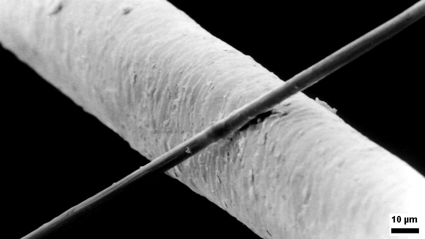
The micrometre (English in the Commonwealth of Nations, Commonwealth English as used by the International Bureau of Weights and Measures; SI symbol: μm) or micrometer (American English), also commonly known by the non-SI term micron, is a unit of length in the International System of Units (SI) equalling (SI standard prefix "micro-" = ); that is, one millionth of a metre (or one thousandth of a millimetre, , or about ). The nearest smaller common SI Unit, SI unit is the nanometre, equivalent to one thousandth of a micrometre, one millionth of a millimetre or one billionth of a metre (). The micrometre is a common unit of measurement for wavelengths of infrared radiation as well as sizes of biological cell (biology), cells and bacteria, and for grading wool by the diameter of the fibres. The width of a single human hair ranges from approximately 20 to . Examples Between 1 μm and 10 μm: * 1–10 μm – length of a typical bacterium * 3–8 μm – width of str ... [...More Info...] [...Related Items...] OR: [Wikipedia] [Google] [Baidu] |
Flagellum
A flagellum (; : flagella) (Latin for 'whip' or 'scourge') is a hair-like appendage that protrudes from certain plant and animal sperm cells, from fungal spores ( zoospores), and from a wide range of microorganisms to provide motility. Many protists with flagella are known as flagellates. A microorganism may have from one to many flagella. A gram-negative bacterium '' Helicobacter pylori'', for example, uses its flagella to propel itself through the stomach to reach the mucous lining where it may colonise the epithelium and potentially cause gastritis, and ulcers – a risk factor for stomach cancer. In some swarming bacteria, the flagellum can also function as a sensory organelle, being sensitive to wetness outside the cell. Across the three domains of Bacteria, Archaea, and Eukaryota, the flagellum has a different structure, protein composition, and mechanism of propulsion but shares the same function of providing motility. The Latin word means " whip" to describe its ... [...More Info...] [...Related Items...] OR: [Wikipedia] [Google] [Baidu] |
Anterior (anatomy)
Standard anatomical terms of location are used to describe unambiguously the anatomy of humans and other animals. The terms, typically derived from Latin or Greek roots, describe something in its standard anatomical position. This position provides a definition of what is at the front ("anterior"), behind ("posterior") and so on. As part of defining and describing terms, the body is described through the use of anatomical planes and axes. The meaning of terms that are used can change depending on whether a vertebrate is a biped or a quadruped, due to the difference in the neuraxis, or if an invertebrate is a non-bilaterian. A non-bilaterian has no anterior or posterior surface for example but can still have a descriptor used such as proximal or distal in relation to a body part that is nearest to, or furthest from its middle. International organisations have determined vocabularies that are often used as standards for subdisciplines of anatomy. For example, '' Terminologia A ... [...More Info...] [...Related Items...] OR: [Wikipedia] [Google] [Baidu] |
Manchomonas
The apusomonads (family Apusomonadidae) are a group of protozoan zooflagellates that glide on surfaces, and mostly consume prokaryotes. They are of particular evolutionary interest because they appear to be the sister group to the Opisthokonts, the clade that includes both animals and fungi. Together with the Breviatea, these form the Obazoa clade. Characteristics Apusomonads are small gliding heterotrophic biflagellates (i.e. with two flagella) that possess a proboscis, formed partly or entirely by the anterior flagellum surrounded by a membranous sleeve. There is a pellicle under the dorsal cell membrane that extends into the proboscis sleeve and into a skirt that covers the sides of the cell. Apusomonads present two different cell plans: *Derived cell plan, represented by ''Apusomonas'', with a round cell body and a mastigophore, a projection of the cell containing both basal bodies at its end. *"''Amastigomonas''-like" cell plan, with an oval or oblong cell that generally f ... [...More Info...] [...Related Items...] OR: [Wikipedia] [Google] [Baidu] |


