|
Placental Expulsion
Placental expulsion (also called afterbirth) occurs when the placenta comes out of the birth canal after childbirth. The time between the expulsion of the baby and the expulsion of the placenta is called the third stage of labor. The third stage of labor can be managed actively with several standard procedures, or it can be managed expectantly, with physiological management or passive management. The latter allows for the placenta to be expelled without medical assistance. Although uncommon, in some countries, such as the United States, Germany, France, Australia, and the United Kingdom, the placenta is kept and consumed by the mother over the weeks following the birth. This practice is termed human placentophagy and can be harmful. Physiology Hormone induction of placental separation As the fetal hypothalamus matures, the activation of the hypothalamic–pituitary–adrenal (HPA) axis initiates labor through two hormonal mechanisms. The end pathway of both mechanisms lead ... [...More Info...] [...Related Items...] OR: [Wikipedia] [Google] [Baidu] |
Placenta Held
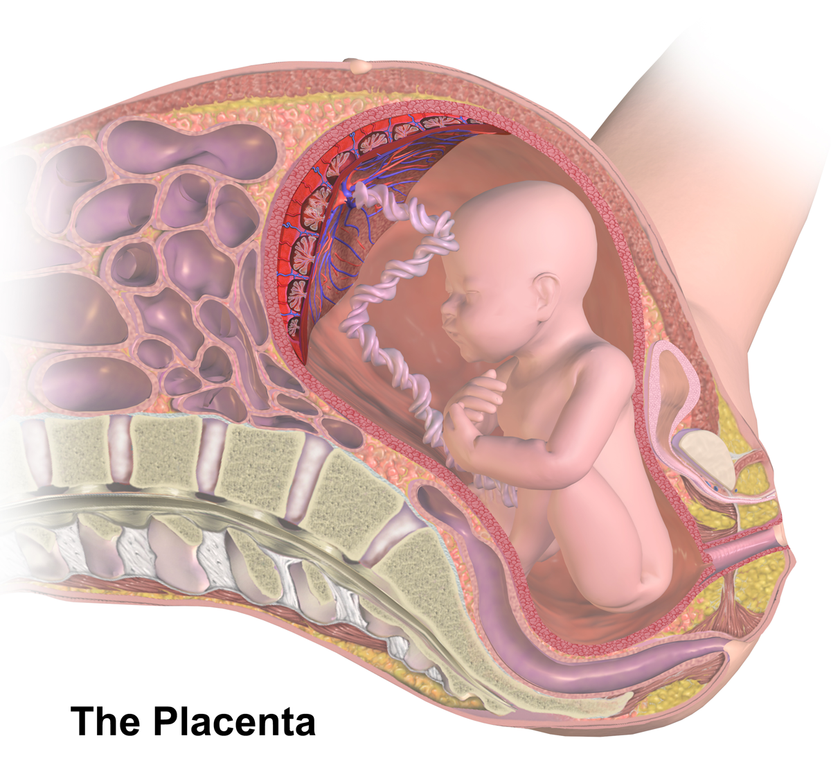
The placenta (: placentas or placentae) is a temporary embryonic and later fetal organ that begins developing from the blastocyst shortly after implantation. It plays critical roles in facilitating nutrient, gas, and waste exchange between the physically separate maternal and fetal circulations, and is an important endocrine organ, producing hormones that regulate both maternal and fetal physiology during pregnancy. The placenta connects to the fetus via the umbilical cord, and on the opposite aspect to the maternal uterus in a species-dependent manner. In humans, a thin layer of maternal decidual (endometrial) tissue comes away with the placenta when it is expelled from the uterus following birth (sometimes incorrectly referred to as the 'maternal part' of the placenta). Placentas are a defining characteristic of placental mammals, but are also found in marsupials and some non-mammals with varying levels of development. Mammalian placentas probably first evolved about 150 mil ... [...More Info...] [...Related Items...] OR: [Wikipedia] [Google] [Baidu] |
Estrogen
Estrogen (also spelled oestrogen in British English; see spelling differences) is a category of sex hormone responsible for the development and regulation of the female reproductive system and secondary sex characteristics. There are three major endogenous estrogens that have estrogenic hormonal activity: estrone (E1), estradiol (E2), and estriol (E3). Estradiol, an estrane, is the most potent and prevalent. Another estrogen called estetrol (E4) is produced only during pregnancy. Estrogens are synthesized in all vertebrates and some insects. Quantitatively, estrogens circulate at lower levels than androgens in both men and women. While estrogen levels are significantly lower in males than in females, estrogens nevertheless have important physiological roles in males. Like all steroid hormones, estrogens readily diffuse across the cell membrane. Once inside the cell, they bind to and activate estrogen receptors (ERs) which in turn modulate the expression of many ... [...More Info...] [...Related Items...] OR: [Wikipedia] [Google] [Baidu] |
Oxytocin
Oxytocin is a peptide hormone and neuropeptide normally produced in the hypothalamus and released by the posterior pituitary. Present in animals since early stages of evolution, in humans it plays roles in behavior that include Human bonding, social bonding, love, reproduction, childbirth, and the Postpartum period, period after childbirth. Oxytocin is released into the bloodstream as a hormone in response to Human sexual activity, sexual activity and during childbirth. It is also available in Oxytocin (medication), pharmaceutical form. In either form, oxytocin stimulates uterine contractions to speed up the process of childbirth. In its natural form, it also plays a role in maternal bonding and lactation, milk production. Production and secretion of oxytocin is controlled by a positive feedback mechanism, where its initial release stimulates production and release of further oxytocin. For example, when oxytocin is released during a contraction of the uterus at the start of c ... [...More Info...] [...Related Items...] OR: [Wikipedia] [Google] [Baidu] |
Hematoma
A hematoma, also spelled haematoma, or blood suffusion is a localized bleeding outside of blood vessels, due to either disease or trauma including injury or surgery and may involve blood continuing to seep from broken capillaries. A hematoma is benign and is initially in liquid form spread among the tissues including in sacs between tissues where it may coagulate and solidify before blood is reabsorbed into blood vessels. An ecchymosis is a hematoma of the skin larger than 10 mm. They may occur among and or within many areas such as skin and other organs, connective tissues, bone, joints and muscle. A collection of blood (or even a hemorrhage) may be aggravated by anticoagulant medication (blood thinner). Blood seepage and collection of blood may occur if heparin is given via an intramuscular route; to avoid this, heparin must be given intravenously or subcutaneously. Signs and symptoms Some hematomas are visible under the surface of the skin (commonly called bruise ... [...More Info...] [...Related Items...] OR: [Wikipedia] [Google] [Baidu] |
Uterine Contraction
Uterine contractions are muscle contractions of the uterine smooth muscle that can occur at various intensities in both the non-pregnant and pregnant uterine state. The non-pregnant uterus undergoes small, spontaneous contractions in addition to stronger, coordinated contractions during the menstrual cycle and orgasm. Throughout gestation, the uterus enters a state of uterine quiescence due to various neural and hormonal changes. During this state, the uterus undergoes little to no contractions, though spontaneous contractions still occur for the uterine myocyte cells to experience hypertrophy. The pregnant uterus only contracts strongly during orgasms, labour, and in the postpartum stage to return to its natural size. Throughout menstrual cycle Uterine contractions that occur throughout the menstrual cycle, also termed ''endometrial waves'' or ''contractile waves'', appear to involve only the sub- endometrial layer of the myometrium. Follicular and luteal phase In the early ... [...More Info...] [...Related Items...] OR: [Wikipedia] [Google] [Baidu] |
Umbilical Cord
In Placentalia, placental mammals, the umbilical cord (also called the navel string, birth cord or ''funiculus umbilicalis'') is a conduit between the developing embryo or fetus and the placenta. During prenatal development, the umbilical cord is physiologically and genetically part of the fetus and (in humans) normally contains two arteries (the umbilical arteries) and one vein (the umbilical vein), buried within Wharton's jelly. The umbilical vein supplies the fetus with oxygenated, nutrient-rich blood from the placenta. Conversely, the fetal heart pumps low-oxygen, nutrient-depleted blood through the umbilical arteries back to the placenta. Structure and development The umbilical cord develops from and contains remnants of the yolk sac and allantois. It forms by the fifth week of human embryogenesis, development, replacing the yolk sac as the source of nutrients for the embryo. The cord is not directly connected to the mother's circulatory system, but instead joins the pla ... [...More Info...] [...Related Items...] OR: [Wikipedia] [Google] [Baidu] |
Umbilical Cord Clamping
In placental mammals, the umbilical cord (also called the navel string, birth cord or ''funiculus umbilicalis'') is a conduit between the developing embryo or fetus and the placenta. During prenatal development, the umbilical cord is physiologically and genetically part of the fetus and (in humans) normally contains two arteries (the umbilical arteries) and one vein (the umbilical vein), buried within Wharton's jelly. The umbilical vein supplies the fetus with oxygenated, nutrient-rich blood from the placenta. Conversely, the fetal heart pumps low-oxygen, nutrient-depleted blood through the umbilical arteries back to the placenta. Structure and development The umbilical cord develops from and contains remnants of the yolk sac and allantois. It forms by the fifth week of development, replacing the yolk sac as the source of nutrients for the embryo. The cord is not directly connected to the mother's circulatory system, but instead joins the placenta, which transfers materials t ... [...More Info...] [...Related Items...] OR: [Wikipedia] [Google] [Baidu] |
Placental Cotyledon
The placenta of humans, and certain other mammals contains structures known as cotyledons, which transmit fetal blood and allow exchange of oxygen and nutrients with the maternal blood. Ruminants The Artiodactyla have a cotyledonary placenta. In this form of placenta, the chorionic villi form a number of separate circular structures (''cotyledons'') which are distributed over the surface of the chorionic sac. Sheep, goats and cattle have between 72 and 125 cotyledons whereas deer have 4-6 larger cotyledons. Human The form of the human placenta is generally classified as a discoid placenta. Within this, the ''cotyledons'' are the approximately 15-25 separations of the decidua basalis of the placenta, separated by placental septa.Page 565 in:/ref> Each cotyledon consists of a main stem of a chorionic villus as well as its branches and sub-branches. Vasculature The cotyledons receive fetal blood from chorionic vessels Chorionic (plate) vessels, also fetal surface vessels are blo ... [...More Info...] [...Related Items...] OR: [Wikipedia] [Google] [Baidu] |
Macrophage
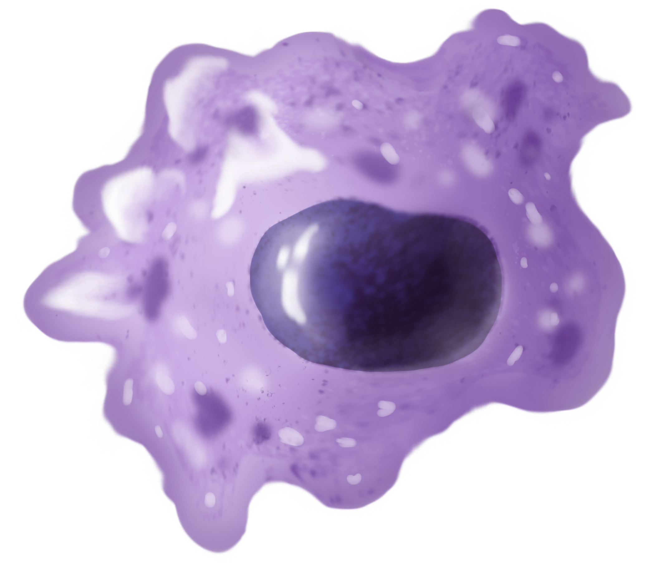
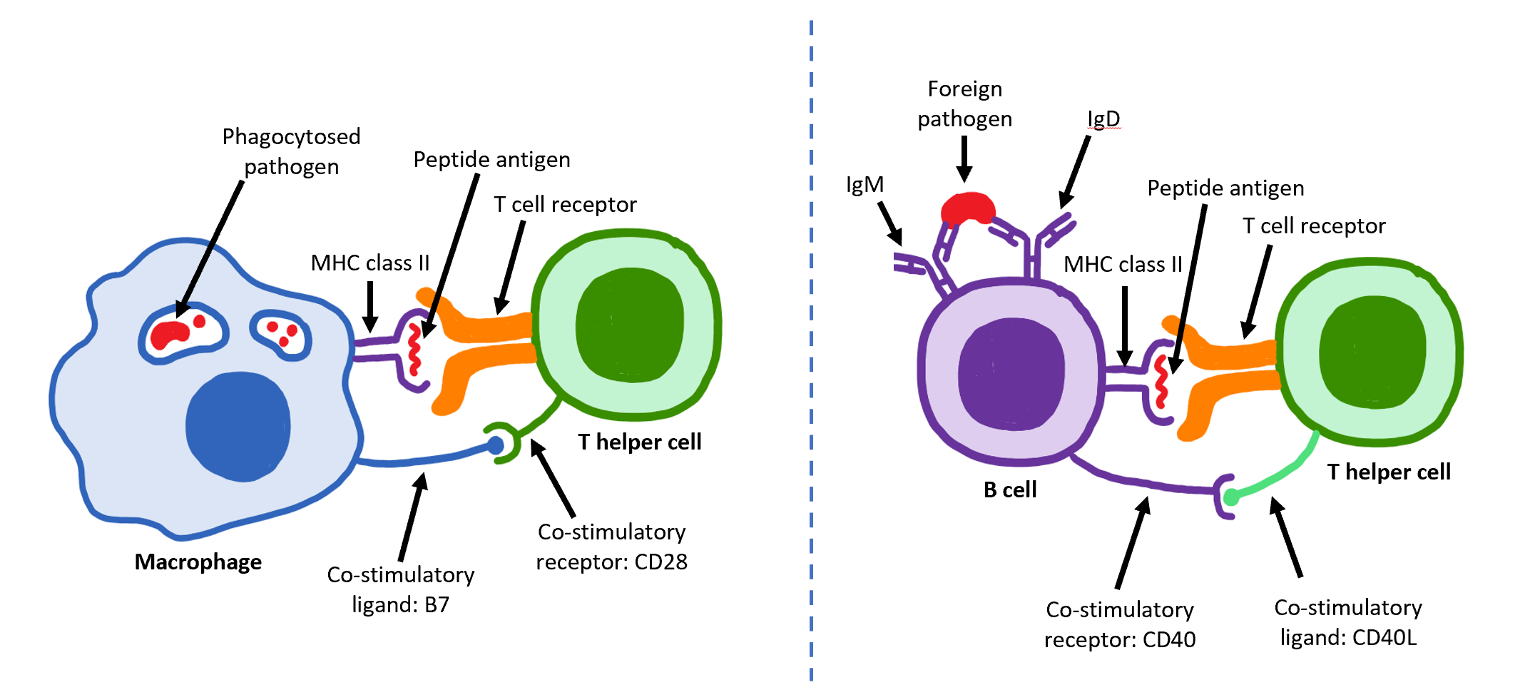
Macrophages (; abbreviated MPhi, φ, MΦ or MP) are a type of white blood cell of the innate immune system that engulf and digest pathogens, such as cancer cells, microbes, cellular debris and foreign substances, which do not have proteins that are specific to healthy body cells on their surface. This self-protection method can be contrasted with that employed by Natural killer cell, Natural Killer cells. This process of engulfment and digestion is called phagocytosis; it acts to defend the host against infection and injury. Macrophages are found in essentially all tissues, where they patrol for potential pathogens by amoeboid movement. They take various forms (with various names) throughout the body (e.g., histiocytes, Kupffer cells, alveolar macrophages, microglia, and others), but all are part of the mononuclear phagocyte system. Besides phagocytosis, they play a critical role in nonspecific defense (innate immunity) and also help initiate specific defense mechanisms (adapti ... [...More Info...] [...Related Items...] OR: [Wikipedia] [Google] [Baidu] |
Helper T Cells
The T helper cells (Th cells), also known as CD4+ cells or CD4-positive cells, are a type of T cell that play an important role in the adaptive immune system. They aid the activity of other immune cells by releasing cytokines. They are considered essential in B cell antibody class switching, breaking cross-tolerance in dendritic cells, in the activation and growth of cytotoxic T cells, and in maximizing bactericidal activity of phagocytes such as macrophages and neutrophils. CD4+ cells are mature Th cells that express the surface protein CD4. Genetic variation in regulatory elements expressed by CD4+ cells determines susceptibility to a broad class of autoimmune diseases. Structure and function Th cells contain and release cytokines to aid other immune cells. Cytokines are small protein mediators that alter the behavior of target cells that express receptors for those cytokines. These cells help polarize the immune response depending on the nature of the immunological ins ... [...More Info...] [...Related Items...] OR: [Wikipedia] [Google] [Baidu] |
Endometrial Lining
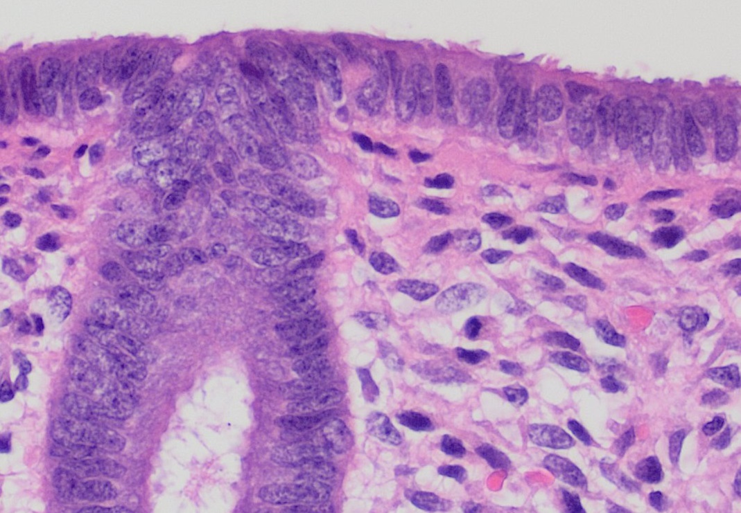
The endometrium is the inner epithelial layer, along with its mucous membrane, of the mammalian uterus. It has a basal layer and a functional layer: the basal layer contains stem cells which regenerate the functional layer. The functional layer thickens and then is shed during menstruation in humans and some other mammals, including other apes, Old World monkeys, some species of bat, the elephant shrew and the Cairo spiny mouse. In most other mammals, the endometrium is reabsorbed in the estrous cycle. During pregnancy, the glands and blood vessels in the endometrium further increase in size and number. Vascular spaces fuse and become interconnected, forming the placenta, which supplies oxygen and nutrition to the embryo and fetus.Blue Histology - Female Reproductive System . Schoo ... [...More Info...] [...Related Items...] OR: [Wikipedia] [Google] [Baidu] |
MHC Class I
MHC class I molecules are one of two primary classes of major histocompatibility complex (MHC) molecules (the other being MHC class II) and are found on the cell surface of all nucleated cells in the bodies of vertebrates. They also occur on platelets, but not on red blood cells. Their function is to display peptide fragments of proteins from within the cell to cytotoxic T cells; this will trigger an immediate response from the immune system against a particular non-self antigen displayed with the help of an MHC class I protein. Because MHC class I molecules present peptides derived from cytosolic proteins, the pathway of MHC class I presentation is often called ''cytosolic'' or ''endogenous pathway''. In humans, the HLAs corresponding to MHC class I are HLA-A, HLA-B, and HLA-C. Function Class I MHC molecules bind peptides generated mainly from the degradation of cytosolic proteins by the proteasome. The MHC I: peptide complex is then inserted via the endoplasmic re ... [...More Info...] [...Related Items...] OR: [Wikipedia] [Google] [Baidu] |

