|
Piriformis Syndrome
Piriformis syndrome is a condition which is believed to result from nerve compression at the sciatic nerve by the piriformis muscle. It is a specific case of deep gluteal syndrome. The largest and most bulky nerve in the human body is the sciatic nerve. Starting at its origin it is 2 cm wide and 0.5 cm thick. The sciatic nerve forms the roots of L4-S3 segments of the lumbosacral plexus. The nerve will pass inferiorly to the piriformis muscle, in the direction of the lower limb where it divides into common tibial and fibular nerves. Symptoms may include pain and numbness in the buttocks and down the leg. Often symptoms are worsened with sitting or running. Causes may include trauma to the gluteal muscle, spasms of the piriformis muscle, anatomical variation, or an overuse injury. Few cases in athletics, however, have been described. Diagnosis is difficult as there is no definitive test. A number of physical exam maneuvers can be supportive. Medical imaging is typ ... [...More Info...] [...Related Items...] OR: [Wikipedia] [Google] [Baidu] |
Orthopedics
Orthopedic surgery or orthopedics (American and British English spelling differences, alternative spelling orthopaedics) is the branch of surgery concerned with conditions involving the musculoskeletal system. Orthopedic surgeons use both surgical and nonsurgical means to treat musculoskeletal Physical trauma, trauma, Spinal disease, spine diseases, Sports injury, sports injuries, degenerative diseases, infections, tumors and congenital disorders. Etymology Nicholas Andry coined the word in French as ', derived from the Ancient Greek words ("correct", "straight") and ("child"), and published ''Orthopedie'' (translated as ''Orthopædia: Or the Art of Correcting and Preventing Deformities in Children'') in 1741. The word was Assimilation (linguistics), assimilated into English as ''orthopædics''; the Typographic ligature, ligature ''æ'' was common in that era for ''ae'' in Greek- and Latin-based words. As the name implies, the discipline was initially developed with atte ... [...More Info...] [...Related Items...] OR: [Wikipedia] [Google] [Baidu] |
Medical Imaging
Medical imaging is the technique and process of imaging the interior of a body for clinical analysis and medical intervention, as well as visual representation of the function of some organs or tissues (physiology). Medical imaging seeks to reveal internal structures hidden by the skin and bones, as well as to diagnose and treat disease. Medical imaging also establishes a database of normal anatomy and physiology to make it possible to identify abnormalities. Although imaging of removed organ (anatomy), organs and Tissue (biology), tissues can be performed for medical reasons, such procedures are usually considered part of pathology instead of medical imaging. Measurement and recording techniques that are not primarily designed to produce images, such as electroencephalography (EEG), magnetoencephalography (MEG), electrocardiography (ECG), and others, represent other technologies that produce data susceptible to representation as a parameter graph versus time or maps that contain ... [...More Info...] [...Related Items...] OR: [Wikipedia] [Google] [Baidu] |
Greater Trochanter
The greater trochanter of the femur is a large, irregular, quadrilateral eminence and a part of the skeletal system. It is directed lateral and medially and slightly posterior. In the adult it is about 2–4 cm lower than the femoral head.Standring, Susan, editor. ''Gray’s Anatomy: The Anatomical Basis of Clinical Practice''. Forty-First edition, Elsevier Limited, 2016, p. 1327. Because the pelvic outlet in the female is larger than in the male, there is a greater distance between the greater trochanters in the female. It has two surfaces and four borders. It is a traction epiphysis. Surfaces The ''lateral surface'', quadrilateral in form, is broad, rough, convex, and marked by a diagonal impression, which extends from the postero-superior to the antero-inferior angle, and serves for the insertion of the tendon of the gluteus medius. Above the impression is a triangular surface, sometimes rough for part of the tendon of the same muscle, sometimes smooth for the interp ... [...More Info...] [...Related Items...] OR: [Wikipedia] [Google] [Baidu] |
Insertion (anatomy)
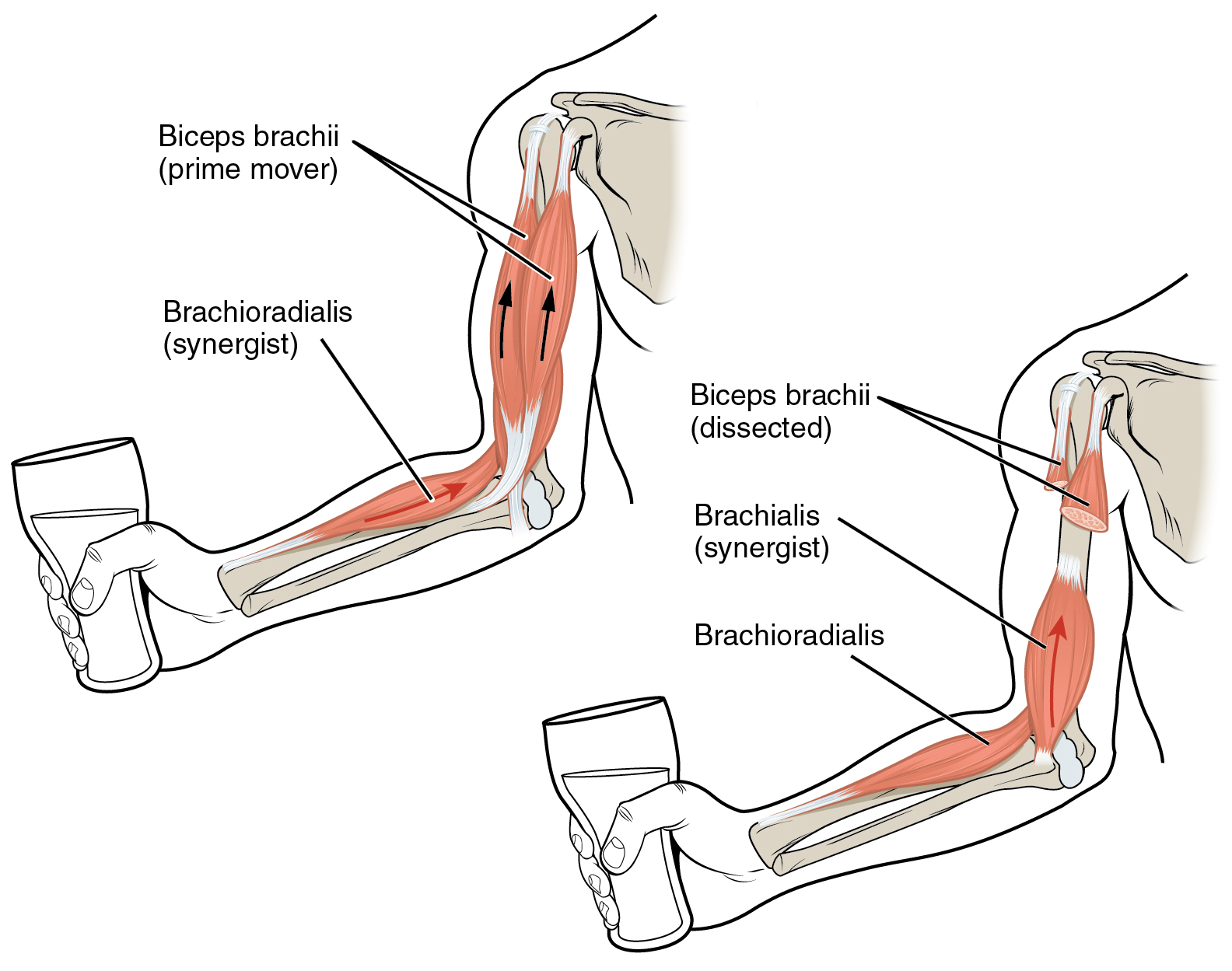
Anatomical terminology is used to uniquely describe aspects of skeletal muscle, cardiac muscle, and smooth muscle such as their actions, structure, size, and location. Types There are three types of muscle tissue in the body: skeletal, smooth, and cardiac. Skeletal muscle Skeletal muscle, or "voluntary muscle", is a striated muscle tissue that primarily joins to bone with tendons. Skeletal muscle enables movement of bones, and Proprioception#Reflexes, maintains posture. The widest part of a muscle that pulls on the tendons is known as the belly. Muscle slip A muscle slip is a slip of muscle that can either be an anatomical variant, or a branching of a muscle as in rib connections of the serratus anterior muscle. Smooth muscle Smooth muscle is involuntary and found in parts of the body where it conveys action without conscious intent. The majority of this type of muscle tissue is found in the digestive system, digestive and urinary systems where it acts by propelling forward foo ... [...More Info...] [...Related Items...] OR: [Wikipedia] [Google] [Baidu] |
Greater Sciatic Foramen
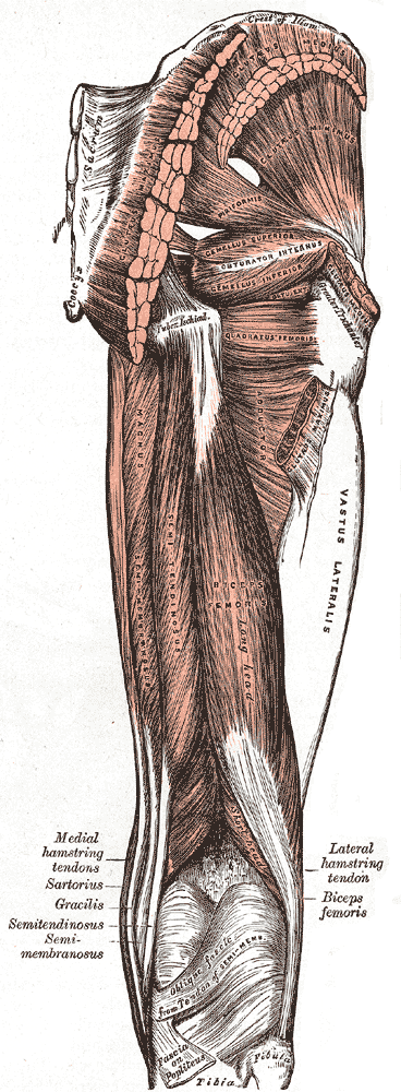
The greater sciatic foramen is an opening (:wikt:foramen, foramen) in the posterior human pelvis. It is formed by the sacrotuberous ligament, sacrotuberous and sacrospinous ligaments. The piriformis muscle passes through the foramen and occupies most of its volume. The greater sciatic foramen is wider in women than in men. Structure It is bounded as follows: * anterolaterally by the greater sciatic notch of the Ilium (bone), ilium. * posteromedially by the sacrotuberous ligament. * inferiorly by the sacrospinous ligament and the ischial spine. * superiorly by the anterior sacroiliac ligament. Function The piriformis, which exits the pelvis through the foramen, occupies most of its volume. The following structures also exit the pelvis through the greater sciatic foramen: See also *Lesser sciatic foramen References External links * * (, ) {{Authority control Anatomy Bones of the pelvis ... [...More Info...] [...Related Items...] OR: [Wikipedia] [Google] [Baidu] |
Sacrum
The sacrum (: sacra or sacrums), in human anatomy, is a triangular bone at the base of the spine that forms by the fusing of the sacral vertebrae (S1S5) between ages 18 and 30. The sacrum situates at the upper, back part of the pelvic cavity, between the two wings of the pelvis. It forms joints with four other bones. The two projections at the sides of the sacrum are called the alae (wings), and articulate with the ilium at the L-shaped sacroiliac joints. The upper part of the sacrum connects with the last lumbar vertebra (L5), and its lower part with the coccyx (tailbone) via the sacral and coccygeal cornua. The sacrum has three different surfaces which are shaped to accommodate surrounding pelvic structures. Overall, it is concave (curved upon itself). The base of the sacrum, the broadest and uppermost part, is tilted forward as the sacral promontory internally. The central part is curved outward toward the posterior, allowing greater room for the pelvic cavity. In a ... [...More Info...] [...Related Items...] OR: [Wikipedia] [Google] [Baidu] |
Origin (anatomy)
Anatomical terminology is used to uniquely describe aspects of skeletal muscle, cardiac muscle, and smooth muscle such as their actions, structure, size, and location. Types There are three types of muscle tissue in the body: skeletal, smooth, and cardiac. Skeletal muscle Skeletal muscle, or "voluntary muscle", is a striated muscle tissue that primarily joins to bone with tendons. Skeletal muscle enables movement of bones, and maintains posture. The widest part of a muscle that pulls on the tendons is known as the belly. Muscle slip A muscle slip is a slip of muscle that can either be an anatomical variant, or a branching of a muscle as in rib connections of the serratus anterior muscle. Smooth muscle Smooth muscle is involuntary and found in parts of the body where it conveys action without conscious intent. The majority of this type of muscle tissue is found in the digestive and urinary systems where it acts by propelling forward food, chyme, and feces in the former and ur ... [...More Info...] [...Related Items...] OR: [Wikipedia] [Google] [Baidu] |
Abduction (anatomy)
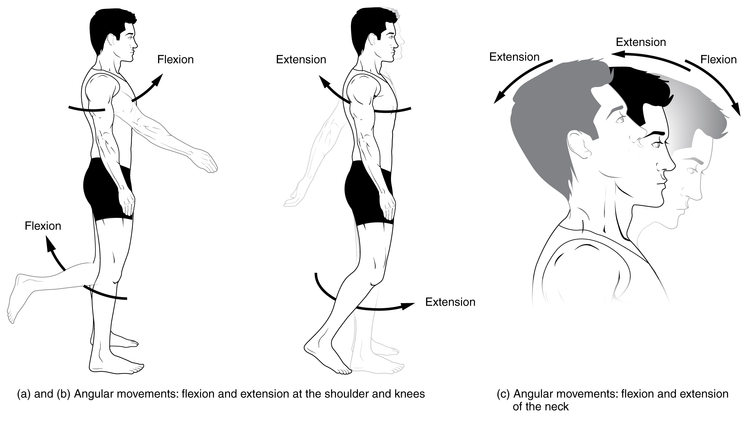
Motion, the process of movement, is described using specific anatomical terms. Motion includes movement of organs, joints, limbs, and specific sections of the body. The terminology used describes this motion according to its direction relative to the anatomical position of the body parts involved. Anatomists and others use a unified set of terms to describe most of the movements, although other, more specialized terms are necessary for describing unique movements such as those of the hands, feet, and eyes. In general, motion is classified according to the anatomical plane it occurs in. ''Flexion'' and ''extension'' are examples of ''angular'' motions, in which two axes of a joint are brought closer together or moved further apart. ''Rotational'' motion may occur at other joints, for example the shoulder, and are described as ''internal'' or ''external''. Other terms, such as ''elevation'' and ''depression'', describe movement above or below the horizontal plane. Many anatomica ... [...More Info...] [...Related Items...] OR: [Wikipedia] [Google] [Baidu] |
Flexion
Motion, the process of movement, is described using specific anatomical terminology, anatomical terms. Motion includes movement of Organ (anatomy), organs, joints, Limb (anatomy), limbs, and specific sections of the body. The terminology used describes this motion according to its direction relative to the anatomical position of the body parts involved. Anatomy, Anatomists and others use a unified set of terms to describe most of the movements, although other, more specialized terms are necessary for describing unique movements such as those of the hands, feet, and eyes. In general, motion is classified according to the anatomical plane it occurs in. ''Flexion'' and ''extension'' are examples of ''angular'' motions, in which two axes of a joint are brought closer together or moved further apart. ''Rotational'' motion may occur at other joints, for example the shoulder, and are described as ''internal'' or ''external''. Other terms, such as ''elevation'' and ''depression'', descri ... [...More Info...] [...Related Items...] OR: [Wikipedia] [Google] [Baidu] |
Extension (anatomy)
Motion, the process of movement, is described using specific anatomical terms. Motion includes movement of organs, joints, limbs, and specific sections of the body. The terminology used describes this motion according to its direction relative to the anatomical position of the body parts involved. Anatomists and others use a unified set of terms to describe most of the movements, although other, more specialized terms are necessary for describing unique movements such as those of the hands, feet, and eyes. In general, motion is classified according to the anatomical plane it occurs in. ''Flexion'' and ''extension'' are examples of ''angular'' motions, in which two axes of a joint are brought closer together or moved further apart. ''Rotational'' motion may occur at other joints, for example the shoulder, and are described as ''internal'' or ''external''. Other terms, such as ''elevation'' and ''depression'', describe movement above or below the horizontal plane. Many anatomical ... [...More Info...] [...Related Items...] OR: [Wikipedia] [Google] [Baidu] |
Lateral Rotator Group
The lateral rotator group is a group of six small muscles of the hip which all Anatomical terms of motion#Rotation, externally (laterally) rotate the femur in the hip, hip joint. It consists of the following muscles: Piriformis muscle, piriformis, Superior gemellus muscle, gemellus superior, Internal obturator muscle, obturator internus, Inferior gemellus muscle, gemellus inferior, Quadratus femoris muscle, quadratus femoris and the External obturator muscle, obturator externus. All muscles in the lateral rotator group Anatomical terms of muscle#Origin, originate from the hip bone and Anatomical terms of muscle#Insertion, insert on to the Upper extremity of femur, upper extremity of the femur. The muscles are innervated by the sacral plexus (Lumbar nerves#Fourth lumbar nerve, L4-Sacral spinal nerve 2, S2), except the External obturator muscle, obturator externus muscle, which is innervated by the lumbar plexus. Individual muscles Other lateral rotators This group does not includ ... [...More Info...] [...Related Items...] OR: [Wikipedia] [Google] [Baidu] |
Medial (anatomy)
Standard anatomical terms of location are used to describe unambiguously the anatomy of humans and other animals. The terms, typically derived from Latin or Greek roots, describe something in its standard anatomical position. This position provides a definition of what is at the front ("anterior"), behind ("posterior") and so on. As part of defining and describing terms, the body is described through the use of anatomical planes and axes. The meaning of terms that are used can change depending on whether a vertebrate is a biped or a quadruped, due to the difference in the neuraxis, or if an invertebrate is a non-bilaterian. A non-bilaterian has no anterior or posterior surface for example but can still have a descriptor used such as proximal or distal in relation to a body part that is nearest to, or furthest from its middle. International organisations have determined vocabularies that are often used as standards for subdisciplines of anatomy. For example, '' Terminolog ... [...More Info...] [...Related Items...] OR: [Wikipedia] [Google] [Baidu] |


