|
Pinocytosis
In cellular biology, pinocytosis, otherwise known as fluid endocytosis and bulk-phase pinocytosis, is a mode of endocytosis in which small molecules dissolved in extracellular fluid are brought into the cell through an invagination of the cell membrane, resulting in their containment within a small vesicle (biology), vesicle inside the cell. These pinocytotic vesicles then typically fuse with endosome, early endosomes to hydrolyze (break down) the particles. Pinocytosis is variably subdivided into categories depending on the molecular mechanism and the fate of the internalized molecules. Function In humans, this process occurs primarily for absorption of fat droplets. In endocytosis the cell plasma membrane extends and folds around desired extracellular material, forming a pouch that pinches off creating an internalized vesicle. The invaginated pinocytosis vesicles are much smaller than those generated by phagocytosis. The vesicles eventually fuse with the lysosome, whereupon the ... [...More Info...] [...Related Items...] OR: [Wikipedia] [Google] [Baidu] |
Macropinosome
Macropinosomes are a type of cellular compartment that form as a result of macropinocytosis. Formation Macropinosomes have been described to form via a wave-like mechanism or via a tent-pole formation both of which processes require rapid polymerisation of actin-rich structures that rise up from the cell surface before collapsing back down into a macropinosome. Function Macropinosomes serve primarily in the uptake of solutes from the extracellular fluid. Once inside the cell, macropinosomes undergo a process of maturation characterized by increasing expression of Rab7 as they progress through the endocytic pathway, until they fuse with lysosomes where the contents of the macropinosome are degraded. Regulation PI3K and phosphoinositide phospholipase C activation have been shown to be necessary for macropinosome formation in fibroblasts. Members of the SNX family have also been shown to be important in macropinosome formation. Conversely, cyclic AMP has been shown to pro ... [...More Info...] [...Related Items...] OR: [Wikipedia] [Google] [Baidu] |
Endocytosis
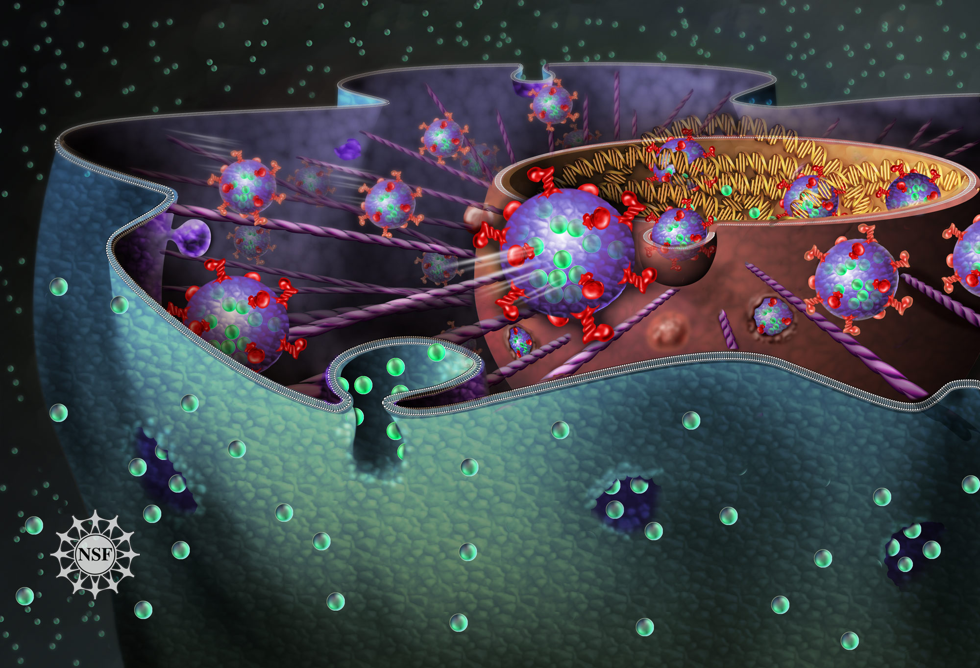
Endocytosis is a cellular process in which Chemical substance, substances are brought into the cell. The material to be internalized is surrounded by an area of cell membrane, which then buds off inside the cell to form a Vesicle (biology and chemistry), vesicle containing the ingested materials. Endocytosis includes pinocytosis (cell drinking) and phagocytosis (cell eating). It is a form of active transport. History The term was proposed by Christian de Duve, De Duve in 1963. Phagocytosis was discovered by Élie Metchnikoff in 1882. Pathways Endocytosis pathways can be subdivided into four categories: namely, receptor-mediated endocytosis (also known as clathrin-mediated endocytosis), caveolae, pinocytosis, and phagocytosis. * Clathrin-mediated endocytosis is mediated by the production of small (approx. 100 nm in diameter) vesicles that have a morphologically characteristic coat made up of the cytosolic protein clathrin. Clathrin-coated vesicles (CCVs) are found in vir ... [...More Info...] [...Related Items...] OR: [Wikipedia] [Google] [Baidu] |
Cytosis
-Cytosis is a suffix that either refers to certain aspects of cells ie cellular process or phenomenon or sometimes refers to predominance of certain type of cells. It essentially means "of the cell". Sometimes it may be shortened to -osis (necrosis, apoptosis) and may be related to some of the processes ending with -esis (eg diapedesis, or emperipolesis, cytokinesis) or similar suffixes. There are three main types of cytosis: endocytosis (into the cell), exocytosis (out of the cell), and transcytosis (through the cell, in and out). Etymology and pronunciation The word ''cytosis'' () uses combining forms of '' cyto-'' and '' -osis'', reflecting a cellular process. The term was coined by Novikoff in 1961. Processes related to subcellular entry or exit Endocytosis Endocytosis is when a cell absorbs a molecule, such as a protein, from outside the cell by engulfing it with the cell membrane. It is used by most cells, because many critical substances are large polar molecul ... [...More Info...] [...Related Items...] OR: [Wikipedia] [Google] [Baidu] |
Receptor-mediated Endocytosis
Receptor-mediated endocytosis (RME), also called clathrin-mediated endocytosis, is a process by which cells absorb metabolites, hormones, proteins – and in some cases viruses – by the inward budding of the plasma membrane (invagination). This process forms vesicles containing the absorbed substances and is strictly mediated by receptors on the surface of the cell. Only the receptor-specific substances can enter the cell through this process. Process Although receptors and their ligands can be brought into the cell through a few mechanisms (e.g. caveolin and lipid raft), clathrin-mediated endocytosis remains the best studied. Clathrin-mediated endocytosis of many receptor types begins with the ligands binding to receptors on the cell plasma membrane. The ligand and receptor will then recruit adaptor proteins and clathrin triskelions to the plasma membrane around where invagination will take place. Invagination of the plasma membrane then occurs, forming a clathrin-coated p ... [...More Info...] [...Related Items...] OR: [Wikipedia] [Google] [Baidu] |
Filopodia
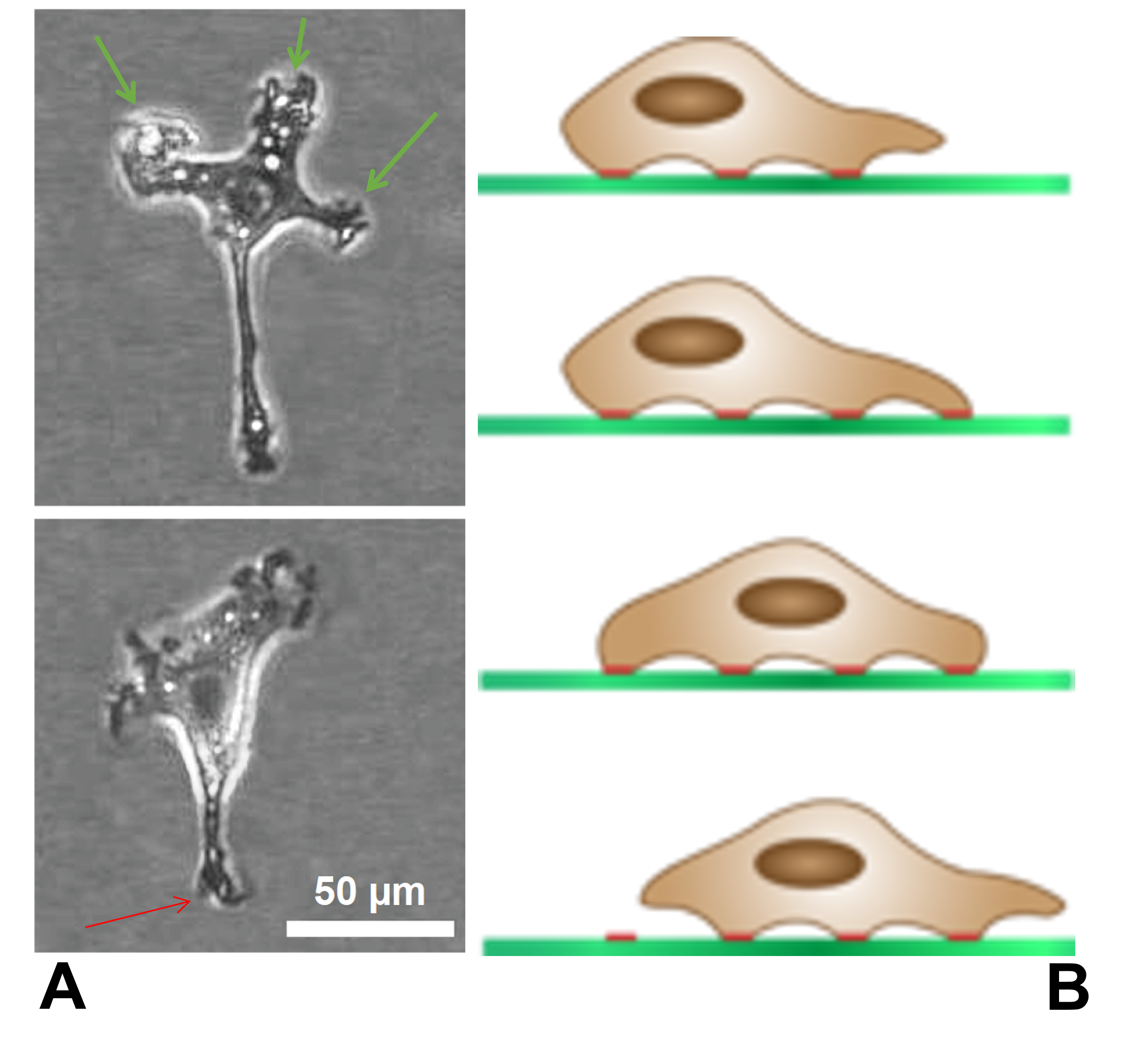
Filopodia (: filopodium) are slender cytoplasmic projections that extend beyond the leading edge of lamellipodia in migrating cells. Within the lamellipodium, actin ribs are known as ''microspikes'', and when they extend beyond the lamellipodia, they're known as filopodia. They contain microfilaments (also called actin filaments) cross-linked into bundles by actin-bundling proteins, such as fascin and fimbrin. Filopodia form focal adhesions with the substratum, linking them to the cell surface. Many types of migrating cells display filopodia, which are thought to be involved in both sensation of chemotropic cues, and resulting changes in directed locomotion. Activation of the Rho family of GTPases, particularly Cdc42 and their downstream intermediates, results in the polymerization of actin fibers by Ena/Vasp homology proteins. Growth factors bind to receptor tyrosine kinases resulting in the polymerization of actin filaments, which, when cross-linked, make up the support ... [...More Info...] [...Related Items...] OR: [Wikipedia] [Google] [Baidu] |
Warren Harmon Lewis
Warren Harmon Lewis (June 17, 1870 – July 3, 1964) was an American embryologist and cell biologist. He became professor of physiological anatomy at the Johns Hopkins University School of Medicine in 1913 and from 1919 to 1940, he worked along with his wife Margaret Reed Lewis at the Carnegie Institute of Washington. Life and work Lewis was born in Suffield to John Lewis and Adelaide Eunice Harmon. The family moved to Chicago where his father practiced law. Lewis went to Oak Park public schools and also attended the Chicago Manual Training School. He was interested in botany as a boy and collected plants. He joined the University of Michigan in 1890 where he studied zoology as also French, German and mathematics. He also took an interest in sports and music. In 1894 he obtained a microscope and was influenced by Jacob Reighard to study medicine. In 1896 he joined Johns Hopkins University in the school of medicine. He spent summers at the Marine Biological Laboratory at Woo ... [...More Info...] [...Related Items...] OR: [Wikipedia] [Google] [Baidu] |
Vacuole
A vacuole () is a membrane-bound organelle which is present in Plant cell, plant and Fungus, fungal Cell (biology), cells and some protist, animal, and bacterial cells. Vacuoles are essentially enclosed compartments which are filled with water containing inorganic and organic molecules including enzymes in Solutes, solution, though in certain cases they may contain solids which have been engulfed. Vacuoles are formed by the fusion of multiple membrane Vesicle (biology), vesicles and are effectively just larger forms of these. The organelle has no basic shape or size; its structure varies according to the requirements of the cell. Discovery Antonie van Leeuwenhoek described the plant vacuole in 1676. Contractile vacuoles ("stars") were first observed by Spallanzani (1776) in protozoa, although mistaken for respiratory organs. Félix Dujardin, Dujardin (1841) named these "stars" as ''vacuoles''. In 1842, Matthias Jakob Schleiden, Schleiden applied the term for plant cells, to dist ... [...More Info...] [...Related Items...] OR: [Wikipedia] [Google] [Baidu] |
Cell Membrane
The cell membrane (also known as the plasma membrane or cytoplasmic membrane, and historically referred to as the plasmalemma) is a biological membrane that separates and protects the interior of a cell from the outside environment (the extracellular space). The cell membrane consists of a lipid bilayer, made up of two layers of phospholipids with cholesterols (a lipid component) interspersed between them, maintaining appropriate membrane fluidity at various temperatures. The membrane also contains membrane proteins, including integral proteins that span the membrane and serve as membrane transporters, and peripheral proteins that loosely attach to the outer (peripheral) side of the cell membrane, acting as enzymes to facilitate interaction with the cell's environment. Glycolipids embedded in the outer lipid layer serve a similar purpose. The cell membrane controls the movement of substances in and out of a cell, being selectively permeable to ions and organic mole ... [...More Info...] [...Related Items...] OR: [Wikipedia] [Google] [Baidu] |
Cell (biology)
The cell is the basic structural and functional unit of all life, forms of life. Every cell consists of cytoplasm enclosed within a Cell membrane, membrane; many cells contain organelles, each with a specific function. The term comes from the Latin word meaning 'small room'. Most cells are only visible under a light microscope, microscope. Cells Abiogenesis, emerged on Earth about 4 billion years ago. All cells are capable of Self-replication, replication, protein synthesis, and cell motility, motility. Cells are broadly categorized into two types: eukaryotic cells, which possess a Cell nucleus, nucleus, and prokaryotic, prokaryotic cells, which lack a nucleus but have a nucleoid region. Prokaryotes are single-celled organisms such as bacteria, whereas eukaryotes can be either single-celled, such as amoebae, or multicellular organism, multicellular, such as some algae, plants, animals, and fungi. Eukaryotic cells contain organelles including Mitochondrion, mitochondria, which ... [...More Info...] [...Related Items...] OR: [Wikipedia] [Google] [Baidu] |
Extracellular Fluid
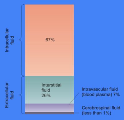
In cell biology, extracellular fluid (ECF) denotes all body fluid outside the cells of any multicellular organism. Total body water in healthy adults is about 50–60% (range 45 to 75%) of total body weight; women and the obese typically have a lower percentage than lean men. Extracellular fluid makes up about one-third of body fluid, the remaining two-thirds is intracellular fluid within cells. The main component of the extracellular fluid is the interstitial fluid that surrounds cells. Extracellular fluid is the internal environment of all multicellular animals, and in those animals with a blood circulatory system, a proportion of this fluid is blood plasma. Plasma and interstitial fluid are the two components that make up at least 97% of the ECF. Lymph makes up a small percentage of the interstitial fluid. The remaining small portion of the ECF includes the transcellular fluid (about 2.5%). The ECF can also be seen as having two components – plasma and lymph as a ... [...More Info...] [...Related Items...] OR: [Wikipedia] [Google] [Baidu] |
Lamellipodium
The lamellipodium (: lamellipodia) (from Latin ''lamella'', related to ', "thin sheet", and the Greek radical ''pod-'', "foot") is a cytoskeletal protein actin projection on the leading edge of the cell. It contains a quasi-two-dimensional actin mesh; the whole structure propels the cell across a substrate. Within the lamellipodia are ribs of actin called microspikes, which, when they spread beyond the lamellipodium frontier, are called filopodia. The lamellipodium is born of actin nucleation in the plasma membrane of the cell and is the primary area of actin incorporation or microfilament formation of the cell. Description Lamellipodia are found primarily in all mobile cells, such as the keratinocytes of fish and frogs, which are involved in the quick repair of wounds. The lamellipodia of these keratinocytes allow them to move at speeds of 10–20 μm / min over epithelial surfaces. When separated from the main part of a cell, a lamellipodium can still crawl a ... [...More Info...] [...Related Items...] OR: [Wikipedia] [Google] [Baidu] |


