|
Photosensitive Ganglion Cell
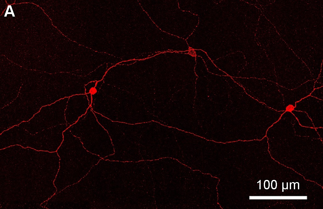
Intrinsically photosensitive retinal ganglion cells (ipRGCs), also called photosensitive retinal ganglion cells (pRGC), or melanopsin-containing retinal ganglion cells (mRGCs), are a type of neuron in the retina of the mammalian eye. The presence of an additional photoreceptor was first suspected in 1927 when mice lacking rod and cone cells still responded to changing light levels through pupil constriction; this suggested that rods and cones are not the only light-sensitive tissue. However, it was unclear whether this light sensitivity arose from an additional retinal photoreceptor or elsewhere in the body. Recent research has shown that these retinal ganglion cells, unlike other retinal ganglion cells, are intrinsically photosensitive due to the presence of melanopsin, a light-sensitive protein. Therefore, they constitute a third class of photoreceptors, in addition to rod and cone cells. Overview Compared to the rods and cones, the ipRGCs respond more sluggishly and ... [...More Info...] [...Related Items...] OR: [Wikipedia] [Google] [Baidu] |
Overview Of The Retina Photoreceptors (a)
Overview may refer to: * Overview article, an article that summarizes the current state of understanding on a topic * Overview map, generalised view of a geographic area See also * Summary (other) * Outline (list) * ''A Brief Overview'' * Overview and Scrutiny * Overview effect * * {{disambiguation ... [...More Info...] [...Related Items...] OR: [Wikipedia] [Google] [Baidu] |
Hypothalamus
The hypothalamus (: hypothalami; ) is a small part of the vertebrate brain that contains a number of nucleus (neuroanatomy), nuclei with a variety of functions. One of the most important functions is to link the nervous system to the endocrine system via the pituitary gland. The hypothalamus is located below the thalamus and is part of the limbic system. It forms the Basal (anatomy), basal part of the diencephalon. All vertebrate brains contain a hypothalamus. In humans, it is about the size of an Almond#Nut, almond. The hypothalamus has the function of regulating certain metabolic biological process, processes and other activities of the autonomic nervous system. It biosynthesis, synthesizes and secretes certain neurohormones, called releasing hormones or hypothalamic hormones, and these in turn stimulate or inhibit the secretion of hormones from the pituitary gland. The hypothalamus controls thermoregulation, body temperature, hunger (physiology), hunger, important aspects o ... [...More Info...] [...Related Items...] OR: [Wikipedia] [Google] [Baidu] |
Melanopsin
Melanopsin is a type of photopigment belonging to a larger family of light-sensitive retinylidene protein, retinal proteins called opsins and encoded by the gene ''Opn4''. In the mammalian retina, there are two additional categories of opsins, both involved in the formation of visual images: rhodopsin and photopsin (types I, II, and III) in the Rod cell, rod and Cone cell, cone photoreceptor cells, respectively. In humans, melanopsin is found in intrinsically photosensitive retinal ganglion cells (ipRGCs). It is also found in the iris of mice and primates. Melanopsin is also found in rats, amphioxus, and other chordates. ipRGCs are photoreceptor cells which are particularly sensitive to the absorption of short-wavelength (blue) visible light and communicate information directly to the area of the brain called the suprachiasmatic nucleus (SCN), also known as the central "body clock", in mammals. Melanopsin plays an important non-image-forming role in the Entrainment (chronobiology ... [...More Info...] [...Related Items...] OR: [Wikipedia] [Google] [Baidu] |
Lateral Geniculate Nucleus
In neuroanatomy, the lateral geniculate nucleus (LGN; also called the lateral geniculate body or lateral geniculate complex) is a structure in the thalamus and a key component of the mammalian visual pathway. It is a small, ovoid, Anatomical terms of location#Dorsal_and_ventral, ventral projection of the thalamus where the thalamus connects with the optic nerve. There are two LGNs, one on the left and another on the right side of the thalamus. In humans, both LGNs have six layers of neurons (grey matter) alternating with optic fibers (white matter). The LGN receives information directly from the ascending retinal ganglion cells via the optic tract and from the reticular activating system. Neurons of the LGN send their axons through the optic radiation, a direct pathway to the primary visual cortex. In addition, the LGN receives many strong feedback connections from the primary visual cortex. In humans as well as other mammals, the two strongest pathways linking the eye to the bra ... [...More Info...] [...Related Items...] OR: [Wikipedia] [Google] [Baidu] |
Superior Colliculus
In neuroanatomy, the superior colliculus () is a structure lying on the tectum, roof of the mammalian midbrain. In non-mammalian vertebrates, the Homology (biology), homologous structure is known as the optic tectum or optic lobe. The adjective form ''tectum, tectal'' is commonly used for both structures. In mammals, the superior colliculus forms a major component of the midbrain. It is a paired structure and together with the paired inferior colliculi forms the corpora quadrigemina. The superior colliculus is a layered structure, with a pattern that is similar in all mammals. The layers can be grouped into the superficial layers (retinal nerve fiber layer, stratum opticum and above) and the deeper remaining layers. Neurons in the superficial layers receive direct input from the retina and respond almost exclusively to visual stimuli. Many neurons in the deeper layers also respond to other modalities, and some respond to stimuli in multiple modalities. The deeper layers also conta ... [...More Info...] [...Related Items...] OR: [Wikipedia] [Google] [Baidu] |
Rhabdom
The compound eyes of arthropods like insects, crustaceans and millipedes are composed of units called ommatidia (: ommatidium). An ommatidium contains a cluster of photoreceptor cells surrounded by support cells and pigment cells. The outer part of the ommatidium is overlaid with a transparent cornea. Each ommatidium is innervated by one axon bundle (usually consisting of 6–9 axons, depending on the number of #rhabdomere , rhabdomeres) and provides the brain with one picture element. The brain forms an image from these independent picture elements. The number of ommatidia in the eye depends upon the type of arthropod and range from as low as five as in the Antarctic isopod ''Glyptonotus antarcticus'', or a handful in the primitive Zygentoma, to around 30,000 in larger Anisoptera dragonflies and some Sphingidae moths. Description Ommatidia are typically hexagonal in cross-section and approximately ten times longer than wide. The diameter is largest at the surface, tapering towar ... [...More Info...] [...Related Items...] OR: [Wikipedia] [Google] [Baidu] |
Phototransduction
Visual phototransduction is the sensory transduction process of the visual system by which light is detected by photoreceptor cells ( rods and cones) in the vertebrate retina. A photon is absorbed by a retinal chromophore (each bound to an opsin), which initiates a signal cascade through several intermediate cells, then through the retinal ganglion cells (RGCs) comprising the optic nerve. Overview Light enters the eye, passes through the optical media, then the inner neural layers of the retina before finally reaching the photoreceptor cells in the outer layer of the retina. The light may be absorbed by a chromophore bound to an opsin, which photoisomerizes the chromophore, initiating both the visual cycle, which "resets" the chromophore, and the phototransduction cascade, which transmits the visual signal to the brain. The cascade begins with graded polarisation (an analog signal) of the excited photoreceptor cell, as its membrane potential increases from a resting potential of - ... [...More Info...] [...Related Items...] OR: [Wikipedia] [Google] [Baidu] |
Farhan H
Farhan or Farhaan is an Arabic name which means "happy, joyful, blessed, delightful, rejoicing, merry, inclined to hopefulness". The name is the male variant from the female stem given name Farah, and is widely used in West Asia, North Africa and South Asia. It is a variant of Farhang as well. Notable people with these names include: Farhan *Farhan Akhtar (born 1974), Indian film director, screenwriter, producer, actor, playback singer and television host * Farhan Khan (actor) (born 1983), Indian actor *Farhan Lalji, Canadian sports reporter * Farhan Nizami, South Asian historian *Farhan Saeed (born 1984), Pakistani singer-songwriter and actor * Farhan Saleh (born 1947), Lebanese writer and militant *Farhan Zaidi (born 1976), Canadian-American baseball executive of Pakistani descent *Farhan Alanazi Farhan or Farhaan is an Arabic name which means "happy, joyful, blessed, delightful, rejoicing, merry, inclined to hopefulness". The name is the male variant from the female stem giv ... [...More Info...] [...Related Items...] OR: [Wikipedia] [Google] [Baidu] |
Melatonin
Melatonin, an indoleamine, is a natural compound produced by various organisms, including bacteria and eukaryotes. Its discovery in 1958 by Aaron B. Lerner and colleagues stemmed from the isolation of a substance from the pineal gland of cows that could induce skin lightening in common frogs. This compound was later identified as a hormone secreted in the brain during the night, playing a crucial role in regulating the sleep-wake cycle, also known as the circadian rhythm, in vertebrates. In vertebrates, melatonin's functions extend to Entrainment (chronobiology), synchronizing sleep-wake cycles, encompassing sleep-wake timing and blood pressure regulation, as well as controlling seasonal rhythmicity (circannual cycle), which includes reproduction, fattening, molting, and hibernation. Its effects are mediated through the activation of melatonin receptors and its role as an antioxidant. In plants and bacteria, melatonin primarily serves as a defense mechanism against oxidative ... [...More Info...] [...Related Items...] OR: [Wikipedia] [Google] [Baidu] |
Pupil
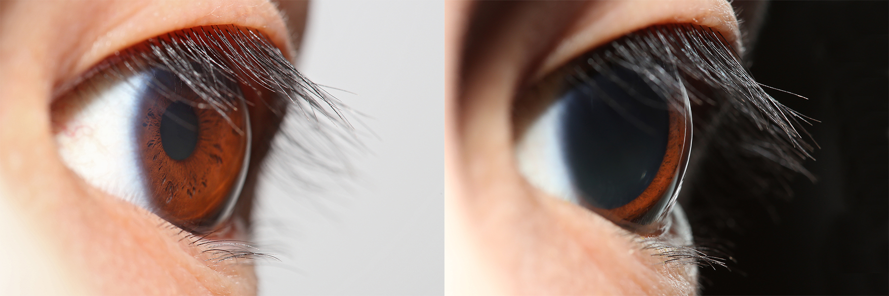
The pupil is a hole located in the center of the iris of the eye that allows light to strike the retina.Cassin, B. and Solomon, S. (1990) ''Dictionary of Eye Terminology''. Gainesville, Florida: Triad Publishing Company. It appears black because light rays entering the pupil are either absorbed by the tissues inside the eye directly, or absorbed after diffuse reflections within the eye that mostly miss exiting the narrow pupil. The size of the pupil is controlled by the iris, and varies depending on many factors, the most significant being the amount of light in the environment. The term "pupil" was coined by Gerard of Cremona. In humans, the pupil is circular, but its shape varies between species; some cats, reptiles, and foxes have vertical slit pupils, goats and sheep have horizontally oriented pupils, and some catfish have annular types. In optical terms, the anatomical pupil is the eye's aperture and the iris is the aperture stop. The image of the pupil as seen from o ... [...More Info...] [...Related Items...] OR: [Wikipedia] [Google] [Baidu] |
Midbrain
The midbrain or mesencephalon is the uppermost portion of the brainstem connecting the diencephalon and cerebrum with the pons. It consists of the cerebral peduncles, tegmentum, and tectum. It is functionally associated with vision, hearing, motor control, sleep and wakefulness, arousal (alertness), and temperature regulation.Breedlove, Watson, & Rosenzweig. Biological Psychology, 6th Edition, 2010, pp. 45-46 The name ''mesencephalon'' comes from the Greek ''mesos'', "middle", and ''enkephalos'', "brain". Structure The midbrain is the shortest segment of the brainstem, measuring less than 2cm in length. It is situated mostly in the posterior cranial fossa, with its superior part extending above the tentorial notch. The principal regions of the midbrain are the tectum, the cerebral aqueduct, tegmentum, and the cerebral peduncles. Rostral and caudal, Rostrally the midbrain adjoins the diencephalon (thalamus, hypothalamus, etc.), while Rostral and caudal, cau ... [...More Info...] [...Related Items...] OR: [Wikipedia] [Google] [Baidu] |
Olivary Pretectal Nucleus
In neuroanatomy, the pretectal area, or pretectum, is a midbrain structure composed of seven nuclei and comprises part of the subcortical visual system. Through reciprocal bilateral projections from the retina, it is involved primarily in mediating behavioral responses to acute changes in ambient light such as the pupillary light reflex, the optokinetic reflex, and temporary changes to the circadian rhythm. In addition to the pretectum's role in the visual system, the anterior pretectal nucleus has been found to mediate somatosensory and nociceptive information. Location and structure The pretectum is a bilateral group of highly interconnected nuclei located near the junction of the midbrain and forebrain. The pretectum is generally classified as a midbrain structure, although because of its proximity to the forebrain it is sometimes classified as part of the caudal diencephalon (forebrain). Within vertebrates, the pretectum is located directly anterior to the superior colliculus ... [...More Info...] [...Related Items...] OR: [Wikipedia] [Google] [Baidu] |


