|
Philip Leverhulme Equine Hospital
The University of Liverpool School of Veterinary Science was the first veterinary school in the United Kingdom to be incorporated into a university. The school's teaching, treatment and research facilities are on the main campus and at Leahurst on the Wirral Peninsula, approximately 12 miles outside Liverpool. History Foundation The foundations for the vet school at Liverpool were laid in the early 1900s when William Owen Williams, principal of the now-defunct New Veterinary College in Edinburgh (not to be confused with the Royal (Dick) Vet School, with which it was in competition), was invited to transfer his institution to Liverpool. The emerging science of veterinary medicine was of particular relevance both to the busy port city itself, which depended upon heavy horses to drive its docks and associated industry, and to the economy of the surrounding countryside, which at the time boasted the highest stocking density of cattle in the UK. Initially, there was conside ... [...More Info...] [...Related Items...] OR: [Wikipedia] [Google] [Baidu] |
Public University
A public university or public college is a university or college that is in state ownership, owned by the state or receives significant government spending, public funds through a national or subnational government, as opposed to a private university. Whether a national university is considered public varies from one country (or region) to another, largely depending on the specific education landscape. Africa Egypt In Egypt, Al-Azhar University was founded in 970 AD as a madrasa; it formally became a public university in 1961 and is one of the oldest institutions of higher education in the world. In the 20th century, Egypt opened many other public universities with government-subsidized tuition fees, including Cairo University in 1908, Alexandria University in 1912, Assiut University in 1928, Ain Shams University in 1957, Helwan University in 1959, Beni-Suef University in 1963, Zagazig University in 1974, Benha University in 1976, and Suez Canal University in 1989. Kenya ... [...More Info...] [...Related Items...] OR: [Wikipedia] [Google] [Baidu] |
Equine
Equinae is a subfamily of the family Equidae, which have lived worldwide (except Indonesia and Australia) from the Hemingfordian stage of the Early Miocene (16 million years ago) onwards. They are thought to be a monophyletic grouping.B. J. MacFadden. 1998. Equidae. In C. M. Janis, K. M. Scott, and L. L. Jacobs (eds.), Evolution of Tertiary Mammals of North America Members of the subfamily are referred to as equines; the only extant equines are the horses, asses, and zebras of the genus ''Equus''. The subfamily contains two tribes, the Equini and the Hipparionini, as well as two unplaced genera, ''Merychippus'' and ''Scaphohippus''. Sister taxa * Anchitheriinae * Hyracotheriinae ''Hyracotherium'' ( ; " hyrax-like beast") is an extinct genus of very small (about 60 cm in length) perissodactyl ungulates that was found in the London Clay formation. This small, fox-sized animal was once considered to be the earliest kn ... References Miocene horses Pliocene odd-t ... [...More Info...] [...Related Items...] OR: [Wikipedia] [Google] [Baidu] |
Medical Ultrasonography
Medical ultrasound includes diagnostic techniques (mainly imaging techniques) using ultrasound, as well as therapeutic applications of ultrasound. In diagnosis, it is used to create an image of internal body structures such as tendons, muscles, joints, blood vessels, and internal organs, to measure some characteristics (e.g. distances and velocities) or to generate an informative audible sound. Its aim is usually to find a source of disease or to exclude pathology. The usage of ultrasound to produce visual images for medicine is called medical ultrasonography or simply sonography. The practice of examining pregnant women using ultrasound is called obstetric ultrasonography, and was an early development of clinical ultrasonography. Ultrasound is composed of sound waves with frequencies which are significantly higher than the range of human hearing (>20,000 Hz). Ultrasonic images, also known as sonograms, are created by sending pulses of ultrasound into tissue usin ... [...More Info...] [...Related Items...] OR: [Wikipedia] [Google] [Baidu] |
Magnetic Resonance Imaging
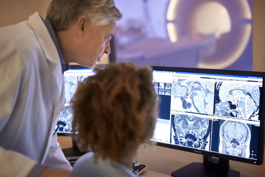
Magnetic resonance imaging (MRI) is a medical imaging technique used in radiology to form pictures of the anatomy and the physiological processes inside the body. MRI scanners use strong magnetic fields, magnetic field gradients, and radio waves to generate images of the organs in the body. MRI does not involve X-rays or the use of ionizing radiation, which distinguishes it from computed tomography (CT) and positron emission tomography (PET) scans. MRI is a medical application of nuclear magnetic resonance (NMR) which can also be used for imaging in other NMR applications, such as NMR spectroscopy. MRI is widely used in hospitals and clinics for medical diagnosis, staging and follow-up of disease. Compared to CT, MRI provides better contrast in images of soft tissues, e.g. in the brain or abdomen. However, it may be perceived as less comfortable by patients, due to the usually longer and louder measurements with the subject in a long, confining tube, although "open ... [...More Info...] [...Related Items...] OR: [Wikipedia] [Google] [Baidu] |
X-ray Computed Tomography
X-rays (or rarely, ''X-radiation'') are a form of high-energy electromagnetic radiation. In many languages, it is referred to as Röntgen radiation, after the German scientist Wilhelm Conrad Röntgen, who discovered it in 1895 and named it ''X-radiation'' to signify an unknown type of radiation.Novelline, Robert (1997). ''Squire's Fundamentals of Radiology''. Harvard University Press. 5th edition. . X-ray wavelengths are shorter than those of ultraviolet rays and longer than those of gamma rays. There is no universally accepted, strict definition of the bounds of the X-ray band. Roughly, X-rays have a wavelength ranging from 10 nanometers to 10 picometers, corresponding to frequencies in the range of 30 petahertz to 30 exahertz ( to ) and photon energies in the range of 100 eV to 100 keV, respectively. X-rays can penetrate many solid substances such as construction materials and living tissue, so X-ray radiography is widely used in medica ... [...More Info...] [...Related Items...] OR: [Wikipedia] [Google] [Baidu] |
Nuclear Medicine
Nuclear medicine or nucleology is a medical specialty involving the application of radioactive substances in the diagnosis and treatment of disease. Nuclear imaging, in a sense, is "radiology done inside out" because it records radiation emitting from within the body rather than radiation that is generated by external sources like X-rays. In addition, nuclear medicine scans differ from radiology, as the emphasis is not on imaging anatomy, but on the function. For such reason, it is called a physiological imaging modality. Single photon emission computed tomography (SPECT) and positron emission tomography (PET) scans are the two most common imaging modalities in nuclear medicine. Diagnostic medical imaging Diagnostic In nuclear medicine imaging, radiopharmaceuticals are taken internally, for example, through inhalation, intravenously or orally. Then, external detectors ( gamma cameras) capture and form images from the radiation emitted by the radiopharmaceuticals. This ... [...More Info...] [...Related Items...] OR: [Wikipedia] [Google] [Baidu] |
Radiology
Radiology ( ) is the medical discipline that uses medical imaging to diagnose diseases and guide their treatment, within the bodies of humans and other animals. It began with radiography (which is why its name has a root referring to radiation), but today it includes all imaging modalities, including those that use no electromagnetic radiation (such as ultrasonography and magnetic resonance imaging), as well as others that do, such as computed tomography (CT), fluoroscopy, and nuclear medicine including positron emission tomography (PET). Interventional radiology is the performance of usually minimally invasive medical procedures with the guidance of imaging technologies such as those mentioned above. The modern practice of radiology involves several different healthcare professions working as a team. The radiologist is a medical doctor who has completed the appropriate post-graduate training and interprets medical images, communicates these findings to other physicians b ... [...More Info...] [...Related Items...] OR: [Wikipedia] [Google] [Baidu] |
Digital X-ray
Digital radiography is a form of radiography that uses x-ray–sensitive plates to directly capture data during the patient examination, immediately transferring it to a computer system without the use of an intermediate cassette. Advantages include time efficiency through bypassing chemical processing and the ability to digitally transfer and enhance images. Also, less radiation can be used to produce an image of similar contrast to conventional radiography. Instead of X-ray film, digital radiography uses a digital image capture device. This gives advantages of immediate image preview and availability; elimination of costly film processing steps; a wider dynamic range, which makes it more forgiving for over- and under-exposure; as well as the ability to apply special image processing techniques that enhance overall display quality of the image. Detectors Flat panel detectors 250px, Flat panel detector used in digital radiography Flat panel detectors (FPDs) are the most common ... [...More Info...] [...Related Items...] OR: [Wikipedia] [Google] [Baidu] |
Ovariectomy
Oophorectomy (; from Greek , , 'egg-bearing' and , , 'a cutting out of'), historically also called ''ovariotomy'' is the surgical removal of an ovary or ovaries. The surgery is also called ovariectomy, but this term is mostly used in reference to animals, e.g. the surgical removal of ovaries from laboratory animals. Removal of the ovaries of females is the biological equivalent of castration of males; the term ''castration'' is only occasionally used in the medical literature to refer to oophorectomy of women. In veterinary medicine, the removal of ovaries and uterus is called ovariohysterectomy ( spaying) and is a form of sterilization. The first reported successful human oophorectomy was carried out by (Sir) Sydney Jones at Sydney Infirmary, Australia, in 1870. Partial oophorectomy or ovariotomy is a term sometimes used to describe a variety of surgeries such as ovarian cyst removal, or resection of parts of the ovaries. This kind of surgery is fertility-preserving, alth ... [...More Info...] [...Related Items...] OR: [Wikipedia] [Google] [Baidu] |
Horse Colic
Colic in horses is defined as abdominal pain, but it is a clinical symptom rather than a diagnosis. The term colic can encompass all forms of gastrointestinal conditions which cause pain as well as other causes of abdominal pain not involving the gastrointestinal tract. The most common forms of colic are gastrointestinal in nature and are most often related to colonic disturbance. There are a variety of different causes of colic, some of which can prove fatal without surgical intervention. Colic surgery is usually an expensive procedure as it is major abdominal surgery, often with intensive aftercare. Among domesticated horses, colic is the leading cause of premature death. The incidence of colic in the general horse population has been estimated between 4 and 10 percent over the course of the average lifespan. Clinical signs of colic generally require treatment by a veterinarian. The conditions that cause colic can become life-threatening in a short period of time. Pathophysiolo ... [...More Info...] [...Related Items...] OR: [Wikipedia] [Google] [Baidu] |
Laparoscopic Surgery
Laparoscopy () is an operation performed in the abdomen or pelvis using small incisions (usually 0.5–1.5 cm) with the aid of a camera. The laparoscope aids diagnosis or therapeutic interventions with a few small cuts in the abdomen.MedlinePlus > Laparoscopy Update Date: 21 August 2009. Updated by: James Lee, MD // No longer valid Laparoscopic surgery, also called minimally invasive procedure, bandaid surgery, or keyhole surgery, is a modern surgical technique. There are a number of advantages to the patient with laparoscopic surgery versus an exploratory laparotomy. These include reduced pain due to smaller incisions, reduced hemorrhaging, and shorter recovery time. The key element is the use of a laparoscope, a long fiber optic cable system that allows viewing of the affected area by snaking the cable from a more distant, but more easily accessible location. Laparoscopic surgery includes operations within the abdominal or pelvic cavities, whereas keyhole surgery p ... [...More Info...] [...Related Items...] OR: [Wikipedia] [Google] [Baidu] |
Neurology
Neurology (from el, νεῦρον (neûron), "string, nerve" and the suffix -logia, "study of") is the branch of medicine dealing with the diagnosis and treatment of all categories of conditions and disease involving the brain, the spinal cord and the peripheral nerves. Neurological practice relies heavily on the field of neuroscience, the scientific study of the nervous system. A neurologist is a physician specializing in neurology and trained to investigate, diagnose and treat neurological disorders. Neurologists treat a myriad of neurologic conditions, including stroke, seizures, movement disorders such as Parkinson's disease, autoimmune neurologic disorders such as multiple sclerosis, headache disorders like migraine and dementias such as Alzheimer's disease. Neurologists may also be involved in clinical research, clinical trials, and basic or translational research. While neurology is a nonsurgical specialty, its corresponding surgical specialty is neurosurgery ... [...More Info...] [...Related Items...] OR: [Wikipedia] [Google] [Baidu] |


